Congenital scoliosis in non-identical twins:
case reports and literature review
Dean Greenwood,
DC, MS, D.A.C.O.1William Bogar,
DC, D.A.C.B.R.21 Private practice of Chiropractic
Vancouver Spine Care Centre
102-1678 W. Broadway, Vancouver, BC, Canada, V6J 1X6
2 Chief of Diagnostic Imaging and Residency
National University of Health Sciences
200 East Roosevelt Rd, Lombard, Illinois, USA, 60146 Corresponding author: Dr. Dean Greenwood
Email: [email protected] Phone: 778-986-6029 ©JCCA 2014
Congenital scoliosis due to vertebral anomalies may occur in less than 0.1% of the population. Several different theories have been put forth in the literature to account for the etiology of congenital scoliosis and the vertebral anomalies which contribute to its development. The study of scoliosis in twins has contributed to the understanding of causative factors including genetics, environment and in utero events during embryologic development. Case reports of fraternal (non-identical) juvenile male twins with congenital scoliosis associated with differing congenital vertebral anomalies are presented. Both children were asymptomatic at the time of the initial consultation and showed no signs of neurologic compromise. Rapidly progressive, severe genetic scoliosis requires prudent observation and referral to a pediatric orthopedic surgeon to determine appropriate options for care and to screen for potentially life threatening disorders. Chiropractors may be seen as
La scoliose congénitale due à des anomalies vertébrales peut se produire chez moins de 0,1 % de la population. Plusieurs théories différentes ont été avancées dans la recherche scientifique pour expliquer l’étiologie de la scoliose congénitale et les anomalies vertébrales qui contribuent à son développement. L’étude de la scoliose chez les jumeaux a contribué à la compréhension des facteurs étiologiques, dont la génétique,
l’environnement, et les événements in utero au cours du développement embryonnaire. On présente des rapports de cas de frères jumeaux (non identiques) mineurs atteints de scoliose congénitale associée à différentes anomalies vertébrales congénitales. Les deux enfants étaient asymptomatiques au moment de la consultation initiale et n’ont montré aucun signe d’atteinte
Introduction
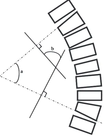
Scoliosis is a lateral curvature and twisting of the spine measuring 10 degrees or more. The Cobb method of men-suration determines the degree of scoliotic curvature by the angle created between lines drawn on endplates of the end vertebrae (superior endplate of upper end vertebra; inferior end plate of lower end vertebra). (Figure 1) This method has been adopted and standardized by the Scolio-sis Research Society, which also classifies the severity of scoliosis. (Table 1) The Nash and Moe method measures vertebral rotation on a frontal radiograph using the
dis-placement of the pedicle on the vertebral body. (Figure 2) Juvenile idiopathic scoliosis is defined as a spinal curva-ture diagnosed between 3 years and 9 years 11 months of age, whereas congenital scoliosis is associated with bony abnormalities of the spine present at birth.1 Juvenile onset
scoliosis has been reported to account for 8% to 21% of patients with scoliosis, although these numbers are based on studies with small numbers of participants and may not be statistically accurate.2,3 The incidence of
congen-ital scoliosis in the juvenile population is unknown since many spinal anomalies go undetected due to the presence
gatekeepers for scoliosis and a thorough understanding of appropriate standards of care is required.
(JCCA. 2014;58(3):291-299)
k e y w o r d s : scoliosis, twins, congenital, hemivertebra, chiropractic
une connaissance approfondie des normes appropriées de soins est nécessaire.
(JCCA. 2014;58(3):291-299)
m o t s c l é s : scoliose, jumeaux, congénital, hémivertèbre, chiropratique
Figure 1. Cobb method of scoliosis mensuration
(Reproduced with permission of the Radiological Society of North America (RSNA). Kim H, Kim HS, Moon ES et al. Scoliosis imaging: What radiologists should know. RadioGraphics. 2010;30:1823-1842)
Table 1.
Lippman-Cobb Classification of Scoliotic Curvature
Group Angle of Curvature in Degrees
I <20
II 21-30
III 31-50
IV 51-75
V 76-10O
VI 101-125
VII >125
Figure 2. Nash and Moe pedicle method for determining
vertebral rotation
(Reproduced with permission of the Radiological Society of North America (RSNA). Kim H, Kim HS, Moon ES et
of minimal spinal deformity. The incidence of vertebral anomalies has been estimated to be 0.05-0.1% of live births.4 Several theories exist as to the etiology of
congen-ital scoliosis. The overall impression among researchers is that the cause is multifactoral. 5-7
These case reports detail the presentation of a pair of fra-ternal juvenile twins with dissimilar scoliotic curve char-acteristics in a private chiropractic practice. A literature review was conducted to appreciate the etiology of con-genital and juvenile onset scoliosis particularly in twins, as well as to briefly outline current standards of care.
Case Report
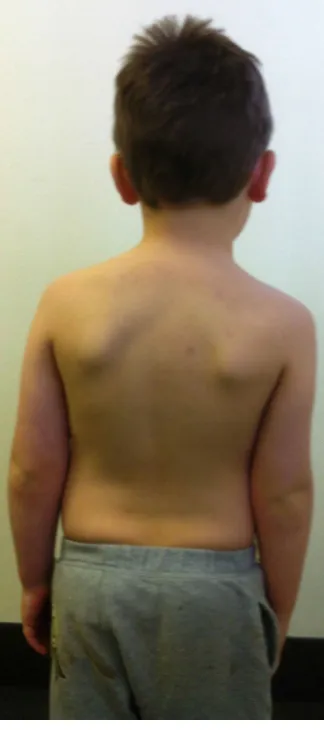
Twin males aged 5 years 9 months presented to a private chiropractic office on referral from the family medical physician for evaluation of juvenile scoliosis. They were escorted by their birth mother who was interviewed with respect to family history, birth history and the twin’s hist-ory to date. At the time of this presentation the twins were asymptomatic, apart from visible signs of truncal asym-metry and postural imbalance. Twin A was 119.5 cm tall and weighed 24.5 kg, while twin B measured 115.5 cm in height and weighed 21.8 kg. Both boys were of nor-Figure 3A.
Photograph of Twin A at 5 years 9 months
Figure 3B.
Twin A-AP standing radiograph at 4 years 11 months. 10 degree right lumbar scoliosis with mild compensatory left thoracic curve associated with decreased vertical interpediculate distance between the left L4 and left L5 pedicles as compared to the corresponding pedicles on the right, and low right hemipelvis
Figure 3C.
mal intellectual development for age and were able to fol-low directions when asked. They were active throughout the interview and examination and appeared to possess equal strength, co-ordination and physical ability. During standing postural assessment, twin A demonstrated pel-vic and shoulder unlevelling, lower on the right side, with left head tilt. (Figure 3A) No rib humping was observed during forward trunk flexion (Adam’s test). Twin B dem-onstrated trunk rotation left posterior and right head tilt (Figure 4A). Slight left lower rib humping was evident on forward trunk flexion. Lateral bending and extension
ranges were tested in twin A and were found to be within normal limits, while twin B exhibited mild segmental re-striction in left lateral bending at the thoraco-lumbar junc-tion. No pain was elicited during examination in either twin. Babinski sign was absent when tested, and gait and balance was normal in both twins.
The mother’s family history is unremarkable for scoli-osis, congenital spinal anomaly or other serious disease. She has two other older female children, neither having any evidence of scoliosis. She denies smoking, drug use, or illness prior to conception or during pregnancy. The Figure 4A.
Photograph of Twin B at 5 years 9 months
Figure 4B.
Twin B AP standing radiograph at 22 months
Hemivertebra at T10 with a 30 degree right thoraco-lumbar scoliosis
Figure 4C.
father’s family history is also negative for scoliosis or spinal anomaly, and there is no consanguinity with the twin’s mother. The boys were full term at delivery and were determined to be fraternal, not identical twins. At birth, twin A was 4.2 kg and 52.5 cm in length while twin B was 3.7 kg and 54 cm in length. They were considered to be very large babies at birth for twins, and gestational diabetes was ruled out as a contributing factor to their size. The twin’s siblings were also born with similarly high birth weights.
The mother was questioned about her health and life-style during the early stages of pregnancy with the twins. She recalled that she had been breast feeding her second child and had been menstruating for one year following a one year absence of her monthly cycle following the delivery of this child. She normally has a 6-8 week men-strual cycle and was unaware that she was pregnant until approximately 6 weeks of gestation. She had been exer-cising regularly and enjoyed very good health during this time. She was not aware that she was carrying twins until 5 months of gestation.
Radiographs of the twins were provided and reviewed during consultation. Twin A had a single AP standing radiograph taken at 4 years 11 months of age which dem-onstrated a 10 degree right lumbar scoliosis measured using the Cobb method between L2 and L5. There was also a mild compensatory left thoracic curve present. (Figure 3B) This view also demonstrated pelvic unlevel-ling, lower on the right which was possibly associated with a lower limb discrepancy due to a shorter right leg. A decreased vertical interpedicular distance was noted at L4-5 suggestive of a failure of segmentation. A lateral lumbar film was recommended and taken at a later time to confirm this anomaly. (Figure 3C) An independent chiro-practic radiologist was consulted to review these films. The anomaly was reported as “L4 hypoplastic facet de-velopment with anterolisthesis of L4 on L5”. “The find-ings suggest congenital etiology scoliosis”. (Addendum) Twin B had serial AP standing radiographs taken at 22 months and again at 4 years 11 months. The initial view demonstrated a hemivertebra at T-10 with incomplete de-velopment of the right side of this anomalous segment. This was associated with a 30 degree right thoraco-lumbar scoliosis measured using the Cobb method between T12 and L4 which had developed below the anomaly. (Fig-ure 4B) The second radiograph demonstrated progression
of the thoraco-lumbar scoliosis to 35 degrees associated with the hemivertebra. (Figure 4C)
Discussion
The study of scoliosis in twins, particularly adolescent idiopathic scoliosis is well documented. 8-21. Grauers et
al published their findings on “Heritability of Scoliosis” following a survey of 64,578 twins in the Swedish twin registry and concluded that genetic factors were respon-sible for 38% of scoliosis cases as compared to 62% en-vironmental association with the development of scolio-sis. This study also concluded that in monozygotic twins, concordance for idiopathic scoliosis is much higher than in same sex dizygotic twins.22 There were three studies
published regarding juvenile scoliosis in twins.23-25
Congenital scoliosis is a lateral curvature of the spine associated with vertebral anomalies such as block verte-bra, wedge verteverte-bra, single hemiverteverte-bra, two unilateral hemivertebrae, a unilateral unsegmented bar, or a unilat-eral unsegmented bar with contralatunilat-eral hemivertebrae at the same level. These represent the five classifications of vertebral anomaly as described by McMaster and Ohts-uka.26 A single hemivertebra, which is classified as a
fail-ure of formation is a common vertebral anomaly found in congenital scoliosis and depending on the location, will contribute to scoliotic progression in the growing child. A fully segmented hemivertebra may be associated with rapid scoliotic progression and may be resistant to conservative management.27-31Genetic signaling in the
embryological development stage of somitogenesis, as well as temporary vascular insufficiency of the growing fetus may contribute to failure of ossification of a vertebra or vertebrae, osseous metaplasia of the annulus fibrosus or persistent notochord. These proposed theories in the formation of vertebral anomalies, such as hemivertebrae and other structural malformations suggest a role for en-vironmental and genetic contributors.32-37
Juvenile scoliosis is unique in that progression may occur during a period of time where growth is dor-mant.2,36,37 Progression of the spinal curvature may
However some patients within these studies showed gradual regression of their curves with time as they ap-proached puberty, further confounding the theories of the natural course of scoliotic progression and complicating clinical decision making with respect to treatment.40,41
Unrecognized physical activity, particularly in male pa-tients may lead to curve regression. Symmetric loading of vertebral structures during weight bearing exercise may be a contributing factor in gradual regression. Prepubes-cent curve regression is not a guarantee that a scoliotic curve will not progress rapidly during adolescence.42,43
Congenital scoliosis due to hemivertebra is more likely to show rapid progression at a younger age than juvenile idiopathic scoliosis and a referral for a surgical opinion at an early age is necessary.30,31,42 Anomalies of the
neuro-logic or visceral structures, especially of the genitourin-ary system, may also occur when errors of formation or segmentation of the spine exist. During the fifth week of embryonic development, the vertebral column and the genitourinary system may be subject to embryonic insult which could lead to abnormalities.44,45 This may present
challenges when considering surgical intervention to minimize the progression of scoliosis. A complete evalua-tion of the surgical candidate with vertebral anomalies, including spinal and abdominal MRI, as well as diag-nostic ultrasound and occasionally voiding cystourethro-grams may be necessary to minimize surgical risk as well as to diagnose and treat potentially life threatening disor-ders.46-49 Neither Twin A or Twin B has yet been assessed
for other developmental anomalies.
Winter and Lonstein published a retrospective case series of 1250 patients with congenital spinal deform-ities and found that only seven patients with scoliosis secondary to a hemivertebra showed gradual improve-ment without treatimprove-ment. This is not a favorable prognosis for children with congenital scoliosis due to a vertebral anomaly and points to the importance of identifying and determining the classification of the anomaly at the earli-est possible age. One of the children in Winter and Lon-stein’s study was a twin with a hemivertebra at L1 while his twin brother had no vertebral anomaly. The child with hemivertebra was followed from age 15 months to age 16 years and showed a reduction of the spinal curvature from 42 degrees to 31 degrees without intervention, and was asymptomatic at all times.50
Non-identical twins are dichorionic, diamniotic twins
and the differences in their genetic makeup would be sim-ilar to siblings born as a result of pregnancies separated by time. However, the twins in these case studies offer a unique opportunity to observe the progression of this challenging clinical condition for the practicing chiro-practor. These case studies and review of the literature suggest that while there is an understanding of the etiol-ogy of scoliosis, accurate prognosis cannot be made on a case by case basis as to the likelihood of progression or regression of scoliosis. There are many and varied contributing factors during fetal development and during childhood which affect the progression of scoliosis. It is incumbent on the practitioner monitoring patients with scoliosis to diligently follow and to carefully observe subtle changes that may foretell progression and to make clinical decisions regarding appropriate standards of care. Examinations should be conducted at 2-3 to 36-60 months intervals according to the specific clinical situation, and standing frontal full spine radiographs including the oc-ciput and pelvis should be obtained when progression is apparent.51 A scoliometer, which is a variant of a
carpen-ter’s level, can be incorporated in the examination process to measure the severity of the rib hump and lumbar bulge. Raster stereography may also be considered to document the shape of the spine using reflected light beams without the use of ionizing radiation.52
Pediatric orthopedic referral would be the most pru-dent course of action for twin B with congenital scoliosis. Hemivertebra resection and transpedicular instrumenta-tion or contralateral hemiepiphysiodesis are opinstrumenta-tions for surgical intervention in young children and should be per-formed early to prevent severe local deformities and sec-ondary structural changes. Adequate post surgical bracing is essential to prevent failure of instrumentation, which has been reported as a frequent occurrence. 28,29,31,40,43-47
Twin A shows signs of a mild congenital scoliosis as-sociated with a subtle vertebral anomaly. Some evidence in the literature supports the use of chiropractic spinal manipulation and rehabilitative exercises for the manage-ment of scoliosis, although long term trials have not yet been conducted.53-56 None of the studies cited were
Limitations
These case studies represent one pair of non-identical twins.
Conclusion
The twins in these case studies were both born with con-genital vertebral anomalies which contribute to dissimilar scoliotic characteristics. Referral and screening for pot-entially life threatening disorders including other CNS or genitourinary anomalies as well as potential referral for surgical intervention is important. By gaining a better understanding of the etiology and time of onset of scolio-sis, improved screening methods and standards of care may be developed in the management of this enigmatic childhood disorder, particularly in twins. These cases demonstrate the importance of the role of the chiropractor in examination, monitoring, discussing risks of progres-sion, and in making an appropriate referral to a pediatric orthopedic surgeon for follow up care in severe cases of congenital scoliosis. Educational resources should be pro-vided to parents of twins for the purpose of monitoring scoliosis progression, especially in families where scolio-sis or congenital structural anomalies are present. Fund-ing for long term studies of conservative management of juvenile idiopathic scoliosis and congenital scoliosis including spinal manipulation should be considered to de-termine effectiveness.
References
1. Grivas T. Terminology-glossary including acronyms and quotations in use for the conservative spinal derformities treatment:8th SOSORT consensus paper. Scoliosis. 2010; 5:23.
2. Dobbs M, Weinstein S. Infantile and juvenile scoliosis. Orthopedic Clinics of North America. 1999; 3: 331-41. 3. Robinson C, McMaster M. Juvenile idiopathic scoliosis:
J Bone Joint Surg. 1996; 78-A: 1140-48.
4. Shands AR, Eisberg HB. The incidence of scoliosis in the state of Delaware. A study of 50,000 minifilms of the chest made during a survey for tuberculosis. J Bone Joint Surg Am. 1955; 47: 1243-55.
5. Lowe TD, Edgar M, et al. Etiology of idiopathic scoliosis: Current Trends in Research. J Bone Joint Surg Am. 2000; Aug; 82-A(8):1157-68.
6. Azegami H, Murachi S, Kitoh J, Ishida Y, Kawakami N, Makino M. Etiology of idiopathic scoliosis:
A computational study. Clin Orthop Relat Res. 1998; Dec; (357):229-36.
7. Wise CA, Xiaochong G et al. Understanding genetic
factors in idiopathic scoliosis, a complex disease of childhood. Current Genomics. 2008;9: 51-59. 8. Pool RD. Congenital scoliosis in monozygotic twins:
genetically determined or acquired in utero? J Bone Joint Surg. 1986; 68-B: 194-96.
9. Weiss H-R. Idiopathic scoliosis: how much of a genetic disorder? Report of five pairs of monozygotic twins. Developmental Neurorehabilitation. 2007; 10(1): 67-73. 10. van Rhijn LW, Jansen E et al. Curve characteristics I
monozygotic twins with adolescent idiopathic scoliosis: 3 new twin pairs and a review of the literature. Acta Orthop Scand. 2001; 72(6): 621-25.
11. Andersen MO, Thomsen K, Kyvik KO. Adolescent idiopathic scoliosis in twins: A population based survey. Spine. 2007; 32(8): 927-30.
12. Kesling, KL, Reinker KA. Scoliosis in twins: A meta-analysis of the literature and report of six cases. Spine. 1997; 22(17): 2009-14.
13. Carr AJ. Adolescent scoliosis in identical twins. J Bone Joint Surg (Br). 1990; 72-B: 1077.
14. Akbarnia BA, Heydarian K, Ganjavian MS. Concordant congenital spine deformity in mononzygotic twins. J Pediatric Orthopedics. 1983; 3: 502-4.
15. Gaertner RL. Idiopathic scoliosis in identical twins. Southern Medical J. 1979; 2: 231.
16. Inoue M, Minami S et al. Idiopathic scoliosis in twins studied by DNA fingerprinting: The incidence and type of scoliosis. J Bone Joint Surg (Br). 1998; 80-B: 221-7. 17. Kaspiris A, Grivas TB, Weiss HR. Congenital scoliosis
in monozygotic twins: case report and review of possible factors contributing to its development. Scoliosis. 2008; 3:17.
18. Ogden JA, Southwick WO. Contraposed curve patterns in monozygotic twins. Clin Orthop Relat Res. 1976;116: 35-7. 19. Cordornui A. Idiopathic scoliosis of congenital origin.
J Bone Joint Surg. 1958; 40 B: 94-6.
20. Murdoch G. Scoliosis in twins. J Bone Joint Surg. 1959; 41B(4): 736-7.
21. McKinley LM, Leatherman KD. Idiopathic and congenital scoliosis in twins. Spine. 1976; 3: 227-9.
22. Grauers A, Rahman I, Gerhem P. Heritability of scoliosis. Eur Spine J. 2011; published online.
23. Sturm PF, Chung R, Bomze SR. Hemivertebra in monozygotic twins. Spine. 2001; 26(12): 1389-91. 24. Hattaway GL. Congential scoliosis in one of monozygotic
twins: a case report. J Bone and Joint Surg. 1977; 59(6): 837-8.
25. Hermus JPS, van Rhijn LW, van Ooij A. Non-genetic expression of adolescent idiopathic scoliosis: a case report and review of the literature. Eur Spine J. 2007; 16(S3): 338-41.
26. McMaster MJ, Ohtsuka K. The natural history of
27. Shahcheraghi GH, Hobbi MH. Patterns and progression of congenital scoliosis. J Pediatric Orthopedics. 1999; 19(6):766-75.
28. Nasca RJ, Stelling FH, Steel HH. Progression of congenital scoliosis due to hemivertebrae and hemivertebrae with bars. J Bone Joint Surg. 1975; 57-A(4): 456-66.
29. Wiggins GC, Shaffrey CI et al. Pediatric spinal deformities. Neruosurg Focus. 2003; 14(1).
30. Mo F, Cunningham ME. Pediatric scoliosis. Curr Rev Musculoskeletal Med. 2011; 4(4): 175-82.
31. McMaster MJ, David CV. Hemivertebra as a cause of scoliosis. J Bone Joint Surg. 1986; 68 B(4): 588-95. 32. Beales RK, Robbins JR, Rolfe B. Anomalies associated
with vertebral malformations. Spine. 1993;18:1329-32. 33. Kusumi K, Turnpenny PD. Formation errors of the vertebral
column. J Bone Joint Surg. 2007; 89-A(S1):64-71.
34. Giampietro PF, Blank RD et al. Congenital and idiopathic scoliosis: Clinical and genetic aspects. Clinical Medicine and Research. 2003; 1(2): 125-36.
35. Hensinger RD. Congenital scoliosis: etiology and associations. Spine. 2009; 34(17): 1745-50.
36. Beals RK. Nosologic and genetic aspects of scoliosis. Clin Orthop Relat Res. 1973; 93: 23-30.
37. Rivard CH. Effects of hypoxia on the embryogenesis of congenital vertebral malformations in the mouse. Clin Orthop Relat Res. 1986: 126-30.
38. Bradford DS, Lonstein JE, Moe JH et al. Moe’s Textbook of Scoliosis and Other Spinal Deformities, ed 2.
Philadelphia, WB Saunders, 1978.
39. Bunnell WP. The natural history of scoliosis. Clin Orthop Relat Res. 1988; 229: 20-25.
40. Masso PD, Meeropol E, Lennon E. Juvenile-onset scoliosis followed up to adulthood: orthopaedic and functional outcomes. J Ped Orthoped. 2002; 22(3): 279-84. 41. Figueiredo UM, James JIP. Juvenile idiopathic scoliosis.
J Bone Joint Surg. 1981; 64-B(1): 61-66.
42. Stehbens WE, Cooper RL. Regression of juvenile idiopathic scoliosis. Experimental and Molecular Pathology. 2003; 74: 326-35.
43. Charles YP, Daures JP, deRosa V, Dimeglio A. Progression risk of idiopathic juvenile scoliosis during pubertal growth. Spine. 2006; 31(17): 1933-42.
44. Vitko RJ, Cass AS, Winter RB. Anomalies of the
genitourinary tract associated with congenital scoliosis and congenital kyphosis. J Urol. 1972; 108: 655-9.
45. Cowell HR, MacEwen GD, Hubben C. Incidence of abnormalities of the kidney and ureter in congenital scoliosis. Birth Defects Orig Artic Ser. 1974; 10: 142-5.
46. Ruf M, Jensen R, et al. Halbwirbel-resektionbei
kongenitaler skoliose-Fruhzeitige korrektur im kindelsalter ( Hemivertebra reseciton in congenital scoliosis-early correction in young children). Kinderorthopadie. 2006; 144(1): 74-79.
47. Jalanko T, Rintala R, et al. Hemivertebra resection for congenital scoliosis in young children: comparison of clinical, radiographic, and health related quality of life outcomes between the anteroposterior and posterolateral approaches. Spine. 2011; 36(1): 41-49.
48. Chen YT, Wang ST, Liu CL. Treatment of congenital scoliosis with single-level hemivertebrae. Arch Orthop Trauma Surg. 2009; 129: 431-38.
49. Kaspiris A, Grivas TB, Wiess HR, Turnbull D. Surgical and conservative treatments of patients with congenital scoliosis: a search for long-term results. Scoliosis. 2011; 6:12.
50. Winter RB, Lonstein JE. Scoliosis secondary to hemivertebra: seven patients with gradual improvement without treatment. Spine. 2010; 35: E49-52.
51. Negrini S et al. 2011 SOSORT Guidelines. Scoliosis. 2012; 7:3
52. Schulte TE et al. Raster stereograph versus radiography in the long term follow up of idiopathic scoliosis. J Spinal Disord Tech. 2008; 21(1): 23-8.
53. Chen KC, Chiu EHH. Adolescent idiopathic scoliosis treated by spinal manipulation: a case study. J Alternative Comp Med. 2008; 14(6): 749-51.
54. Morningstar MW, Woggon D, Lawrence G. Scoliosis treatment using a combination of manipulative and rehabilitative therapy: a retrospective case series. BMC Musculoskelet Disord. 2004; 5:32.
55. Morningstar MW, Joy T. Scoliosis treatment using spinal manipulation and the Pettibon weighting system: a summary of 23 atypical presentations. Chiropractic and Osteopathy. 2006; 14: 1.
56. Lantz CA, Chen J. Effect of chiropractic intervention on small scoliotic curves in younger subjects: a time-series cohort design. J Manip Physiol Thera. 2001; 24(6): 385-93.
57. Coillard C, Circo AB, Rivard CH. Spinecor treatment for juvenile idiopathic scoliosis. Scoliosis. 2010; 5: 25. 58. Culik J, Marik I, Cerny P. Treatment of children
scoliosis by corrective brace with regulated force effect. J Musculoskelet Neuronal Interact. 2011; 11(2): 203-7. 59. Jarvis J, Garbedian S, Swamy G. Juvenile idiopathic
Addendum
Independent Chiropractic Radiologist’s report for Twin A radiographs
11-28-1013
This is review of a two view series of a five year old twin for scoliosis
Anteroposterior upright thoracic, lumbar spine and pelvic and lateral lumbar spine views reveal Lovett positive dextroscoliosis of the lumbar spine, apex at the L3-L4 level measuring less than ten degree Cobb angle. The right hemipelvis is inferior to the left as seen at the iliac crest and femoral head levels. No compensatory levoscoliosis of the thoracic spine is noted. On the anteroposterior view the left sided disc space is unilaterally less than the right and the pedicle length development at this site is noted on lateral projection to be mildly decreased compared to the superior adjacent L3-L4 level. The L5 pedicle formation is less in sagittal measurement than the L4 or L3 levels. The inferior facet development is hypoplastic at the L4 level and the McNabb line drawn under the L5 inferior ver-tebral body shows the first sacral facet to fall markedly superior to it. There is hyperextension of L5 on the sacrum. This creates a facet syndrome of imbrication of the first sacral facet into the upper intervertebral foramen of L4-L5. The L4-L5 osseoligamentous canal area is also diminished in comparison to the superior L3-L4 level. The L4 ver-tebral body is anterior on the fifth lumbar body as seen both at the anterior and posterior verver-tebral body alignment. No pars interarticulares deformity is detected on this two film study.
Impression:
1. Dextroscoliosis of the lumbar spine
2. L4 hypoplastic facet development with anterolisthesis of L4 on L5 and intervertebral foraminal nar-rowing at the L4-L5 level
3. L5 hyperextension and facet syndrome of L5-S1 resulting in imbrication of the first sacral facet into the L5-S1 intervertebral foramen
4. Low right hemipelvis as noted above
Comment:
The findings suggest congenital etiology scoliosis. Oblique views with CT scanning would render further detailed anatomical structural confirmation of the impressions given in this report.
James M. Cox, DC, DACBR