Chromosomal phenotypes and
submicroscopic abnormalities
Koen Devriendt and Joris R. Vermeesch*
Center for Human Genetics, University Hospital Leuven, Herestraat 49, B-3000 Leuven, Belgium *Correspondence to:E-mail: [email protected]
Date received (in revised form): 17th October 2003
Abstract
The finding, during the last decade, that several common, clinically delineated syndromes are caused by submicroscopic deletions or, more rarely, by duplications, has provided a powerful tool in the annotation of the human genome. Since most microdeletion/microduplication syndromes are defined by a common deleted/duplicated region, abnormal dosage of genes located within these regions can explain the phenotypic similarities among individuals with a specific syndrome. As such, they provide a unique resource towards the genetic dissection of complex phenotypes such as congenital heart defects, mental and growth retardation and abnormal behaviour. In addition, the study of phenotypic differences in individuals with the same microdeletion syndrome may also become a treasury for the identification of modifying factors for complex phenotypes. The molecular analysis of these chromosomal anomalies has led to a growing understanding of their mechanisms of origin. Novel tools to uncover additional submicroscopic chromosomal anomalies at a higher resolution and higher speed, as well as the novel tools at hand for deciphering the modifying factors and epistatic interactors, are ‘on the doorstep’ and will, besides their obvious diagnostic role, play a pivotal role in the genetic dissection of complex phenotypes.
Keywords:microdeletion, low copy repeat, syndrome, microarray
Clinical features of
microdeletion syndromes
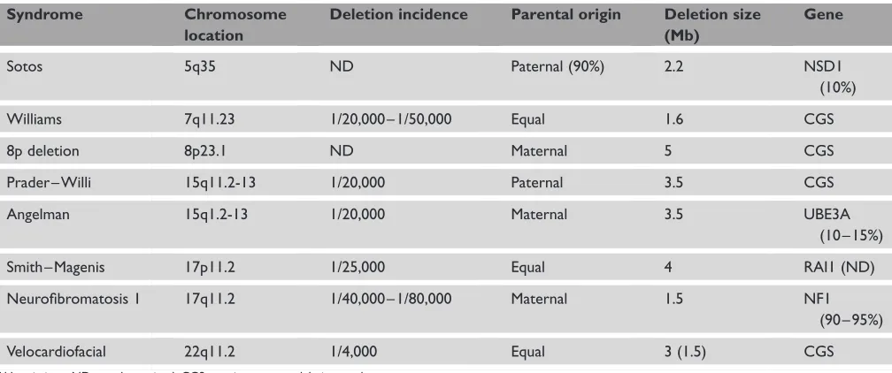
The clinical features of the microdeletion syndromes are highly diverse and complex (Table 1). Microduplications appear to be rarer, and will not be discussed in detail here. For some syndromes, patients present with a major organ malformation, such as a congenital heart defect. Characteristic heart defects are seen for each microdeletion syndrome, eg supravalvular aortic stenosis (SVAS) and peripheral pulmonary stenosis (PPS) in the Williams syndrome (7q11 deletion), conotruncal heart malformations (such as tetralogy of Fallot, truncus arteriosus and interrupted aortic arch) in the 22q11.2 deletion (causing the DiGeorge/velocardiofacial [VCF] syndrome) and atrioventricular septal defect or pulmonary stenosis with atrial septal defect in the 8p23.1 deletion. Characteristic facial features are frequently present and may aid in the diagnosis. As with most chromosomal aberrations, retarded physical growth is also commonly observed — with the notable exception of Sotos syndrome — where somatic overgrowth is a key feature. Besides the physical findings, many individuals with a microdeletion come to medical attention because of developmental delay. The delay ranges from borderline (eg in VCF syndrome) to severe (as in the Angelman syndrome; AS). It is of interest that these learning
difficulties often have a characteristic profile, most typically seen in the Williams syndrome, where individuals have a large discrepancy between their verbal and performance intelli-gence.1Moreover, several microdeletions cause a characteristic behavioural phenotype.2 The most studied example is the Williams syndrome, where individuals are described as ‘over-friendly-though-anxious’ and lack social judgement skills.1In the Smith–Magenis syndrome (SMS; del17p11.2) and in the chromosome 8p23.1 deletion, severe sleeping disturbances and self-injury are common.3,4The Prader-Willi syndrome (PWS; del15q11-13), gives rise to an insatiable appetite, leading to morbid obesity, with frequent temper tantrums.2 It is also recognised that individuals with either VCF syndrome or PWS are at increased risk for psychiatric disease, specifically psychoses.5,6
Given the wide range of phenotypic manifestations, clinical recognition of the microdeletion syndromes has become part of general medical practice, beyond just paediatrics and clinical genetics.
Cause of microdeletions
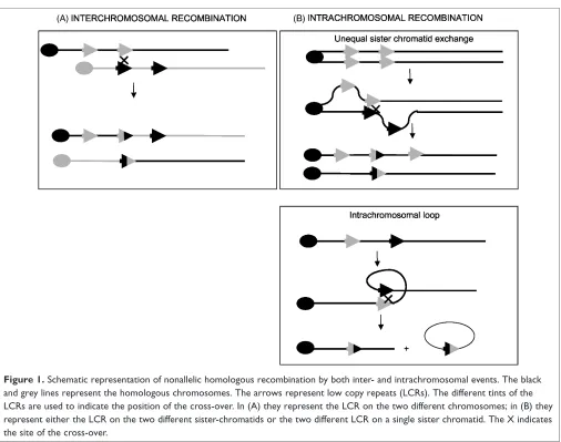
an underlying chromosomal structural feature in the region that confers a predisposition to the loss of small chromosomal fragments. Subsequently, it was proven that region-specific low-copy repeats (LCRs) flank all the microdeletions mentioned in Table 1. The deletion breakpoints cluster within these flanking LCRs. It is now well established that non-allelic homologous recombination by both inter- and intrachromo-somal events cause the deletions.7 – 10 The interchromosomal rearrangements arise by an unequal crossing-over during meiosis I through paralogous LCRs between homologous chromosomes (Figure 1). Intrachromosomal rearrangements may occur by several mechanisms, including LCR mispairing-mediated unequal sister chromatid exchange and the for-mation of an intrachromosomal loop that is also mediated by LCRs within a single chromatid.11,12Intrachromosomal rearrangements can occur either during meiosis or post-zygotically. Since mosaics for microdeletion syndromes have only rarely been detected, however, it is most likely that the majority of the intrachromosomal rearrangements occur during the meiotic period.
The reciprocal product of the deletion caused by hom-ologous recombination by misaligned flanking LCRs is a duplication. The frequency of duplications should equal the frequency of microdeletions. This paradigm is exemplified by two syndromes, Charcot – Marie – Tooth disease type 1A (CMT1A) and hereditary neuropathy with liability to press-ure palsies (HNPP), with an incidence of 1/3,000 and 1/ 7,000, respectively. While CMT1A is caused by a microdu-plication of 17p11.2, HNPP is caused by a microdeletion in the same region. Surprisingly, however, reciprocal micro-deletion/microduplication syndromes like CMT1A/HNPP are rare, or even never observed, for the other microdeletion
syndromes. Duplications have now been described for the 22q11 region,13 the 15q11-13 region14 and the 17p11.2-12 region.15 It is intriguing that only a few duplications have been detected so far. One possibility is that duplications would be lethal. In general, however, chromosome
duplications cause a less severe phenotype than deletions (in unbalanced translocations, the phenotype is usually
determined more by the deleted chromosome fragment than by the duplication). Alternatively, it may be that there is no distinct phenotype, and that individuals with duplications do not come to medical attention. If this were so, however, one would expect that the duplications would remain present in the general population and that they would be detected during the many tests and screenings that are currently performed. Another possibility is that carriers of duplications may be less reproductively fit, so the duplication may not embed in the population. In conclusion, at present, there is no satisfactory explanation for the apparently low frequency of microduplication syndromes.
While it is now well established that LCRs in the genome induce a susceptibility for the generation of microdeletions, it remains unclear why some rearrangements occur more often than others. It has been estimated that 5 – 10 per cent of the genome is composed of LCRs;16 – 18however, only a fraction of these seem to cause recurrent rearrangements. For Sotos syndrome, a thus far unexplained difference exists in the occurrence of a microdeletion in chromosome 5q35 between Japanese and non-Japanese individuals. While at least 50 per cent of Japanese patients carry a 5q35 microdeletion,19,20these microdeletions appear to be exceptional in non-Japanese patients.21Determining the factors that influence a predispo-sition for rearrangements is currently the topic of active Table 1. Characteristics of common microdeletion syndromes
Syndrome Chromosome
location
Deletion incidence Parental origin Deletion size (Mb)
Gene
Sotos 5q35 ND Paternal (90%) 2.2 NSD1
(10%)
Williams 7q11.23 1/20,000 – 1/50,000 Equal 1.6 CGS
8p deletion 8p23.1 ND Maternal 5 CGS
Prader – Willi 15q11.2-13 1/20,000 Paternal 3.5 CGS
Angelman 15q1.2-13 1/20,000 Maternal 3.5 UBE3A (10 – 15%)
Smith – Magenis 17p11.2 1/25,000 Equal 4 RAI1 (ND)
Neurofibromatosis 1 17q11.2 1/40,000 – 1/80,000 Maternal 1.5 NF1 (90 – 95%)
Velocardiofacial 22q11.2 1/4,000 Equal 3 (1.5) CGS
research. The major elements known to influence the fre-quency of such rearrangements will be discussed below.
The size of the LCRs
In general, LCRs flanking the commonly deleted regions range between 200 and 500 kb in size. Apart from these flanking LCRs, however, smaller paralogous sequences exist within the commonly deleted regions of several syndromes. Such sequences exist within DiGeorge/VCF syndrome (22q11.2),22SMS (del17p11.2),23 PWS/AS (del15q11-13)24 and neurofibromatosis type 1 (NF1) (del17q11.2)25commonly deleted regions. These sequences have been shown to pre-dispose to microdeletions as well, albeit at a lower frequency. Hence, the size of the paralogous sequences appears, not sur-prisingly, to influence the frequency by which meiotic misa-lignment and subsequent uneven cross-overs do occur.
The sequence identity among the LCRs
It seems likely that sequence identity will influence the fre-quency of nonallelic homologous recombination (NAHR).Evolutionary studies and sequence analysis of the LCRs has unveiled some intriguing features. The LCRs seem to have appeared during primate speciation.22,23,26 – 29Why such large homology segments are preserved in the human genome and why different LCRs seem to populate different chromosomes is not obvious. It is also unclear how the homology is pre-served. Sequence analysis in the CMT1A/HNPP flanking LCRs has shown the occurrence of gene conversion flanking the recombination hotspots.8,30 It is possible that gene con-version homogenises LCRs and influences the rate of NAHR. This hypothesis deserves closer scrutiny.
The inherent capacity of the LCR sequence to
initiate homologous recombination
paternally derived chromosomes than in the maternally derived chromosomes.8,33 – 35Microdeletions in theNF1locus and the 8p region are predominantly of maternal origin.10,36,37 These observations are most easily explained by differences in the capacity of certain sequences to initiate homologous recombination. Recombination rates vary greatly across the genome, from zero to 8.8 cM/Mb. Moreover, recombination rates and sites vary between males and females,38and sex-specific recombination hotspots have been mapped.39,40 In fact, the NF1-flanking LCRs and 8p olfactory receptor gene clusters flanking the 8p terminal microdeletion appear to be female-specific recombination sites. Lopez-Correaet al.
found that 46 per cent of the NF1 microdeletion breakpoints cluster in a 2 kb region within the flanking LCRs, supporting the thesis of the presence of a recombination hotspot.41 Similarly, Reiteret al. discovered a 2 kb region where the breakpoints cluster has been found in 75 per cent of CMT1A patients and 84 per cent of HNPP patients.42The CMT1A and Sotos syndrome flanking sequences may thus represent male-specific recombination hotspots.
The orientation of the sequence
between the LCRs
Parental submicroscopic inversion polymorphisms between the LCRs flanking the microdeletion regions have recently been demonstrated to be present in several microdeletion syndromes. An inversion polymorphism on chromosome 8p was present in all mothers whose offspring had a deletion of the corresponding olfactory receptor gene cluster.37,43,44 This polymorphism turned out to be present in the hetero-zygous state in 26 per cent of a population of European des-cent. A comparable inversion polymorphism was present in one-third of parents of origin of probands with the 1.5 Mb deletion at 7q11.23 causing Williams-Beuren syndrome.45 At the Emery Dreifuss muscular dystrophy region, a similar inversion polymorphism has been detected in 33 per cent of females.46Gimelliet al.showed that four out of six mothers of AS patients having the BP2/3 deletion carry an inversion.24 This inversion appears to be present at 9 per cent in the general population. These observations raise the question of whether parental inversion polymorphisms are a common phenomenon in all microdeletion syndromes. A recent report suggests that, at least for the most common deletion syndrome, the 22q11 microdeletion, inversion polymorphisms do not exist or are very rare.47Another question raised is whether the presence of an inversion increases the risk of producing microdeleted offspring. Although the answer is likely to be positive, the risk level is probably only marginally increased. Considering that about one-quarter of the population carries the 8p inversion, the occurrence of del(8p) is extremely rare; however, this risk may be variable for inversion
polymorphisms at different loci, and further studies are needed to address this question.
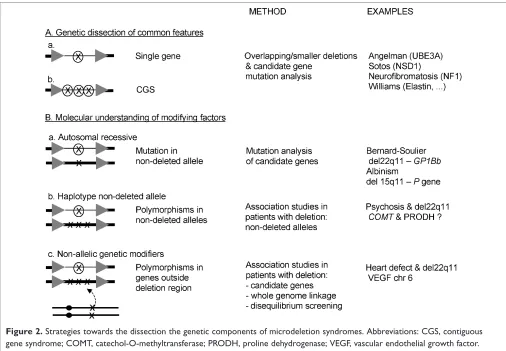
Genetic dissection of the common
features in microdeletion syndromes
The phenotype of a number of microdeletion syndromes is by and large determined by haploinsufficiency of a single gene within the deleted region. In NF1, the majority of patients carry a mutation in a single gene (the NF1 gene), and a microdeletion is found in only about 5 – 10 per cent of cases. The phenotype of deletion patients is more severe, with more pronounced facial dysmorphism and more pronounced developmental delay.25,48Interestingly, these individuals also have a higher number of neurofibromata at a younger age and a higher risk of malignant peripheral nerve sheet tumours,48,49 indicating the presence of one or more genes in this region contributing to this. In AS, patients with a mutation in the UBE3Agene are clinically almost indistin-guishable from those with a microdeletion.50Nevertheless, as a group, deletion patients are more severely affected, with a higher incidence of seizures, microcephaly and more severe developmental delay. This suggests that other genes in the deleted region contribute to the phenotype, and that the GABA receptor genes in 15q11-13 are possible candidate genes for the more pronounced epilepsy.51 In Sotos syn-drome, most features can be explained by haploinsufficiency for the NSD1 gene, whereas in some individuals, additional manifestations are probably related to other genes in the deleted region.19 – 21or overlap the common Williams deletion region, and which manifest only some of the Williams syndrome features.55,56
In addition to explaining the phenotypic manifestations in patients carrying a microdeletion syndrome, the identified genes are also excellent candidate susceptibility genes for the more common multifactorial phenotypes in men, such as organ malformations, intelligence and behaviour. For example, individuals with a del22q11 have an increased risk of developing psychosis. Two candidate genes for psychoses are located within the deleted region on chromosome 22q11.2, the catechol-O-methyltransferase (COMT) gene and the proline dehydrogenase (PRODH) gene. TheCOMTgene plays a role in dopamine neurotransmitter degradation, and abnormal function of dopaminergic pathways is thought to play a role in psychosis.57Thus far, no convincing studies exist that show the association of the common functional val-met polymorphism and the risk for psychosis.57More recently, however, Schifmanet al. have shown the association of a COMT haplotype (but not the val-met polymorphism) with schizophrenia in the general population.58 Mutations in the
PRODHgene have been detected in some patients with schizophrenia;59moreover, polymorphisms near thePRODH
gene were associated with schizophrenia in three independent study samples.60
Genetic dissection of the differences
in microdeletion syndromes
Despite the deletions having a uniform size in the majority of patients with the same microdeletion syndrome, clinical expression can vary widely. The best example to illustrate this with is the phenotype caused by del22q11, which has been described as a number of different syndromes, such as the DiGeorge syndrome (hypoparathyroidism, thymus hypoplasia, conotruncal heart defect and facial dysmorphism), the VCF syndrome (cleft palate, conotruncal heart defect, learning difficulties and characteristic facial features) and, in the Japanese literature, the conotruncal anomaly facies syndrome (CTAFS).
Since haploinsufficiency of the deleted gene(s) alone cannot explain clinical variability in most cases, other modifying factors must exist. As for all complex genetic features or dis-orders, phenotype is influenced by additional genetic and environmental factors. In a microdeletion syndrome, the phenotype is determined by a single gene with major effect and influenced by a limited number of additional genetic and environmental factors with a small effect. This is a less com-plex situation than the more common multifactorial features, where the phenotype is determined by multiple environmental and genetic factors, each with a small effect. Because of this, and given their frequent occurrence, the microdeletion syndromes present a unique opportunity to dissect modifying factors of complex phenotypes.
The most obvious candidate modifiers are mutations/ polymorphisms in the non-deleted allele of genes within the deleted region (Figure 2). The most extreme presentation of this is a recessive disorder, where a microdeletion unmasks a mutation in the non-deleted allele. For example, Bernard-Soulier syndrome (BSS) can be caused by homozygous mutations in theGP1Bbgene, located in chromosome 22q11. This disorder features platelet dysfunction, with elevated platelet volumes and lowered counts. In one patient with a microdeletion in 22q11 and BSS, a mutation was detected in the non-deletedGP1Bballele.61Another example is a patient with AS and recessive oculocutaneous albinism type 2 (OCA2), caused by absence of the Pgene. Fridman et al.found this gene to be located in the deleted region on the maternal chromosome 15q11.2-13 in AS, and a deletion within the
P gene to be present on the paternal chromosome.62 In contrast to mutations, polymorphisms are an almost general feature of most human genes. Therefore, poly-morphisms in the non-deleted allele are excellent candidates for genetic modifiers. As stated above, individuals with del22q11 have an increased risk of developing psychosis, an observation which has been seen in approximately 10 per cent of cases. Polymorphisms in theCOMTandPRODHgenes are obvious candidates for association studies in VCF syndrome individuals with and without psychosis.
Besides mutations or polymorphisms in the non-deleted allele, the phenotype can also be influenced by polymorphisms in genes located elsewhere in the genome. In VCF syndrome, between 50 and 75 per cent of cases have a congenital heart defect, despite the presence of a uniform-sized deletion. Mice lacking the 164 isoform of the vascular endothelial growth factor (vegf) proteins display a phenotype almost identical to the tbx1 knockout mouse.63In addition, in zebrafish, an interaction between tbx1 and vegf could be demonstrated in knockdown experiments.63These data suggested that human VEGF might be a modifier for the heart defects in individuals with del22q11. This was confirmed by showing that functional polymorphisms in theVEGFgene promotor/5’UTR on chromosome 6p12 are associated with decreased VEGF expression and confer an increased risk for the development of a congenital heart defect in individuals with del22q11.63This represents the first known genetic modifier outside the deleted region for a malformation seen in a microdeletion syndrome.
Towards whole genome
microdeletion/microduplication
screens
As discussed above, the study of microdeletions is a powerful tool in the annotation of the human genome. The discovery of these microdeletion syndromes has so far occurred hapha-zardly. It is likely that some of the so far unexplained clinically recognisable syndromes with a ‘chromosomal’ phenotype can be explained by a recurrent microdeletion or microduplica-tion. Moreover, a significant proportion of individuals with unexplained developmental disorders will probably be explained by random microdeletions and/or duplications. Evidence for this hypothesis comes from the many sporadic reports of submicroscopic interstitial chromosomal rearrange-ments. The introduction of a subtelomeric screen enabled a molecular diagnosis to be made for about 5 per cent of the idiopathic mentally retarded population.68These observations raise the expectation that a genome-wide microdeletion screen may well pick up various unknown intrachromosomal aberrations. Techniques that will enable genome-wide aneuploidy analysis are thus needed and are being developed.
The most advanced of these techniques is matrix or array comparative genomic hybridisation (CGH).69 – 71This tech-nique combines the advantages of the resolution power of fluorescent in situhybridisation (FISH) with the screening capacity of the chromosome scans used by classical cytogenetic techniques. In this technique, genomic DNA from patient and control are differentially labelled with two fluorescent dyes. The labelled DNAs are co-hybridised to DNA arrays, which consist of DNA spots derived from clones containing genomic DNA fragments — usually bacteria artificial chromosomes (BACs). Chromosomal deletions are detected by fluorescent intensity ratios of the spots containing less patient dye compared with control dye, and vice versa for duplications. Chromosomal imbalances across the genome can thus be quantified and their position determined. The resolution of array CGH can be controlled and is dependent on a combi-nation of the number, size and map positions of the DNA elements within the array.71 – 73Array CGH has been successfully applied to analyse a variety of constitutional aberrations. Just recently, the technique has been introduced into the clinical genetics laboratory.73 – 76
diagnostic cytogenetic laboratories. Certainly, this will have a major impact on clinical genetics. In addition, correlations of defined phenotypic manifestations with the deletion or duplication of specific genes will provide a unique opportunity to further the annotation of the human genome.
Acknowledgments
Koen Devriendt is a Senior Clinical Investigator in the Fund for Scientific Research-Flanders (FWO, Vlaanderen). He is supported by grants from the University of Leuven (OT) and FWO, Vlaanderen.
References
1. Donnai, D. and Karmiloff-Smith, A. (2000), ‘Williams syndrome: From genotype through to the cognitive phenotype’,Am. J. Med. Genet.Vol. 97, pp. 164 – 171.
2. Cassidy, S.B. and Morris, C.A. (2002), ‘Behavioral phenotypes in genetic syndromes: Genetic clues to human behaviour’,Adv. Pediatr.Vol. 49, pp. 59 – 86.
3. Smith, A.C., Dykens, E. and Greenberg, F. (1998), ‘Behavioral phenotype of Smith-Magenis syndrome (del 17p11.2)’,Am. J. Med. Genet.Vol. 81, pp. 179 – 185.
4. Claeys, I., Holvoet, M., Eyskens, B.et al.(1997), ‘A recognisable beha-vioural phenotype associated with terminal deletions of the short arm of chromosome 8’,Am. J. Med. Genet.Vol. 74, pp. 515 – 520.
5. Clarke, D.J. (1993), ‘Prader-Willi syndrome and psychoses’,Br. J. Psychiatry
Vol. 163, pp. 680 – 684.
6. Murphy, K.C. and Owen, M.J. (2001), ‘Velo-cardio-facial syndrome: A model for understanding the genetics and pathogenesis of schizophrenia’,
Br. J. PsychiatryVol. 179, pp. 397 – 402.
7. Urban, Z., Helms, C., Fekete, G.et al.(1996), ‘7q11.23 deletions in Williams syndrome arise as a consequence of unequal meiotic crossover’,
Am. J. Hum. Genet.Vol. 59, pp. 958 – 962.
8. Lopes, J., Vandenberghe, A., Tardieu, S.et al.(1997), ‘Sex-dependent rearrangements resulting in CMT1A and HNPP’,Nat. Genet.Vol. 17, pp. 136 – 137.
9. Baumer, A., Dutly, F., Balmer, D.et al.(1998), ‘High level of unequal meiotic crossovers at the origin of the 22q11. 2 and 7q11.23 deletions’,
Hum. Mol. Genet.Vol. 7, pp. 887 – 894.
10. Lopez, C.C., Brems, H., Lazaro, C.et al.(2000), ‘Unequal meiotic crossover: A frequent cause of NF1 microdeletions’,Am. J. Hum. Genet.
Vol. 66, pp. 1969 – 1974.
11. Ji, Y., Eichler, E.E., Schwartz, S.et al.(2000), ‘Structure of chromosomal duplicons and their role in mediating human genomic disorders’,Genome Res.Vol. 10, pp. 597 – 610.
12. Stankiewicz, P., Shaw, C.J., Dapper, J.D.et al.(2003), ‘Genome archi-tecture catalyzes nonrecurrent chromosomal rearrangements’,Am. J. Hum. Genet.Vol. 72, pp. 1101 – 1116.
13. Ensenauer, R.E., Adeyinka, A., Flynn, H.C.et al.(2003), ‘Microdupli-cation 22q11.2, an emerging syndrome: Clinical, cytogenetic, and molecular analysis of thirteen patients’,Am. J. Hum. Genet.Vol. 73, pp. 1027 – 1040.
14. Roberts, S.E., Dennis, N.R., Browne, C.E.et al.(2002), ‘Characterisation of interstitial duplications and triplications of chromosome 15q11-q13’,
Hum. Genet.Vol. 110, pp. 227 – 234.
15. Potocki, L., Chen, K.S., Park, S.S.et al.(2000), ‘Molecular mechanism for duplication 17p11.2 — the homologous recombination reciprocal of the Smith-Magenis microdeletion’,Nat. Genet.Vol. 24, pp. 84 – 87. 16. Bailey, J.A., Yavor, A.M., Massa, H.F.et al.(2001), ‘Segmental
dupli-cations: Organization and impact within the current human genome project assembly’,Genome Res.Vol. 11, pp. 1005 – 1017.
17. Bailey, J.A., Gu, Z., Clark, R.A.et al.(2002), ‘Recent segmental dupli-cations in the human genome’,ScienceVol. 297, pp. 1003 – 1007. 18. Lupski, J.R. (2003), 2002 Curt Stern Award Address. ‘Genomic disorders
recombination-based disease resulting from genomic architecture’,Am. J. Hum. Genet.Vol. 72, pp. 246 – 252.
19. Kurotaki, N., Imaizumi, K., Harada, N.et al.(2002), ‘Haploinsufficiency of NSD1 causes Sotos syndrome’,Nat. Genet.Vol. 30, pp. 365 – 366. 20. Nagai, T., Matsumoto, N., Kurotaki, N.et al.(2003), ‘Sotos syndrome and
haploinsufficiency of NSD1: Clinical features of intragenic mutations and submicroscopic deletions’,J. Med. Genet.Vol. 40, pp. 285 – 289. 21. Douglas, J., Hanks, S., Temple, I.K.et al.(2003), ‘NSD1 mutations are the
major cause of Sotos syndrome and occur in some cases of Weaver syn-drome but are rare in other overgrowth phenotypes’,Am. J. Hum. Genet.
Vol. 72, pp. 132 – 143.
22. Shaikh, T.H., Kurahashi, H. and Emanuel, B.S. (2001), ‘Evolutionarily conserved low copy repeats (LCRs) in 22q11 mediate deletions, dupli-cations, translocations and genomic instability: An update and literature review’,Genet. Med.Vol. 3, pp. 6 – 13.
23. Park, S.S., Stankiewicz, P., Bi, W.et al.(2002), ‘Structure and evolution of the Smith-Magenis syndrome repeat gene clusters, SMS-REPs’,Genome Res.Vol. 12, pp. 729 – 738.
24. Gimelli, G., Pujana, M.A., Patricelli, M.G.et al.(2003), ‘Genomic inversions of human chromosome 15q11-q13 in mothers of Angelman syndrome patients with class II (BP2/3) deletions’,Hum. Mol. Genet.
Vol. 12, pp. 849 – 858.
25. Lopez, C.C., Brems, H., Lazaro, C.et al.(1999), ‘Molecular studies in 20 submicroscopic neurofibromatosis type 1 gene deletions’,Hum. Mutat.
Vol. 14, pp. 387 – 393.
26. Kiyosawa, H. and Chance, P.F. (1996), ‘Primate origin of the CMT1A-REP repeat and analysis of a putative transposon-associated recombina-tional hotspot’,Hum. Mol. Genet.Vol. 5, pp. 745 – 753.
27. Christian, S.L., Fantes, J.A., Mewborn, S.K.et al.(1999), ‘Large genomic duplicons map to sites of instability in the Prader-Willi/Angelman syndrome chromosome region (15q11-q13)’,Hum. Mol. Genet.Vol. 8, pp. 1025 – 1037.
28. Valero, M.C., de Luis, O., Cruces, J.et al.(2000), ‘Fine-scale comparative mapping of the human 7q11.23 region and the orthologous region on mouse chromosome 5G: The low-copy repeats that flank the Williams-Beuren syndrome deletion arose at breakpoint sites of an evolutionary inversion(s)’,GenomicsVol. 69, pp. 1 – 13.
29. Inoue, K., Dewar, K., Katsanis, N.et al.(2001), ‘The 1.4 Mb CMT1A duplication/HNPP deletion genomic region reveals unique genome architectural features and provides insights into the recent evolution of new genes’,Genome Res.Vol. 11, pp. 1018 – 1033.
30. Reiter, L.T., Hastings, P.J., Nelis, E.et al.(1998), ‘Human meiotic recombination products revealed by sequencing a hotspot for homologous strand exchange in multiple HNPP deletion patients’,Am. J. Hum. Genet.
Vol. 62, pp. 1023 – 1033.
31. Dutly, F. and Schinzel, A. (1996), ‘Unequal interchromosomal
rearrangements may result in elastin gene deletions causing the Williams-Beuren syndrome’,Hum. Mol. Genet.Vol. 5, pp. 1893 – 1898.
32. Shaw, C.J., Bi, W. and Lupski, J.R. (2002), ‘Genetic proof of unequal meiotic crossovers in reciprocal deletion and duplication of 17p11.2’,Am. J. Hum. Genet.Vol. 71, pp. 1072 – 1081.
33. Palau, F., Lofgren, A., De Jonghe, P.et al.(1993), ‘Origin of the de novo duplication in Charcot-Marie-Tooth disease type 1A: Unequal nonsister chromatid exchange during spermatogenesis’,Hum. Mol. Genet.Vol. 2, pp. 2031 – 2035.
34. Bort, S., Martinez, F. and Palau, F. (1997), ‘Prevalence and parental origin of de novo 1.5 Mb duplication in Charcot-Marie-Tooth disease type 1A’,
Am. J. Hum. Genet.Vol. 60, pp. 230 – 233.
35. Miyake, N., Kurotaki, N. and Sugawara, H.et al.(2003), ‘Preferential paternal origin of microdeletions caused by prezygotic chromosome or chromatid rearrangements in Sotos syndrome’,Am. J. Hum. Genet.Vol. 72, pp. 1331 – 1337.
37. Giglio, S., Broman, K.W., Matsumoto, N.et al.(2001), ‘Olfactory receptor-gene clusters, genomic-inversion polymorphisms and common chromosome rearrangements’,Am. J. Hum. Genet.Vol. 68, pp. 874 – 883. 38. Yu, A., Zhao, C., Fan, Y.et al.(2001), ‘Comparison of human genetic and
sequence-based physical maps’,NatureVol. 409, pp. 951 – 953. 39. Robinson, W.P. and Lalande, M. (1995), ‘Sex-specific meiotic
recombi-nation in the Prader-Willi/Angelman syndrome imprinted region’,Hum. Mol. Genet.Vol. 4, pp. 801 – 806.
40. Badge, R.M., Yardley, J., Jeffreys, A.J.et al.(2000), ‘Crossover breakpoint mapping identifies a subtelomeric hotspot for male meiotic recombina-tion’,Hum. Mol. Genet.Vol. 9, pp. 1239 – 1244.
41. Lopez-Correa, C., Dorschner, M., Brems, H.et al.(2001), ‘Recombi-nation hotspot in NF1 microdeletion patients’,Hum. Mol. Genet.Vol. 10, pp. 1387 – 1392.
42. Reiter, L.T., Murakami, T., Koeuth, T.et al.(1996), ‘A recombination hotspot responsible for two inherited peripheral neuropathies is located near a mariner transposon-like element’,Nat. Genet.Vol. 12, pp. 288 – 297.
43. Giglio, S., Calvari, V., Gregato, G.et al.(2002), ‘Heterozygous sub-microscopic inversions involving olfactory receptor-gene clusters mediate the recurrent t(4;8)(p16;p23) translocation’,Am. J. Hum. Genet.Vol. 71, pp. 276 – 285.
44. Vermeesch, J.R., Thoelen, R. and Salden, I.et al.(2003), ‘Mosaicism del(8p)/inv dup(8p) in a dysmorphic female infant: A mosaic formed by a meiotic error at the 8p OR gene and an independent terminal deletion event’,J. Med. Genet.Vol. 40(8), e93.
45. Osborne, L.R., Li, M., Pober, B.et al.(2001), ‘A 1.5 million-base pair inversion polymorphism in families with Williams-Beuren syndrome’,
Nat. Genet.Vol. 29, pp. 321 – 325.
46. Small, K., Iber, J. and Warren, S.T. (1997), ‘Emerin deletion reveals a common X-chromosome inversion mediated by inverted repeats’,Nat. Genet.Vol. 16, pp. 96 – 99.
47. Gebhardt, G.S., Devriendt, K., Thoelen, R.et al.(2003), ‘No evidence for a parental inversion polymorphism predisposing to rearrangements at 22q11.2 in the DiGeorge/velocardiofacial syndrome’,Eur. J. Hum. Genet.
Vol. 11, pp. 109 – 111.
48. Kayes, L.M., Burke, W., Riccardi, V.M.et al.(1994), ‘Deletions spanning the neurofibromatosis 1 gene: Identification and phenotype of five patients’,Am. J. Hum. Genet.Vol. 54, pp. 424 – 436.
49. De Raedt, T., Brems, H., Wolkenstein, P.et al.(2003), ‘Elevated risk for MPNST in NF1 microdeletion patients’,Am. J. Hum. Genet.Vol. 72, pp. 1288 – 1292.
50. Clayton-Smith, J. and Laan, L. (2003), ‘Angelman syndrome: A review of the clinical and genetic aspects’,J. Med. Genet.Vol. 40, pp. 87 – 95. 51. Minassian, B.A., DeLorey, T.M., Olsen, R.W.et al.(1998), ‘Angelman
syndrome: Correlations between epilepsy phenotypes and genotypes’,
Ann. Neurol.Vol. 43, pp. 485 – 493.
52. Tassabehji, M., Metcalfe, K., Donnai, D.et al.(1997), ‘Elastin: genomic structure and point mutations in patients with supravalvular aortic steno-sis’,Hum. Mol. Genet.Vol. 6, pp. 1029 – 1036.
53. Bi, W., Yan, J., Stankiewicz, P. et al. (2002), ‘Genes in a refined Smith-Magenis syndrome critical deletion interval on chromosome 17p11.2 and the syntenic region of the mouse’, Genome Res. Vol. 12, pp. 713 – 728.
54. Slager, R.E., Newton, T.L., Vlangos, C.N.et al.(2003), ‘Mutations in RAI1 associated with Smith-Magenis syndrome’,Nat. Genet.Vol. 33, pp. 466 – 468.
55. Frangiskakis, J.M., Ewart, A.K., Morris, C.A.et al.(1996), ‘LIM-kinase1 hemizygosity implicated in impaired visuospatial constructive cognition’,
CellVol. 86, pp. 59 – 69.
56. Tassabehji, M., Metcalfe, K., Karmiloff-Smith, A.et al.(1999), ‘Williams syndrome: Use of chromosomal microdeletions as a tool to dissect cognitive and physical phenotypes’,Am. J. Hum. Genet.Vol. 64, pp. 118 – 125.
57. Murphy, K.C., Jones, L.A. and Owen, M.J. (1999), ‘High rates of schizophrenia in adults with velo-cardio-facial syndrome’,Arch. Gen. PsychiatryVol. 56, pp. 940 – 945.
58. Shifman, S., Bronstein, M., Sternfeld, M.et al.(2002), ‘A highly signifi-cant association between a COMT haplotype and schizophrenia’,Am. J. Hum. Genet.Vol. 71, pp. 1296 – 1302.
59. Jacquet, H., Raux, G., Thibaut, F.et al.(2002), ‘PRODH mutations and hyperprolinemia in a subset of schizophrenic patients’,Hum. Mol. Genet.
Vol. 11, pp. 2243 – 2249.
60. Liu, H., Heath, S.C., Sobin, C.et al.(2002), ‘Genetic variation at the 22q11 PRODH2/DGCR6 locus presents an unusual pattern and increases susceptibility to schizophrenia’,Proc. Natl. Acad. Sci. USAVol. 99, pp. 3717 – 3722.
61. Ludlow, L.B., Schick, B.P., Budarf, M.L.et al.(1996), ‘Identification of a mutation in a GATA binding site of the platelet glycoprotein Ibbeta promoter resulting in the Bernard-Soulier syndrome’,J. Biol. Chem.
Vol. 271, pp. 22076 – 22080.
62. Fridman, C., Hosomi, N., Varela, M.C.et al.(2003), ‘Angelman syndrome associated with oculocutaneous albinism due to an intragenic deletion of thePgene’,Am. J. Med. Genet.Vol. 119A, pp. 180 – 183.
63. Stalmans, I., Lambrechts, D., De Smet, F.et al.(2003), ‘VEGF: A modifier of the del22q11 (DiGeorge) syndrome?’,Nat. Med.Vol. 9, pp. 173 – 182. 64. Descheemaeker, M.J., Swillen, A., Plissart, L.et al.(1994), ‘The Prader-Willi syndrome: A self-supporting program for children, youngsters and adults’,Genet. Couns.Vol. 5, pp. 199 – 205.
65. Swillen, A., Devriendt, K., Legius, E.et al.(1997), ‘Intelligence and psychosocial adjustment in velocardiofacial syndrome: a study of 37 children and adolescents with VCFS’,J. Med. Genet.Vol. 34, pp. 453 – 458. 66. Malich, S., Largo, R.H., Schinzel, A.et al.(2000), ‘Phenotypic
hetero-geneity of growth and psychometric intelligence in Prader-Willi syndrome: Variable expression of a contiguous gene syndrome or parent-child resemblance?’,Am. J. Med. Genet.Vol. 91, pp. 298 – 304. 67. Vincent, M.C., Heitz, F., Tricoire, J.et al.(1999), ‘22q11 deletion in
DGS/VCFS monozygotic twins with discordant phenotypes’,Genet. Couns.Vol. 10, pp. 43 – 49.
68. Knight, S.J., Regan, R., Nicod, A.et al.(1999), ‘Subtle chromosomal rearrangements in children with unexplained mental retardation’,Lancet
Vol. 354, pp. 1676 – 1681.
69. Bentz, M., Plesch, A., Stilgenbauer, S.et al.(1998), ‘Minimal sizes of deletions detected by comparative genomic hybridization’,Genes Chromosomes CancerVol. 21, pp. 172 – 175.
70. Kirchhoff, M., Rose, H. and Lundsteen, C. (2001), ‘High resolution comparative genomic hybridisation in clinical cytogenetics’,J. Med. Genet.
Vol. 38, pp. 740 – 744.
71. Solinas-Toldo, S., Lampel, S., Stilgenbauer, S.et al.(1997), ‘Matrix-based comparative genomic hybridization: Biochips to screen for genomic imbalances’,Genes Chromosomes CancerVol. 20, pp. 399 – 407.
72. Pinkel, D., Segraves, R., Sudar, D.et al.(1998), ‘High resolution analysis of DNA copy number variation using comparative genomic hybridization to microarrays’,Nat. Genet.Vol. 20, pp. 207 – 211.
73. Fiegler, H., Carr, P., Douglas, E.J.et al.(2003), ‘DNA microarrays for comparative genomic hybridization based on DOP-PCR amplification of BAC and PAC clones’,Genes Chromosomes CancerVol. 36, pp. 361 – 374. 74. Veltman, J.A., Jonkers, Y., Nuijten, I.et al.(2003), ‘Definition of a critical region on chromosome 18 for congenital aural atresia by array CGH’,Am. J. Hum. Genet.Vol. 72, pp. 1578 – 1584.
75. Bruder, C.E., Hirvela, C., Tapia-Paez, I.et al.(2001), ‘High resolution deletion analysis of constitutional DNA from neurofibromatosis type 2 (NF2) patients using microarray-CGH’,Hum. Mol. Genet.Vol. 10, pp. 271 – 282.