R E S E A R C H
Open Access
Contamination of nanoparticles by endotoxin:
evaluation of different test methods
Stijn Smulders
1, Jean-Pierre Kaiser
2, Stefano Zuin
3, Kirsten L Van Landuyt
4, Luana Golanski
5, Jeroen Vanoirbeek
1,
Peter Wick
2and Peter HM Hoet
1*Abstract
Background:Nanomaterials can be contaminated with endotoxin (lipopolysaccharides, LPS) during production or handling. In this study, we searched for a convenientin vitromethod to evaluate endotoxin contamination in nanoparticle samples. We assessed the reliability of the commonly used limulus amebocyte lysate (LAL) assay and an alternative method based on toll-like receptor (TLR) 4 reporter cells when applied with particles (TiO2, Ag, CaCO3and SiO2), or after extraction of the endotoxin as described in the ISO norm 29701.
Results:Our results indicate that the gel clot LAL assay is easily disturbed in the presence of nanoparticles; and that the endotoxin extraction protocol is not suitable at high particle concentrations. The chromogenic-based LAL endotoxin detection systems (chromogenic LAL assay and Endosafe-PTS), and the TLR4 reporter cells were not significantly perturbed.
Conclusion:We demonstrated that nanoparticles can interfere with endotoxin detection systems indicating that a convenient test method must be chosen before assessing endotoxin contamination in nanoparticle samples.
Keywords:Endotoxin, Nanoparticles, LAL assay, TLR4 reporter cells
Background
Nanoparticles are worldwide produced and used in various commercially available applications (cosmetics, paints, tex-tiles) [1] and predictions estimate that by 2014, more than 15% of all products on the global market will have some kind of nanotechnology incorporated into their manufac-turing process [2]. Besides their ubiquitous lucrative effects, also toxic effects have been reported [3]. It cannot be excluded that nanoparticles, especially when they were not kept sterile, can be contaminated with endotoxin during production or handling. Therefore, endotoxin con-tamination should be assessed when evaluating the poten-tial toxicity, to distinguish specific nanoparticles toxicity from the endotoxin effects.
Endotoxins or lipopolysaccharides (LPS) are large (mo-lecular weight: 200 to 1000 kDa), heat-stable (to 100°C) molecules that form the major structural components of the outer cell wall of gram-negative bacteria [4,5]. High levels of endotoxin are omnipresent in our living
environment, and exposure can induce a variety of bio-logical effects such as airway disease, fever, hypotension, coagulopathies, septic shock and even death. Endotoxin consists of a bioactive lipid component, termed lipid A, covalently bound to a hydrophilic heteropolysaccharide of variable length [6]. Induction of a signal transduction cascade evolves binding of endotoxin on CD14 followed by association with the protein MD2 and the transmem-brane TLR4 [7]. This finally results in the release of in-flammatory cytokines, including IL-1β, TNF-α and IL-6, mainly secreted by immune cells such as macrophages and dendritic cells.
Currently, the LAL test is the assay of choice for the determination of endotoxin in medicines, biological products and medical devices [5]. In general, three dif-ferent LAL assays are used worldwide: gel clot, turbidi-metric (increase in turbidity) and chromogenic (color formation) assay. A good overview of the different assays, along with their advantages and disadvantages, can be found in the review article of Hurley [5].
In spite of the new ISO norm published in 2010 on endotoxin test on nanomaterial samples forin vitro sys-tems [8], not much is known in which way nanoparticles * Correspondence:[email protected]
1
Laboratory of Pneumology, Unit for Lung Toxicology, KU Leuven, Leuven, Belgium
Full list of author information is available at the end of the article
interfere with the different types of LAL assays [9]. The
aim of our study is to find a convenient in vitro test
method to evaluate endotoxin contamination in nano-particle samples. Therefore, in this study, we assessed the reliability of a gel clot LAL assay, an endpoint chromogenic LAL assay and a FDA-licensed endotoxin detection system when performed in the presence of nanoparticles, as well as the proposed sample prepar-ation methods of the ISO norm were evaluated. More-over, as an alternative for the LAL assay, we tested another method based on TLR4 reporter cells to meas-ure endotoxin in nanoparticle formulations.
Results
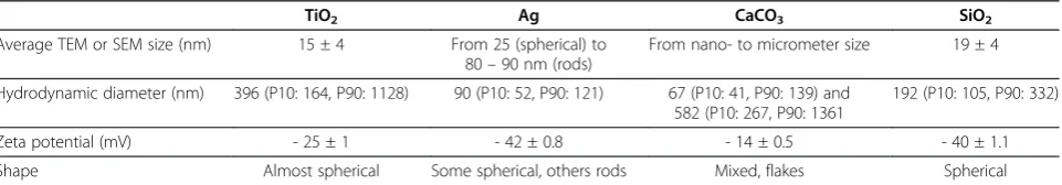
Characteristics of TiO2, Ag, CaCO3 and SiO2 particles
are summarized in Table 1, electron microscopy images are shown in Figure 1. Transmission electron microscopy (TEM) analysis showed average particle sizes of 15 nm (TiO2), 25 to 85 nm (Ag), and 19 nm (SiO2). Scanning
electron microscopy (SEM) analysis of CaCO3revealed a
very heterogeneous composition, showing particles of both nano- and micrometer sizes. Analysis of the parti-cles by dynamic light scattering (DLS) showed single
populations of 396 nm (TiO2), 90 nm (Ag) and 192 nm
(SiO2), and two populations of 67 and 582 nm in the
CaCO3sample. All particles were negatively charged, the
largest electrostatic stabilization was found in the Ag
and SiO2 samples showing zeta potentials of
respect-ively −42 and −40 mV.
The results of the spiking experiments of the different LAL assays (gel clot, endpoint chromogenic, endosafe-Portable Test System (PTS) and endotoxin extraction protocol) are summarized in Figure 2.
In the gel clot LAL assay, negative results (no clot for-mation) were obtained with all particles at each concen-tration (12.5, 50 and 200μg/ml) both for non-baked and baked particle samples (negative controls) (data not shown). In the presence of non-baked particles, spiking at half of assay sensitivity (½λ: 0.0625 EU/ml) resulted not in clot formation (negative result) at all particle concentrations (Figure 2A); spiking at assay sensitivity
(λ: 0.125 EU/ml) leads not to clot formation at the
highest particle concentration (200 μg/ml), while no
inhibition of clot formation was seen at lower
concen-trations (12.5 and 50 μg/ml). After spiking non-baked
particles at double of assay sensitivity (2λ: 0.25 EU/ml) no inhibition of clot formation was observed for the
TiO2 and Ag samples, whereas only an increased
tur-bidity, but no clot, was observed in CaCO3 and SiO2
at 200 μg/ml. The same results were observed for
baked spiked TiO2, Ag and CaCO3samples.
In all samples (baked and non-baked), no (or at least lower than the detection limit) endotoxin contamination was measured in the endpoint chromogenic LAL assay (data not shown). A complete spike recovery was seen for all particle samples (non-baked and baked) at all ap-plied concentrations (Figure 2B). Figure 3 shows the grad-ual increase of background optical density by increasing concentrations of TiO2 particles. A similar increase was
seen in case of the Ag particles (data not shown). The background particle optical density was substracted from the corresponding measured values in the endpoint chromogenic LAL assay to obtain the final results. In the endosafe-PTS, all particle samples showed a complete spike recovery (within 50-200% tolerance limits) at all concentrations (Figure 2C). In all non-spiked samples, no (or at least lower than the detection limit) endotoxin contamination was measured in the endosafe-PTS LAL assay (data not shown).
The endotoxin extraction procedure was performed, as described in the ISO protocol on endotoxin tests on nanomaterial samples with the aim to reduce particle assay interference. Therefore, the endotoxin concentra-tion was measured in the supernatant after
centrifuga-tion with the endpoint chromogenic LAL assay. TiO2,
Ag and CaCO3 samples (baked and non-baked) tested
negative at all applied concentrations (0.2, 2 and 20 mg/ml) (data not shown). After spiking (0.5 EU/ml), no endotoxin could be recovered at the highest particle concentration (20 mg/ml) for all particles (baked and non-baked) (Figure 2D). At lower particle concentra-tions (0.2 and 2 mg/ml), the spike recovery was within the tolerance range of 50-200% for all particles (baked and non-baked), except for the non-baked Ag sample (2 mg/ml) in which we only measured a spike recovery of 22%.
Table 1 Characteristics of TiO2, Ag, CaCO3and SiO2particles
TiO2 Ag CaCO3 SiO2
Average TEM or SEM size (nm) 15 ± 4 From 25 (spherical) to 80–90 nm (rods)
From nano- to micrometer size 19 ± 4
Hydrodynamic diameter (nm) 396 (P10: 164, P90: 1128) 90 (P10: 52, P90: 121) 67 (P10: 41, P90: 139) and 582 (P10: 267, P90: 1361
192 (P10: 105, P90: 332)
Zeta potential (mV) - 25 ± 1 - 42 ± 0.8 - 14 ± 0.5 - 40 ± 1.1
Shape Almost spherical Some spherical, others rods Mixed, flakes Spherical
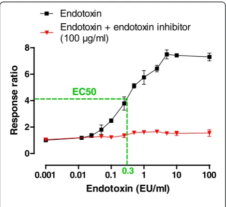
TLR4 reporter cells were used to evaluate the influ-ence of the particles on TLR4-activation by endotoxin. Figure 4 shows the responsiveness of the cells to increas-ing concentrations of endotoxin. TLR4 reporter cells already show an increased response starting at an endotoxin concentration of 0.05 EU/ml. Assuming this dose–response relationship, the half maximum effective concentration (EC50) was determined (0.3 EU/ml),
caus-ing a response ratio of about 4, and this concentration was chosen for spiking (½λmax). Also the biological
func-tionality (TNF-α release) of the cells was assessed. No
increase in TNF-α release was observed after exposure
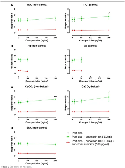
to endotoxin (See in Additional file 1: Figure S1). Exposing the cells to the particles (non-baked and baked), did not result in a measurable response with in-creasing concentrations of particles in all cases (Figure 5 (grey lines)). Spiking (½λmax) the particles (positive
con-trol) resulted in a higher response compared to the re-spective non-spiked samples at all particle concentrations and approximates the response observed at the endotoxin EC50value (response ratio of approximately 4), indicating
a complete spike recovery (Figure 5 (green lines) and Figure 6). Moreover, an increased response was seen in
the spiked TiO2and CaCO3 samples at a concentration
of 200 μg/ml compared to the respective spiked sample
without particles, which was significant in the case of
TiO2(baked) and CaCO3(non-baked and baked). Applying
an endotoxin inhibitor (polymyxin B sulfate, 100μg/ml) in the presence of spiked particles resulted in a complete in-hibition of the response at all particle concentrations (Figure 5 (red lines)). Regarding Ag, significant cytotox-icity was observed at concentrations above 50μg/ml and therefore, measurements were limited in this case.
Discussion
In the present study, we investigated the performance of different LAL assays in the presence of nanoparticles
and evaluated another–non LAL-based–in vitroassay
to assess endotoxin contamination. We have shown that nanoparticles have the potential to interfere with the gel
clot LAL assay at elevated – but not excessive high –
particle concentrations. Likewise, an endotoxin extrac-tion protocol, including shaking and centrifugaextrac-tion of the particle dispersion, seems to be unsatisfactory at high particle concentrations. Furthermore, we also demonstrated that chromogenic-based LAL endotoxin detection systems (chromogenic LAL assay and Endosafe-PTS) and TLR4 reporter cells report no interfering effects at all applied particle concentrations.
Over the past decades, the LAL assay has been found an application in various domains, ranging from the phar-maceutical and aerospace industry [10] to toxicological
A
B
Endpoint chromogenic non-baked samples
Spiking ½λmax(0.5 EU/ml)
NP 0 12.5 50 200
TiO2 100 82 86 81
Ag 100 88 97 92
CaCO3 100 97 102 104
SiO2 100 87 88 92
C
Endosafe-PTS
Spiking (internal spiking)
NP 0 12.5 50 200
TiO2 82 90 98 90
Ag 82 /////// /////// 70
CaCO3 82 /////// /////// 112 SiO2 82 /////// /////// 87
D
Gel Clot – non-baked samples
Spiking ½λ (0.0625 EU/ml) (0.125 EU/ml) 2λ (0.25 EU/ml)
NP 0 12.5 50 200 0 12.5 50 200 0 12.5 50 200
TiO2 Ag CaCO3 SiO2
Gel Clot – baked samples
Spiking ½λ (0.0625 EU/ml) (0.125 EU/ml) 2λ (0.25 EU/ml)
0 12.5 50 200 0 12.5 50 200 0 12.5 50 200
TiO2 Ag CaCO3
Endpoint chromogenic baked samples
Spiking ½λmax(0.5 EU/ml)
NP 0 12.5 50 200
TiO2 100 88 84 81
Ag 100 90 91 85
CaCO3 100 102 99 96
Extraction protocol non-baked samples
Spiking ½λmax(0.5 EU/ml)
NP (mg/ml) 0 0.2 2 20
TiO2 100 97 89 0
Ag 100 98 22 0
CaCO3 100 135 115 0
Extraction protocol baked samples
Spiking ½λmax(0.5 EU/ml)
NP (mg/ml) 0 0.2 2 20
TiO2 100 89 88 7
Ag 100 95 85 0
CaCO3 100 81 78 0
λ
λ
research. The potential interference of biological pro-ducts has been extensively studied and it is known that certain body fluids (e.g. urine and blood) can influence the outcome of the LAL assay [5,11]. A study carried out by the FDA almost 30 years ago showed that out of the 333 drug products tested, 236 (71%) interfered
with the LAL assay when applied prior to any dilution [12]. Surprisingly, almost no research has been done to which extent nanoparticles can interfere with the different LAL assays. Recently, Dobrovolskaia et al.published the first study showing that endotoxin levels can be under- or overestimated due to the presence of nanoparticles [9].
Interference can occur when endotoxin interferes with the particles or when particles interfere with the LAL specific enzymes resulting in the decrease or increase of the sensitivity of the assay. Therefore, appropriate inhib-ition/enhancement controls are essential to recognize whether negative results are due to absence of endo-toxin, or inhibition of the assay. According to the United States, European and Japanese pharmacopeia, a test is considered valid if the measured concentration of endo-toxin added falls within the tolerance range of 50-200% of the known added endotoxin concentration [13-15]. Appropriate spiking concentrations need to be chosen dependent on the applied LAL assay and its associated sensitivity.
The gel clot LAL assay is quite easy to perform, but limitations are the subjective endpoint and the relative lack of sensitivity. In our study, endotoxin spiking concentrations were chosen below (50% assay sensitiv-ity; ½λ: 0.0625 EU/ml), at (100% assay sensitivity; λ:
0.125 EU/ml) and above (200% assay sensitivity; 2λ:
0.25 EU/ml) the sensitivity of the assay. In this way, we were able to assess whether potential interference was situated in- or outside the tolerable limits. Two of four tested particles, SiO2 and CaCO3, fall out of
this range and exert substantial inhibitory effect on the gel clot LAL assay, at the highest particle
concen-tration tested (200 μg/ml). However, the other tested
particles, TiO2 and Ag, show inhibitory effects as well,
but only at spiking concentrations at the sensitivity of
the assay (λ), and fall therefore within the 50-200%
limits. Similarly, in the study of Dobrovolskaia et al., 3 of the 5 tested particle formulation interfered with the gel clot LAL assay [9]. Dilution is the simplest and most widely used technique to overcome interfer-ence, in our experiments no more inhibitory effects were seen after diluting to particle concentrations of 50 and 12.5μg/ml. Important to note is that particle con-centrations of about 200μg/ml are often used in toxico-logical research [16,17], thus adequate endotoxin detection methods at this concentration are indispensable.
The chromogenic-based LAL assays (chromogenic LAL assay and the Endosafe-PTS) are, compared to the semiquantitative gel clot LAL assay, more quantitative. Neither in the chromogenic LAL assay, nor in the Endosafe-PTS, any significant interfering effect due to the presence of the particles were observed at the
highest applied concentration (200 μg/ml), suggesting
that chromogenic-based LAL methods are convenient
0.001 0.01 0.1 1 10 100
0 2 4 6 8
Endotoxin
Endotoxin + endotoxin inhibitor
EC50
0.3
Endotoxin (EU/ml)
R
e
spo
n
se
r
a
ti
o
Figure 4Dose–response curve of TLR4 reporter cells without and with endotoxin inhibitor.Black line shows response ratio of TLR4 reporter cells by increasing concentrations of endotoxin. Red line shows response ratio when applied with endotoxin inhibitor (polymyxin B sulfate, 100μg/ml). EC50: Half maximum effective concentration.
0 50 100 150 200
0.0 0.4 0.8 1.2
Spiking (without particles)
Conc particles (µg/ml)
OD
405
n
m
TiO2 (non-baked)
0 50 100 150 200
0 2 4 6 8
Conc particles (µg/ml)
R
e
spo
n
se
r
a
ti
o
TiO2 (baked)
0 50 100 150 200
0 2 4 6 8
*
Conc particles (µg/ml)
R
e
spo
n
se
r
a
ti
o
Ag (non-baked)
0 50 100 150 200
0 2 4 6 8
Conc particles (µg/ml)
Re
s
p
on
s
e
r
a
ti
o
Ag (baked)
0 50 100 150 200
0 2 4 6 8
Conc particles (µg/ml)
Re
s
p
on
s
e
r
a
ti
o
CaCO3 (non-baked)
0 50 100 150 200
0 2 4 6 8
***
Conc particles (µg/ml)
R
e
spo
n
se
r
a
ti
o
CaCO3 (baked)
0 50 100 150 200
0 2 4 6
8 *
Conc particles (µg/ml)
R
e
spo
n
se
r
a
ti
o
SiO2(non-baked)
0 50 100 150 200
0 2 4 6 8
Conc particles (µg/ml)
R
e
spo
n
se
r
a
ti
o
Particles + endotoxin (0.3 EU/ml)
Particles
Particles + endotoxin (0.3 EU/ml) +
A
B
C
D
for endotoxin testing in particle samples. However, at increasing concentrations of certain particles, in our
study in particular the TiO2 and Ag particles, a
grow-ing background color/optical density has to be taken in account. The increase of optical density, caused by the particles, could therefore be misinterpreted as
in-creasing levels of endotoxin. Oostingh et al. reached
the same conclusion regarding the use of spectophoto-metric detection techniques in the case of nanoparticle samples [18]. They showed that Au nanoparticles, already at very low concentrations (starting from 1 μg/ ml), significantly interfered with the spectophotometric readout in the endpoint chromogenic LAL assay. Therefore, background optical density should always be substracted from their respective measured value. At too high particle concentrations, measurements can become unreliable and in this case another endotoxin detection method needs to be considered.
In 2010, ISO published an international standard on endotoxin test on nanomaterial samples forin vitro sys-tems (LAL test), including an endotoxin extraction method [8]. Comparable procedures are already per-formed to extract endotoxin from air filters and dust particles [19,20]. Usually, water is the extraction medium, other media like polysorbate 20 can be used but these can interfere with the LAL assay. As men-tioned above, particles can interfere with
spectophoto-metric detection methods and therefore, in the
endotoxin extraction experiments, we chose to work with higher particle concentrations (up to 20 mg/ml). In our study, extraction in water lead to a complete spike
recovery at the two lowest particle concentrations (0.2 and 2 mg/ml) in most samples, suggesting this proced-ure is adequate for many nanoparticle suspensions. However, at very high particle concentrations (20 mg/ ml) no endotoxin could be recovered indicating the lim-itations of this technique. Furthermore, particles that do not pellet during classical centrifugation are not suitable for this method.
Knowing that nanoparticles are coated with proteins when entering a biological fluid (e.g. blood, plasma) [21], it cannot be excluded that endotoxin and/or LAL pro-teins bind on the particle surface, possibly resulting in modification/inactivation of those attached molecules. However, as demonstrated interfering effects were only seen in the gel clot LAL assay, not in the chromogenic-based LAL assays, suggesting gel clot specific proteins are probably involved in the inhibitory effects. We hypothesize that interaction between the particles and the coagulogen protein or its activated counterpart (coa-gulin) causes the observed inhibitory effects in the gel clot LAL assay. Likewise, the unsuitability of the endo-toxin extraction protocol at high particle concentrations, in our hand above 2 mg/ml, can be attributed to the attached endotoxin does not wash off during shaking or (a part of ) the free endotoxin ends up in the pellet, cap-tured in a matrix of particles, after centrifugation.
The lipid A component represents the toxic and immunomodulating domain of endotoxin and it is the part of the molecule that is reactive in both the LAL assay andin vivoafter binding on the TLR4 receptor [7]. TLR4 reporter cells are widely used to screen and
(See figure on previous page.)
Figure 5Response ratio of TLR4 reporter cells when applied with particles.The response ratio of TLR4 reporter cells after exposure to increasing concentration of particles (0, 12.5, 50 and 200μg/ml) (grey line), spiked (0.3 EU/ml) particles (green line) and spiked particles + endotoxin inhibitor (polymyxin B sulfate, 100μg/ml) (red line).A: TiO2particles (non-baked and baked);B: CaCO3particles (non-baked and baked);C: Ag particles (non-baked and baked);D: SiO2particles (non-baked).
TLR4 reporter cells non-baked samples
Spiking ½λmax(0.3 EU/ml)
NP (mg/ml) 0 12.5 50 200
TiO2 100 101 100 116
Ag 100 95 95
CaCO3 100 98 107 126
SiO2 100 97 98 93
TLR4 reporter cells baked samples
Spiking ½λmax(0.3 EU/ml)
NP (mg/ml) 0 12.5 50 200
TiO2 100 101 107 123
Ag 100 107 107
CaCO3 100 98 105 134
validate TLR4 agonists and antagonists. To our know-ledge, we are the first that used TLR4 reporter cells as a tool to evaluate endotoxin contamination, and the po-tential interference of nanoparticles. We observed a complete spike recovery in all samples when exposing the TLR4 reporter cells to increasing concentration of spiked particles, indicating that TLR4 reporter cells can be used as a replacement for the commonly used LAL assay. Because TLR reporter cells already show an increased response starting at an endotoxin concentra-tion of 0.05 EU/ml, the sensitivity is comparable to those of the LAL assay. However, nanoparticles can exert cyto-toxic effects on the TLR4 reporter cells, limiting the measurements above particle cytotoxicity.
Furthermore, an increased response was seen in the
spiked TiO2(baked) and CaCO3(non-baked and baked)
samples at the highest concentration compared to the re-spective spiked samples without particles. We reasoned that sedimentation of the particles on the cells during incubation results in higher concentrations of particles, accompanied by higher concentrations of endotoxin bound on the particles and/or endotoxin captured in the matrix of the particles, in close proximity of the cells. Re-cently, Cho et al. demonstrated that particles can sedi-ment, which means that the concentrations of particles on the cell surface at the bottom of a culture plate may be higher than the initial bulk concentration, and this could lead to increased activation or uptake by cells [22].
To produce LAL assays, horseshoe crabs are caught, bled and then returned to the ocean alive. Through-out this process, the crabs are exposed to a variety of potential stressors, such as air exposure, increased temperature, handling and blood loss [23]. Mortality associated with the collection and bleeding procedures may not be neglected, several studies have estimated mortality rates between 8 and 20% [23-26]. From an economic and ethical perspective, alternative endo-toxin detection methods should be considered if avail-able. From this point of view, TLR4 reporter cells can potentially be used as an alternative for the commonly used LAL assay, however, drawbacks are potential cytotoxicity and the relative long measurement time.
Conclusion
In conclusion, our results indicate that nanoparticles (TiO2, Ag, CaCO3, SiO2) can interfere with certain
endo-toxin detection methods (gel clot LAL assay, endoendo-toxin extraction protocol), while other assays (chromogenic-based LAL assay, TLR4 reporter cells) are not hampered. Dependent on the particle and its concentration used, a convenient endotoxin detection test method must be chosen.
Methods
Materials
Ag and CaCO3powders were provided by PPG Europe
BV (The Netherlands), while TiO2powder and SiO2, in
suspension at a concentration of 370 mg/ml, were obtained from respectively Materis Paints Italia (Italy) and Akzo Nobel Coatings S.A. (The Netherlands). The
gel clot PYROGENTW Plus LAL assay and the endpoint
chromogenic QCL-1000W LAL assay were purchased
from Lonza (Verviers, Belgium). The EndosafeW-Portable Test System (PTS) Cartridges were purchased from Charles
River (Wilmington, United States). HEK-Blue™ hTLR4
(TLR4 reporter) cells, QUANTI-Blue™ and 250X
HEK-Blue™Selection were obtained from Invivogen (Toulouse, France), while endotoxin inhibitor (polymyxin B Sulfate) was purchased from Calbiochem (Darmstadt, Germany).
Dulbecco’s Modified Eagle Medium (DMEM), fetal bovine
serum (FBS), penicillin–streptomycin (10,000 U/ml and
10,000μg/ml), fungizone, L-glutamine (200 mM) were pur-chased from Invitrogen (Merelbeke, Belgium). Endotoxin (E. coli strain O111:B4) was purchased from Sigma Aldrich (Bornem, Belgium).
Particle characterization
DLS
TiO2, Ag, CaCO3 and SiO2 particles were diluted in
water to concentrations of respectively 8, 20, 40 and
400 μg/ml. DLS measurements were performed with a
Brookhaven 90 Plus NanoParticle Size Distribution Analyser (scattering angle 90 u, wavelength 659 nm, power 15 mW; Brookhaven Instruments Ltd, Redditch, UK). Correlation functions were analysed using the Clem-entine package (maximum entropy method) for Igor Pro 6.02A (WaveMetrics, Portland, OR, USA). This resulted in intensity-weighted distribution functions versus decay times. By converting the decay times with instrument parameters and physical parameters to hydrodynamic diameters, an intensity-weighted size distribution is obtained. A log-normal fit was applied to each population, resulting in the intensity-weighted average hydrodynamic diameter of the population.
Zeta potential
The zeta potential was measured in distilled water using a Brookhaven 90Plus/ZetaPlus instrument applying elec-trophoretic light scattering. A primary and reference beam (659 nm, 35 mW), modulated optics and a dip-in electrode system were used. The frequency shift of scat-tered light (relative to the reference beam) from a charged particle moving in an electric field is related to
Smoluchowski limit was used to calculate the zeta po-tential from the electrophoretic mobility.
TEM
Suspensions of the SiO2 particles were applied on
formvar-coated cupper mesh grids. After drying over-night, the particles were characterized by transmission electron microscopy (TEM) (JEOL JEM-1200 EX-II, Tokyo, Japan) at a magnification of 20.000-200.000 x.
TiO2 and Ag pristine particles were suspended in
etha-nol, and a 5 μl drop of these dispersions were then
deposited on a holey carbon film supported on 3 mm copper grids for TEM investigations. After solvent evap-orating at room temperature, grids were dried overnight at dark at 25°C. Size and shape of particles were deter-mined by using JEOL JEM - 3010 TEM, operating at 300 kV, with a high-resolution pole piece (0.17 nm point to point resolution) and equipped with Energy Dispersive X-ray Spectroscopy (EDS) detector (Oxford Link ISIS).
SEM
The CaCO3 powder was applied on aluminum stubs
covered with self-adhesive carbon tabs (G3347N, Agar Scientific, Essex, UK) and was subsequently character-ized by SEM (Jeol JSM-6610 LV).
General strategy
To cover a broad spectrum ofin vitroendotoxin detection methods, different types of LAL assays - gel clot, endpoint chromogenic and kinetic chromogenic (EndosafePTS) -were used to evaluate the potential endotoxin contamin-ation in the particle samples. In addition to the LAL assay, a cell-based method (TLR4 reporter cells) was tested on its potential to detect endotoxin in the presence of particles.
The powder samples (TiO2, Ag and CaCO3) were
baked for 4 h at 200°C to remove all endotoxin (negative controls).
To study the effect of the particles on the endotoxin detection, the samples (baked and non-baked) were spiked with different amounts of endotoxin in the different assays (positive controls).
Sample preparation
Powder samples (TiO2, Ag and CaCO3) were prepared
by suspending the particles in endotoxin-free water and a dilution series (0, 12.5, 50 and 200μg/ml) was made in case of the gel clot LAL assay, endpoint chromogenic LAL assay, the Endosafe-PTS LAL assay and the
experi-ments with TLR4 reporter cells. Dilutions of the SiO2
suspension were prepared to reach equal concentrations. In addition, sample suspensions were prepared at con-centrations of 0.2, 2 and 20 mg/ml and subsequently vortexed thoroughly, shaken (10 min) and centrifuged (2 min, 1000 g) to extract endotoxin, according to the
ISO protocol (ISO 29701) [8]. The supernatant served as sample in the endpoint chromogenic LAL assay. In two preliminary tests, we verified whether endotoxins will pellet in particle free samples, possibly resulting in an underestimation of the contamination, and whether par-ticles pellet during centrifugation. This clearly showed that endotoxin in suspension will not simply pellet and
remained easily detectable (data not shown). TiO2, Ag
and CaCO3clearly pellet during centrifugation, while SiO2
particles remained in suspension during centrifugation (see in Additional file 1: Figure S2), and therefore no endotoxin extraction experiments were performed with SiO2particles.
LAL assay
The LAL assay is based on clottable proteins present in the blood cells (amebocytes) of the horseshoe crab (Limulus polyphemus) as described by Levin and Bang [27-29].
Gel Clot assay
In the gel clot LAL assay, activation of a preclotting en-zyme cleaves the coagulogen protein to form a gelatin-ous clot. LAL reagent is added to an equal volume of sample and the formation of a clot is determined. The sensitivity of the gel clot LAL assay we used was 0.125 EU/ml, thus samples were spiked with endotoxin (E.
coli strain O55:B5) concentrations half of assay
sensi-tivity (½λ: 0.0625 EU/ml), assay sensitivity (λ: 0.125 EU/ml) and double of assay sensitivity (2λ: 0.25 EU/ml). Measurements were performed according to the manufacturer’s instructions.
Endpoint chromogenic LAL assay
In the chromogenic LAL assay, the coagulogen protein is replaced by a chromogenic substrate, a small peptide linked to a chromophore (p-nitroaniline) containing amino acid sequence, which can be cleaved by the clot-ting enzyme. The (yellow) color generated by cleavage of the substrate, as measured spectrophotometrically at 405 nm, is proportional to the amount of endotoxin in the sample. In the endpoint chromogenic LAL assay, the endotoxin concentration is measured once after a fixed time. The detection range was from 0.1 to 1.0 EU/ml,
samples were spiked with an endotoxin (E. coli strain
O111:B4) concentration in between this range (½λmax:
0.5 EU/ml). Measurements were performed according to the manufacturer’s instructions.
Endosafe-PTS
the positive control. Readouts between 50% and 200% spike recovery are considered to be acceptable. The sensitivity of the assay we used was 0.05 EU/ml.
TLR4 reporter cells
TLR4 reporter cells were obtained by co-transfection of the human TLR4 (hTLR4) and MD-2/CD14 co-receptor genes and an optimized secrected embryonic alkaline phosphatase (SEAP) reporter gene under the control of a promoter (IL-12 p40) inducible by the transcription
factors NF-ĸB and activator protein 1 (AP-1). TLR4
stimulation causes SEAP production, which can be easily
determined spectophotometrically by QUANTI-BlueTM.
TLR4 reporter cells were grown in DMEM
supplemen-ted with 5% (v/v) FBS, 100 U/ml penicillin, 100 μg/ml
streptomycin, 1.25 μg/ml fungizone, 2 mM L-glutamine
and 1X HEK-Blue™Selection.
HEK-TLR4 cells were exposed to a dilution series of endotoxin (E. coli strain O111:B4) to generate a dose re-sponse curve, the half maximum effective concentration
(EC50) was determined which served as spiking
concentration (½λmax). Measurements were performed
according to the manufacturer’s instructions. Shortly, each sample was added to ~25,000 cells in a flat-bottom 96- well plate. After incubation for 22 h (at 37°C, 5%
CO2), the cell supernatant was added to
QUANTI-Blue™. After incubation for 2 h, SEAP levels were mea-sured spectophotometrically at 655 nm.
In preliminary experiments, we assessed the cytotox-icity of TLR4 reporter cells after exposure to polymyxin B Sulfate (100μg/ml) or nanoparticles. Polymyxin B sul-fate, TiO2, SiO2and CaCO3did not cause significant cell
death, while cytotoxicity was observed after Ag exposure only at highest concentration (200μg/ml).
Data reporting - presentation
According to the United States, European and Japanese pharmacopeia, a test is considered valid if the measured concentration of endotoxin added falls within the tolerance range of 50-200% of the known added endotoxin concentration [13-15].
Additional file
Additional file 1:Figure S1.TNF-αrelease of TLR4 reporter cells after exposure to different concentrations of endotoxin.Figure S2.Pictures of particle samples (20 mg/ml) after centrifugation (2 min, 1000 g).
Competing interests
None of the authors have competing interests.
Authors’contributions
SS, JPK, JV, PW, LG and PHMH were involved in setting up the experiments; SZ and KLVL were involved in the strategy to characterize correctly the materials in view of the research question. SS and JPK performed the experiments assessing endotoxin levels, along with writing the manuscript.
KLVL and SZ performed particle characterization including TEM imaging and thoroughly read the manuscript. PHMH and JV are the supervisors of SS and contributed to the study design and helped to draft the manuscript. PW, the supervisor of JPK, contributed to the study design and thoroughly read the manuscript. All authors read and approved the final manuscript.
Acknowledgements
The work is financially supported by the Seventh Framework Program of the European Commission NanoHouse-Grant (Agreement No. 207816).
Author details
1Laboratory of Pneumology, Unit for Lung Toxicology, KU Leuven, Leuven, Belgium.2Empa, Swiss Federal Laboratories for Materials Science and Technology, Laboratory for Materials-Biology Interactions, St. Gallen CH-9014, Switzerland.3Venice Research Consortium, c/o VEGA Park - Venice Gateway for Science and Technology, Venice, Italy.4KU Leuven BIOMAT, Department of Oral Health Sciences, KU Leuven, Leuven, Belgium.5CEA-Grenoble, Liten, Laboratory of Tracer Technologies, Grenoble, France.
Received: 20 June 2012 Accepted: 31 October 2012 Published: 9 November 2012
References
1. Som C, Wick P, Krug H, Nowack B:Environmental and health effects of nanomaterials in nanotextiles and facade coatings.Environ Int2011,
37:1131–1142.
2. Dawson NG:Sweating the small stuff: Environmental risk and nanotechnology.BioScience2008,58:690.
3. Oberdorster G, Oberdorster E, Oberdorster J:Nanotoxicology: an emerging discipline evolving from studies of ultrafine particles.Environ Health Perspect2005,113:823–839.
4. Doreswamy V, Peden DB:Modulation of asthma by endotoxin.Clin Exp Allergy2011,41:9–19.
5. Hurley JC:Endotoxemia: methods of detection and clinical correlates.Clin Microbiol Rev1995,8:268–292.
6. Rietschel ET, Kirikae T, Schade FU, Mamat U, Schmidt G, Loppnow H, Ulmer AJ, Zähringer U, Seydel U, Di Padova F, Schreier M, Brade H:Bacterial endotoxin: molecular relationships of structure to activity and function.
FASEB J1994,8:217–225.
7. Liebers V, Raulf-Heimsoth M, Bruning T:Health effects due to endotoxin inhalation (review).Arch Toxicol2008,82:203–210.
8. ISO/FDIS 29701:Nanotechnologies - Endotoxin test on nanomaterial samples for in vitro systems - Limulus amebocyte lysate (LAL) test. 2010.
9. Dobrovolskaia MA, Neun BW, Clogston JD, Ding H, Ljubimova J, McNeil SE:
Ambiguities in applying traditional Limulus amebocyte lysate tests to quantify endotoxin in nanoparticle formulations.Nanomedicine (Lond)
2010,5:555–562.
10. Morris HC, Monaco LA, Steele A, Wainwright N:Setting a standard: the limulus amebocyte lysate assay and the assessment of microbial contamination on spacecraft surfaces.Astrobiology2010,10:845–852. 11. Ketchum P, Novitsky T:Assay of Endotoxin by Limulus Amebocyte Lysate.
Methods Mol Med2000,36:3–12.
12. Twohy CW, Duran AP, Munson TE:Endotoxin contamination of parenteral drugs and radiopharmaceuticals as determined by the limulus amebocyte lysate method.J Parenter Sci Technol1984,38:190–201. 13. United States Pharmacopeia-National Formulary (USP-NF):Bacterial
endotoxins test. 2005,85.
14. European Pharmacopoeia 5.0:Bacterial endotoxins. 2005,2.6.14:161–168. 15. The Japanese Pharmacopoeia (JP):Bacterial Endotoxins Test. 2006. 16. Thomassen LC, Napierska D, Dinsdale D, Lievens N, Jammaer J, Lison D,
Kirschhock CE, Hoet PH, Martens JA:Investigation of the cytotoxicity of nanozeolites A and Y.Nanotoxicology2012,6:472–485.
17. Sandberg WJ, Lag M, Holme JA, Friede B, Gualtieri M, Kruszewski M, Schwarze PE, Skuland T, Refsnes M:Comparison of non-crystalline silica nanoparticles in IL-1ss release from macrophages.Part Fibre Toxicol2012,
9:32.
determine nanoparticle-induced immunomodulatory effects.Part Fibre Toxicol2011,8:8.
19. Spaan S, Heederik DJ, Thorne PS, Wouters IM:Optimization of airborne endotoxin exposure assessment: effects of filter type, transport conditions, extraction solutions, and storage of samples and extracts.
Appl Environ Microbiol2007,73:6134–6143.
20. Douwes J, Versloot P, Hollander A, Heederik D, Doekes G:Influence of various dust sampling and extraction methods on the measurement of airborne endotoxin.Appl Environ Microbiol1995,61:1763–1769. 21. Nel AE, Madler L, Velegol D, Xia T, Hoek EM, Somasundaran P, Klaessig F,
Castranova V, Thompson M:Understanding biophysicochemical interactions at the nano-bio interface.Nat Mater2009,8:543–557. 22. Cho EC, Zhang Q, Xia Y:The effect of sedimentation and diffusion on
cellular uptake of gold nanoparticles.Nat Nanotechnol2011,6:385–391. 23. Hurton L, Berkson J:Potential causes of mortality for horseshoe crabs
(Limulus polyphemus) during the biomedical bleeding process.Fish Bull
2006,104:293–298.
24. Rudloe A:The effect of heavy bleeding on mortality of the Horseshoe-crab, Limulus-polyphemus, in the natural-environment.J Invertebr Pathol
1983,42:167–176.
25. Kurz W, James-Pirri MJ:The impact of biomedical bleeding on horseshoe crab, Limulus polyphemus movement patterns on Cape Cod, Massachusetts.Mar Freshw Behav Physiol2002,35:261–268. 26. Walls EA, Berkson J:Effects of blood extraction on horseshoe crabs
(Limulus polyphemus).Fish Bull2003,101:457–459.
27. Levin J, Bang FB:The role of endotoxin in the extracellular coagulation of limulus blood.Bull Johns Hopkins Hosp1964,115:265–274.
28. Levin J, Bang FB:A description of cellular coagulation in the limulus.Bull Johns Hopkins Hosp1964,115:337–345.
29. Levin J, Bang FB:Clottable protein in Limulus; its localization and kinetics of its coagulation by endotoxin.Thromb Diath Haemorrh1968,
19:186–197.
doi:10.1186/1743-8977-9-41
Cite this article as:Smulderset al.:Contamination of nanoparticles by endotoxin: evaluation of different test methods.Particle and Fibre Toxicology20129:41.
Submit your next manuscript to BioMed Central and take full advantage of:
• Convenient online submission
• Thorough peer review
• No space constraints or color figure charges
• Immediate publication on acceptance
• Inclusion in PubMed, CAS, Scopus and Google Scholar
• Research which is freely available for redistribution