R E S E A R C H
Open Access
The carcinogenic effect of various multi-walled
carbon nanotubes (MWCNTs) after intraperitoneal
injection in rats
Susanne Rittinghausen
1*, Anja Hackbarth
1, Otto Creutzenberg
1, Heinrich Ernst
1, Uwe Heinrich
1,
Albrecht Leonhardt
2and Dirk Schaudien
1Abstract
Background:Biological effects of tailor-made multi-walled carbon nanotubes (MWCNTs) without functionalization were investigatedin vivoin a two-year carcinogenicity study. In the past, intraperitoneal carcinogenicity studies in rats using biopersistent granular dusts had always been negative, whereas a number of such studies with different asbestos fibers had shown tumor induction. The aim of this study was to identify possible carcinogenic effects of MWCNTs. We compared induced tumors with asbestos-induced mesotheliomas and evaluated their relevance for humans by immunohistochemical methods.
Methods:A total of 500 male Wistar rats (50 per group) were treated once by intraperitoneal injection with 109or 5 × 10⁹WHO carbon nanotubes of one of four different MWCNTs suspended in artificial lung medium, which was also used as negative control. Amosite asbestos (108WHO fibers) served as positive control. Morbid rats were sacrificed and necropsy comprising all organs was performed. Histopathological classification of tumors and, additionally, immunohistochemistry were conducted for podoplanin, pan-cytokeratin, and vimentin to compare induced tumors with malignant mesotheliomas occurring in humans.
Results:Treatments induced tumors in all dose groups, but incidences and times to tumor differed between groups. Most tumors were histologically and immunohistochemically classified as malignant mesotheliomas, revealing a predominantly superficial spread on the serosal surface of the abdominal cavity. Furthermore, most tumors showed invasion of peritoneal organs, especially the diaphragm. All tested MWCNT types caused
mesotheliomas. We observed highest frequencies and earliest appearances after treatment with the rather straight MWCNT types A and B. In the MWCNT C groups, first appearances of morbid mesothelioma-bearing rats were only slightly later. Later during the two-year study, we found mesotheliomas also in rats treated with MWCNT D–the most curved type of nanotubes. Malignant mesotheliomas induced by intraperitoneal injection of different MWCNTs and of asbestos were histopathologically and immunohistochemically similar, also compared with mesotheliomas in man, suggesting similar pathogenesis.
Conclusion:We showed a carcinogenic effect for all tested MWCNTs. Besides aspect ratio, curvature seems to be an important parameter influencing the carcinogenicity of MWCNTs.
Keywords:Carbon nanotubes, Carcinogenicity, Intraperitoneal injection, Mesothelioma, Nanotoxicology, Rats, MWCNT
* Correspondence:[email protected] 1
Fraunhofer Institute for Toxicology and Experimental Medicine ITEM, Nikolai-Fuchs-Str. 1, 30625 Hannover, Germany
Full list of author information is available at the end of the article
© 2014 Rittinghausen et al.; licensee BioMed Central Ltd. This is an Open Access article distributed under the terms of the Creative Commons Attribution License (http://creativecommons.org/licenses/by/4.0), which permits unrestricted use, distribution, and reproduction in any medium, provided the original work is properly credited. The Creative Commons Public Domain Dedication waiver (http://creativecommons.org/publicdomain/zero/1.0/) applies to the data made available in this article, unless otherwise stated.
Background
Discussions about the hazardous potential of CNTs began very early, with regard to possible toxic and carcinogenic effects well known from persistent natural and man-made mineral fibers. CNTs came under suspicion, because the generally accepted prerequisites for a mineral fiber to be able to exert a carcinogenic effect in the lung and pleura can also be found in some CNTs. These prerequisites include: low solubility, long alveolar macrophage clearance half-time, a fiber length of at least 5μm and a fiber diam-eter of less than 3μm with an aspect ratio (ratio of fiber length to fiber diameter) of at least 3:1. Fibers fulfilling these criteria fall under the definition of the so-called
“WHO fibers” [1]. Experimental animal studies have also shown that the toxic and carcinogenic potency of mineral fibers increases with growing fiber length [2]. Fibers longer than 15 to 20μm exerted stronger toxic and carcinogenic effects than shorter ones, because alveolar macrophages are unable to completely take up such longer fibers [3]. Epidemiological studies and also investigations in experi-mental animals have proven the toxic and carcinogenic effects of natural mineral fibers such as asbestos [4,5]. Fiber-specific tumors are mesotheliomas of the pleura, but lung tumors are caused as well. Man-made mineral fibers (MMMF) displaying low solubility, high biopersistence, and fulfilling the WHO fiber definition caused lung tumors and mesotheliomas in experimental animals in a way very similar to asbestos fibers [6].
Proving or disproving the carcinogenic potential of min-eral fibers in experimental animals after inhalation exposure is very costly and there are only few laboratories worldwide that are able to adequately conduct such inhalation studies with fiber dusts. Therefore, another test was developed and accepted by the regulatory authorities in the EU to investi-gate whether newly developed mineral fibers have a poten-tial to induce tumors originating from the epithelial cells of the pleura or peritoneum [7]. For this test, a suspension of a certain number concentration of WHO fibers of the min-eral fiber under evaluation is injected once into the periton-eum of rats, and the animals thus treated have to be kept for two years and investigated histopathologically for meso-theliomas. The number of WHO fibers that have to be ap-plied according to the regulatory authorities is 109and in a more rigorous test approach can be increased to 5 × 10⁹. As-bestos fibers that caused mesotheliomas in humans as well as asbestos fibers and man-made mineral fibers that caused mesotheliomas in experimental animals after inhalation ex-posure also caused mesotheliomas after intraperitoneal (i.p.) injection in rats [8]. This test in rats is specific for biopersis-tent fibers; biopersisbiopersis-tent granular dusts, in contrast, are not able to induce mesotheliomas in this test system, even when administered at very high doses (4 × 20 mg per rat i.p.) [9].
CNTs can be produced as single- or multi-walled tubes. Diameters of single-walled carbon nanotubes (SWCNTs)
are generally below 10 nm, whereas those of MWCNTs are mostly above 20 nm up to 150 nm [10,11]. In the present study, we investigated only MWCNTs. SWCNTs and also MWCNTs can be very long, reaching dimensions comparable to long mineral fibers, and CNTs are almost insoluble because of their inorganic carbon structure. This means that some MWCNTs also meet the prerequisites for causing fiber-specific toxic effects.
Early pathological effects observed after short-time intraperitoneal administration of CNTs in mice were very similar to effects observed with asbestos fibers [12]. Takagi and co-workers [13] found mesotheliomas in p53-heterozygous mice after intraperitoneal injection of CNTs and asbestos fibers, whereas a different study revealed no tumors in rats after i.p. injection of CNTs shorter than 1μm [14]. In another study in rats, mesothe-liomas were found after i.p. injection of MWCNTs with an average length of 4 to 5μm [15].
In the present study, we also used the i.p. carcinogenicity test in rats to investigate whether four specially designed MWCNTs of different lengths, diameters, and curvatures have the potential to induce mesotheliomas similarly to asbestos fibers. We tested each of the four CNTs used with WHO dimensions at two CNT number concentrations according to the recommendation for the carcinogenicity test with mineral fibers. Fifty male rats were treated in each dose group. The negative control group was treated with suspension medium only and the positive control group with amosite asbestos at a ten-fold lower fiber number. We chose the number concentration used for i.p. administra-tion of amosite asbestos based on published data showing that 1 × 10⁸WHO amosite asbestos fibers will induce about 50% mesothelioma-bearing rats after two years of experi-mental time [16].
Besides the number and percentage of MWCNTs with WHO dimensions, we also determined the percentage of MWCNTs longer than 20μm per injected suspension.
For suspension of the nanotubes and also for the negative control group, we used the fluid according to Porter and co-workers [17] so as to prevent aggregation of nanotubes. Due to the content of 1,2-dipalmitoyl-sn-glycero-3-phos-phocholine this fluid has similarities to surfactant.
Experimental animals were killed when morbid or after two years of experimental time. We performed histopathological examination and determined the numbers of animals with mesotheliomas per experimental group. In addition, we con-ducted immunohistochemical investigations of the tumor tissue in respect of some markers specific for mesothelioma. Furthermore, we also recorded body weight and mortality.
The tumors induced by intrapleural or intraperitoneal injection of asbestos fibers in rats were classified as malig-nant mesotheliomas [18,19] closely resembling human malignant mesothelioma in their biological behavior and histopathology. Human malignant mesotheliomas have
Rittinghausenet al. Particle and Fibre Toxicology2014,11:59 Page 2 of 18
been described to show epithelial, sarcomatoid, or biphasic growth patterns [20,21]. The same categories have been specified for rat mesotheliomas [22]. For mesotheliomas in humans, a variety of immunohistochemical antigens have been described. Key features seem to be the occurrence of vimentin in the majority of mesotheliomas [23,24], a marker for mesenchymal cells and mesothelium, wide-spectrum cytokeratin [25-27] as a marker for epithelial cells and mesothelium, and positivity for podoplanin [28-31], which is a marker for lymphatic endothelium. These markers were also observed in mesotheliomas of rats [32].
In the present study, the tumor material was investi-gated histopathologically and immunohistochemically for the three markers cytokeratin, vimentin, and podoplanin.
All MWCNTs tested in this study showed a strong carcinogenic effect. Besides the diameter and length of MWCNTs, curvature seems to have had an influence on carcinogenic potency.
Results Mortality
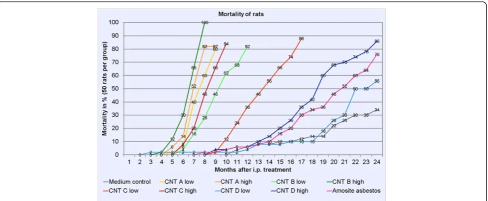
In the MWCNT A and B low- and high-dose groups and in the MWCNT C high-dose group, 80% of the ani-mals died or were humanely killed due to a bad clinical condition (for mortality curves see Figure 1). In the MWCNT C low-dose group, mortality reached 80% 16 months after the intraperitoneal treatment. The MWCNT D high-dose group showed 80% mortality after 23 months. At the end of the experimental time after 24 months, mortality was 56% in the MWCNT D low-dose group, 76% in the amosite asbestos group, and 34% in the negative control group.
Macroscopic findings
At necropsy most animals of the MWCNT treatment groups and the majority of rats of the amosite asbestos group revealed ascites consisting of blood-containing fluid. Most animals showed adhesions and multifocal to coalescing small nodular lesions on the surface of the abdominal organs. Many rats had large nodules or tissue masses especially in the area of the diaphragm, liver, and pancreas, but the small intestine was also frequently in-volved. In some animals, we found aggregates of foreign material covered by a thin layer of serosa, primarily at the diaphragm and liver. The liver and spleen capsules were thickened and firm in numerous rats.
Histopathological findings
Tumor incidences and time dependence
The most frequent tumors in the MWCNT and amosite asbestos treatment groups were malignant mesotheliomas of the peritoneum. In most cases these tumors were the obvious reason for a morbid condition of the rats and trig-gered the decision to perform euthanasia. In addition, we found a few pancreatic islet-cell adenomas, Leydig cell adenomas of the testes, and one renal lipoma. Tumors in other organs were infrequently and their incidences corre-sponded to normal spontaneous background.
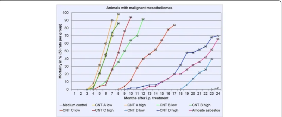
Table 1 shows the incidences of malignant mesothelio-mas including survival times of mesothelioma-bearing rats. The development of malignant mesotheliomas over time is shown in Figure 2.
Rats in the MWCNT A groups developed a mesotheli-oma rate of 98% in the high-dose group and of 90% in the low-dose group. Mesothelioma-bearing animals had a mean survival time of 194 days in the high-dose group
Figure 1Mortality curves in % of all rats of the study comprising all animals which died or were humanely killed due to a bad clinical condition.A group was terminated when 80% mortality was reached.
Rittinghausenet al. Particle and Fibre Toxicology2014,11:59 Page 3 of 18
Table 1 Incidences of malignant mesotheliomas, mean survival times of morbid mesothelioma-bearing rats, and first day of mesothelioma detection
Substance Length (μm) WHO fibers*
Diameter (μm) WHO fibers*
Dose WHO fibers* 109
per rat (actual##) No. ofrats Rats withmesothelioma Mean survival time ofmesothelioma-bearing
rats (days)
Month of first detection of a morbid
mesothelioma-bearing rat Final month**
Medium control - - - 50 1 (2%) 739 24 24
MWCNT A low 8.57 ± 1.51 0.085 ± 1.60 0.48 50 49 (98%) 213 5 9
MWCNT A high 2.39 50 45 (90%) 194 5 8
MWCNT B low 9.30 ± 1.63 0.062 ± 1.71 0.96 50 46 (92%) 294 6 13
MWCNT B high 4.80 50 45 (90%) 207 5 9
MWCNT C low 10.24 ± 1.64 0.040 ± 1.57 0.87 50 42 (84%) 415 10 18
MWCNT C high 4.36 50 47 (94%) 265 6 11
MWCNT D low 7.91 ± 1.40 0.037 ± 1.45 1.51 50 20 (40%) 666 20 24
MWCNT D high 7.54 50 35 (70%) 585 11 24
Long amosite asbestos
13.95 ± 2.10 0.394 ± 1.83 0.14 50 33 (66%) 623 14 24
*WHO fibers: length greater than 5μm, diameter less than 3μm, and a length-to-width ratio (aspect ratio) of at least 3:1.
##
Actual WHO fiber number dose; deviations from target values are due to inhomogeneities during successive synthesis or suspension of test items.
**
Last month when a mesothelioma-bearing rat was found in a group.
Rittingha
usen
et
al.
Particle
and
Fibre
Toxicology
2014,
11
:59
Page
4
o
f
1
8
http://ww
w.particleand
fibretoxicolo
gy.com/cont
and of 231 days in the low-dose group. Mesotheliomas were detected in rats euthanized between 5 and 9 months in the low-dose group and between 5 and 8 months in the high-dose group.
In the MWCNT B groups, we found mesothelioma rates of 90% (high dose) and 92% (low dose). Survival times of mesothelioma-bearing rats were 207 days (high dose) and 294 days (low dose). Mesothelioma-bearing rats were euthanized between 5 and 12 months in the low-dose group and between 5 and 9 months in the high-dose group.
Incidences of mesotheliomas in the MWCNT C groups were 94% after treatment with the high dose and 84% with the low dose. In these groups, mesothelioma-bearing rats had a mean survival time of 265 days with the high dose and of 415 days with the low dose. Ani-mals with mesotheliomas were observed between 10 and 18 months after treatment in the low-dose group and between 5 and 10 months after treatment in the high-dose group.
Animals of the MWCNT D treatment groups developed malignant mesotheliomas in 70% (high dose) and 40% (low dose). Survival times of the mesothelioma-bearing rats were 585 days with the high dose and 666 days with the low dose. We detected mesotheliomas in rats eutha-nized between 20 and 24 months in the low-dose group and between 11 and 24 months in the high-dose group.
In the amosite asbestos group, the mesothelioma inci-dence was 66%, with a mean survival time of 623 days. We observed mesotheliomas in rats euthanized between 14 and 24 months after intraperitoneal injection.
In the rats of the medium control group, we found one mesothelioma (2%) during final necropsies.
Other non-neoplastic MWCNT-induced histopathological findings in the abdominal cavity
Most of the MWCNT-treated rats showed multiple granulomas on the peritoneal surface of the abdominal organs and diaphragm. The granulomas frequently consisted of a central hyaline area which mostly included a few single fibers, surrounded by macrophages and lymphocytes. An outer layer of fibrocytes and collagen, covered by serosa, was mostly visible (see Figure 3). Organs such as the liver and spleen were engulfed by a thick layer of collagen-rich con-nective tissue intermingled with granulomas. In many cases, the liver showed adhesions to the diaphragm by fibrotic if not tumor tissue. We found agglomerates of foreign material underneath the serosa in many animals. Multifocal papillary projections of mesothelial hyperplasia were observed espe-cially on the diaphragm and diaphragmatic liver surface.
Large aggregates occurred infrequently, whereas we observed single nanotubes in every slide (see Figure 3). Similar lesions were found in asbestos-treated animals (see Figure 3).
Histologic appearance of malignant mesotheliomas
Malignant mesotheliomas showed predominantly superfi-cial growth on the serosal surface of the peritoneum of the liver, spleen, intestine, and abdominal wall. We frequently observed invasion into the mesentery, pancreatic tissue, and diaphragm. All in all, 320 of the 329 MWCNT-related malignant mesotheliomas had invaded the diaphragm, and 174 of these tumors showed invasion of the thoracic cavity. In the amosite asbestos group, which displayed 33 (66%) mesotheliomas, 29 mesotheliomas had invaded the dia-phragm, and 5 of these tumors showed invasion of the thoracic cavity.
Figure 2Animals with malignant mesotheliomas over time in %.A group was terminated when 80% mortality was reached.
Rittinghausenet al. Particle and Fibre Toxicology2014,11:59 Page 5 of 18
In the majority of cases, the liver was completely encap-sulated, but it was frequently invaded as well. In contrast, invasion of the spleen along the vascular blood supply was rare. All malignant mesotheliomas showed some cellular pleomorphism; some of the tumors exhibited partly ana-plastic round cells, especially those occurring early in the study. A couple of mesotheliomas developed areas with mucus production, others showed microcystic areas.
Based on their appearance in the H & E stain, we differen-tiated the malignant mesotheliomas into epithelioid, sarco-matoid, and biphasic types (see Figure 4). Malignant mesotheliomas of the epithelioid type occurred occasionally in the MWCNT groups B high, C low and high, D low and high, and in the amosite asbestos group. The malignant mesothelioma found in the medium control group displayed an epithelioid appearance. Epithelioid-type malignant meso-theliomas showed papillary structures or poorly formed glandular structures and round nuclei, and they infiltrated the adjacent connective tissue. All epithelioid-type malignant mesotheliomas were immunohistochemically positive for the markers pan-cytokeratin, podoplanin, and vimentin.
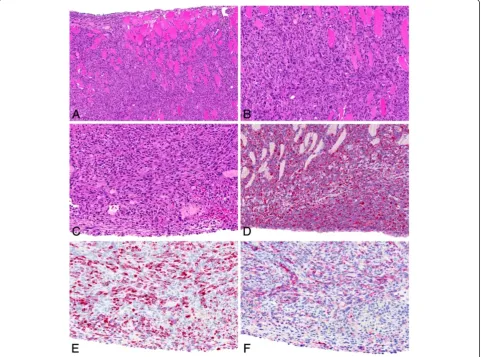
Malignant mesotheliomas of the sarcomatoid type (see Figure 5) were frequent in all MWCNT and amosite asbes-tos groups. The sarcomatoid-type mesotheliomas consisted of spindle-shaped cells with elongated nuclei forming bun-dles or whorls of cells. We observed osseous differentiation in a number of cases in each group. All sarcomatoid-type
Figure 4Histological typing of malignant mesotheliomas occurring in the study.The most frequent type of mesothelioma was the sarcomatoid type, followed by mesotheliomas of the biphasic type. Mesotheliomas of the epithelioid type were rare. Figure 3Non-neoplastic histopathological findings in the abdominal cavity. A: High-power view of anti-podoplanin immunohistochemistry showing single MWCNT A (high dose) nanotubes in the tissue (arrows).B: High-power view of anti-podoplanin immunohistochemistry showing single asbestos fibers in the tissue (arrows).C: H & E, high-power view of granuloma induced by MWCNT A (low dose) nanotubes including single nanotube (arrow, 25×).D: H & E, high-power view of granuloma induced by asbestos including single fiber (arrow, 40×).
Rittinghausenet al. Particle and Fibre Toxicology2014,11:59 Page 6 of 18
malignant mesotheliomas were immunohistochemically positive for the markers vimentin and podoplanin.
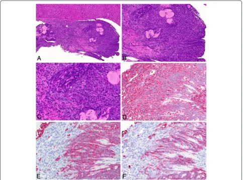
Malignant mesotheliomas of the biphasic type, compris-ing both epithelioid and sarcomatoid patterns, were also seen in animals of all MWCNT groups (see Figure 6) and the amosite asbestos group (see Figure 7). Several of these tumors exhibited osseous differentiation in parts of the lesion. All biphasic-type malignant mesotheliomas were immunohistochemically positive for the markers vimentin, podoplanin, and pan-cytokeratin.
Mesotheliomas occurring in the early phase of the study more frequently showed a higher number of poorly differen-tiated cells or areas of anaplastic cells.
Malignant mesotheliomas in the amosite asbestos group developed the same histological appearances and reactions to immunohistological markers as the MWCNT-induced mesotheliomas.
Overall, the study revealed highest incidences for the sarcomatoid type of mesothelioma, followed by the biphasic
type. Mesotheliomas of the epithelioid type were rare in our study.
Discussion
The hypothesis of a mesothelioma-inducing potential of multi-walled carbon nanotubes, assumed because of their fiber-like dimensions and biopersistence comparable to asbestos fibers, was confirmed by our experimental investi-gations with rats treated intraperitoneally with tailor-made MWCNTs which were suspended in fluid according to Por-ter and co-workers [17] similar to surfactant-containing lung fluid. Amosite asbestos fibers served as positive con-trol. A dose-dependent difference in the carcinogenic response, however, is difficult to deduce from our study. Tumor incidences in all MWCNT dose groups treated with 109or 5 × 10⁹nanotubes were 40 to 100%. Our experimen-tal results thus clearly provide proof of the mesothelioma-inducing potential of these MWCNTs in rats. However, the data cannot be used for risk assessment. This is especially
Figure 5Sarcomatoid-type malignant mesothelioma observed in the MWCNT B high-dose group. A: H & E, low magnification of tumor tissue invading the diaphragm (20×).BandC: H & E, high-power view (40×).D: High-power view of anti-vimentin immunohistochemistry showing a strongly positive result (40×).E: High-power view of anti-pan-cytokeratin immunohistochemistry showing several positive cells (40×). F: High-power view of anti-podoplanin immunohistochemistry revealing positively stained cytoplasm of many cells (40×).
Rittinghausenet al. Particle and Fibre Toxicology2014,11:59 Page 7 of 18
true because we have no data on the carcinogenic potential of MWCNTs in rats after inhalation exposure and in par-ticular also no information on the exposure concentration needed to reach a sufficient dose of MWCNTs at the pleura to induce mesotheliomas.
As positive control, we injected a long type of amosite asbestos, dosed at a concentration of 1 × 10⁸WHO fibers per animal. Using this dose, a mesothelioma incidence of ap-proximately 50% could be expected. Davis and co-workers [16], who investigated the UICC standard reference of amos-ite asbestos, reported the following data: At doses of 0.5, 2.5 mg, and 15 mg amosite, corresponding to 1.7 × 10⁷, 8.5 × 10⁷, and 5.1 × 10⁸ fibers with a length greater than 5 μm, respectively, they found mesothelioma incidences of 46.9%, 59.4%, and 79.2%. At a dose of 1 × 10⁸fibers greater than 5μm, a mesothelioma incidence of approximately 50% can be predicted [16]. In the present study, the incidence of mesotheliomas caused by asbestos was 66%.
There are various hypotheses on the mechanisms that might contribute to the induction of tumors by biopersis-tent fibrous particles. The carcinogenic effect of the tested nanotubes might be due to frustrated phagocytosis followed by chronic inflammation, which has been described for asbestos fibers [33,34] and for MWCNTs as well [35]. An exaggerated inflammatory response of the peritoneal meso-thelium with granuloma formation was observed by Poland and co-workers [12] after injection of long but not short amosite asbestos fibers and also after injection of MWCNTs with a large proportion of long fibers. Poorly soluble but otherwise non-toxic particles that are non-digestible and non-degradable can harm macrophages when they take up a large volume of this material or when this material activates and damages the macrophages due to its long fiber shape. These macrophages will no longer be able to move properly and eliminate the foreign material. Activated and apoptotic macrophages and interaction of the foreign material with
Figure 6Biphasic-type malignant mesothelioma observed in the MWCNT D low-dose group. A: H & E, low magnification of tumor tissue found on the surface of the omentum (20×).BandC: H & E, high-power view (40×).D: High-power view of anti-vimentin immunohistochemistry showing a strongly positive result (40×).E: High-power view of anti-pan-cytokeratin immunohistochemistry showing positive staining of the majority of cells (40×).F: High-power view of anti-podoplanin immunohistochemistry revealing positively stained cytoplasm of many cells (40×).
Rittinghausenet al. Particle and Fibre Toxicology2014,11:59 Page 8 of 18
surrounding epithelial cells then causes an inflammation that becomes chronic because of the continuing irritant and damaging effect of the foreign material. This particle-induced chronic inflammation and the high concentration of reactive oxygen and nitrogen species generated in the course of the inflammatory process can also lead to oxida-tive damage and genotoxic effects. A significant increase in reactive oxygen species was measured in monocyte-derived macrophage monocultures after administration of surfac-tant pre-coated MWCNTs, whereas uncoated MWCNTs caused only a minor reaction [36]. In our study, the carbon nanotubes tested were suspended in a fluid [17] contain-ing 1,2-dipalmitoyl-sn-glycero-3-phosphocholine (DPPC), a major component of surfactant. We used this fluid for dispersion, because it prevented agglomeration of the MWCNTs during dosing of the animals. The surface of the nanotubes in our study was otherwise free of chemi-cals and unloaded, so that they were hydrophobic. Func-tionalization of carbon nanotubes affects their bioactivity
and changes surface reactivity, surface loading, stability, dispersability, and agglomeration [37]. A short-term
in-vivo test [38] demonstrated a different behavior of functionalized MWCNTs, interpreted as an influence on nanotube size and agglomeration. Our non-functionalized hydrophobic MWCNTs dispersed well in the DPPC-containing medium, avoiding aggregation of nanotubes. Aggregated nanotubes would have produced a different reaction because of the resulting change in bioavailability, width, length, surface area, shape, and other factors. The hydrophobicity of carbon nanotubes enables them to pene-trate cell membranes quite easily. Penetration of uncoated MWCNTs with a diameter of 49 nm and a length of 3.86μm into lung epithelial cells has been demonstrated [39].
Furthermore, uncontrolled cell proliferation may be the result of CNT interaction with the mitotic spindle apparatus, which has been describedin vitrofor asbestos fibers [40], for 1- to 4-nm-thin single-walled CNTs [41], and for 10- to 20-nm-thin MWCNT fibers [42]. The
Figure 7Biphasic-type malignant mesothelioma observed in the amosite asbestos group. A: H & E, low magnification of tumor tissue found on the liver surface (10×).B: H & E, higher magnification (20×).C: H & E, high-power view (40×).D: High-power view of anti-vimentin immunohistochemistry showing a strongly positive result (40×).E: High-power view of anti-pan-cytokeratin immunohistochemistry showing positive staining of many cells (40×).F: High-power view of anti-podoplanin immunohistochemistry revealing positivity in distinct parts of the tumor.
Rittinghausenet al. Particle and Fibre Toxicology2014,11:59 Page 9 of 18
impact of MWCNT treatment on the cell cycle resulted in an increase in cells in the proliferation phase. The MWCNTs in our study were more than twice the diam-eter of the nanotubes used by Siegrist and co-workers [42], therefore, interaction with the mitotic spindle cell apparatus may be less likely.
The results of our study are in contrast to the findings by Varga and Szendi [43], who found induction of granu-lomas but not mesotheliomas in Fischer 344 rats. They used single-walled CNTs with a diameter of less than 2 nm and lengths of 4 to 15 μm and short multi-walled CNTs with diameters of 10 to 30 nm and lengths of 1 to 2μm. Furthermore, their experimental time was only one year. Their negative finding in respect of tumors after treatment with MWCNTs may be due to the short nano-tube length of only 1 to 2μm. At least, the carcinogenic potential of mineral fibers with a length below 5μm was questioned. The single-walled CNTs were less than 2 nm in diameter, leading presumably to a curled shape getting tangled, resulting in the occurrence of almost no single CNT but rather a particle-like appearance and behavior. Another difference from our study was the mode of administration. Varga and Szendi implanted the CNT fi-bers, encapsulated in hard gelatin capsules, into the Kertai fold located on the inner liver surface, probably leading to reduced deposition of nanotubes at the serosa. In our study, we injected the fibers directly into the abdominal cavity, suspended in a fluid.
No mesotheliomas were observed after intraperitoneal injection of multi-walled carbon nanotubes measuring 11 nm in diameter and with a length of most nanotubes below 5μm into Wistar rats [14]. Nanotubes with diame-ters of less than 20 nm most likely have a curled shape and are tangled, and together with the relatively short length of most fibers this is responsible for the negative outcome in respect of tumors. The thinnest nanotubes used in our study, which had more than twice the width of the nanotubes tested by Muller and co-workers [14], showed a later onset of tumor morbidity compared with our other MWCNTs, but the induced tumor incidence was still quite high.
In another study, MWCNTs were injected subcutane-ously in rasH2 mice [44]. The authors implanted the test material, which had a mean diameter of 100 nm and a mean length of 10 micrometers, into the subcutaneous tissue. They evaluated the tumor outcome after 26 weeks, but observed neither tumors nor inflammatory cells at the injection site. There are no data in the literature showing tumors after subcutaneous injection of mineral fibers, except the report about mesothelioma induction in mice by subcutaneous injection of asbestos fibers by Roe and co-workers [45], and even for a tumor response based on a foreign-body effect the experimental time was too short to allow final conclusions to be drawn.
Mitsui MWCNT-7 carbon nanotubes with an average width of 100 nm and a length of 1 to 20 μm (median of 2μm) administered by intraperitoneal injection into het-erozygous p53 mice [13,46] and by intrascrotal injection into Fischer 344 rats [47] caused mesotheliomas as well. Mitsui MWCNT-7 show a needle-like straight structure, probably due to their relatively large diameter, and this kind of MWCNTs is structurally very similar to carcino-genic mineral fibers. This is in accordance with our observations on our tailor-made MWCNTs. The more needle-like MWCNTs A and B with mean widths of 85 and 62 nm, respectively, seemed to have a stronger car-cinogenic potency than MWCNTs of type C with a diameter of 40 nm and a more bent structure than MWCNTs A and B. At least the low-dose MWCNT C group showed a slightly later onset of tumor-related morbidity compared with MWCNTs A and B. The later onset of tumor-related morbidity and moderate meso-thelioma incidences in the MWCNT D groups may be due to the same reason. In the scanning electron micros-copy photographs this type of nanotube shows a more curved shape compared with MWCNTs A, B, and also C. The bent nanotube material D had the highest por-tion of WHO fibers with a length greater than 5μm, but the lowest portion of WHO fibers with a length greater than 20 μm. It is known that the carcinogenic potency of biopersistent mineral fibers increases with growing fiber length. Frustrated phagocytosis and long clearance half-times in the pleural space may play a role in this context [3].There must be other factors than length and diameter influencing the effect of MWCNTs. The kinetic behavior of bent MWCNTs is most probably different from that of straight MWCNTs, which might influence clearance half-times.
Nagai and co-workers [15] demonstrated cytotoxicity of thin and rigid MT50a-MWCNTs with a diameter of 50 nm and a length of 5.29 μm in vitro. In contrast, thick NT115- and NT145-MWCNTs with a diameter of 150 nm and a length of 4.88 or 4.34 μm, respectively, and tangled NTtngl nanotubes with diameters of 2 to 20 nm and a length of 3μm were less toxic.
The same authors observed mesotheliomas in rats after i.p. injection of MWCNTs. The diameters of these nano-tubes were 50, 116, and 143 nm, but their average length was only between 4 and 5μm. The study report provided no information as to whether the various types of nano-tubes used differed in shape (straight, needle-like, or rather curved or wavy). The authors treated rats with 107to 108 nanotubes per animal and observed the effects for one year. The number of rats available for histopathological investi-gations was quite different per experimental group, ranging from 6 to 43. Incidences of mesotheliomas observed per group were mostly quite high, reaching 80 to 100%. Nano-tubes of 50 nm in diameter caused more tumor-bearing
Rittinghausenet al. Particle and Fibre Toxicology2014,11:59 Page 10 of 18
animals after one year than nanotubes of 143 nm diameter after treatment of the rats with identical number concen-trations of nanotubes. Based on this result, the authors suggested reduced carcinogenic potency for nanotubes with lower diameters. No tumors were found in 6 rats treated with tangled nanotubes with a diameter of 15 nm, even after up to three years of observation time [48]. No tumors were found either in 15 rats treated with the same tangled nanotubes after one year of experimental time. The average length of the nanotubes used in this experi-ment was quite low, but the frequency distribution in the lengths of the various nanotubes used showed some differ-ences, especially in the length range of 7 to 13μm. We think that a major effect of the length of nanotubes on their carcinogenic potency cannot be ruled out. Tangled nanotubes behave like granular particles, with almost no single nanotubes available to induce mesotheliomas.
The observations by Nagai and co-workers [15] were in concordance with the results of our study, where all
MWCNTs with mean diameters between 37 and 85 μm
and mean lengths between 8 and 10 μm (the length distribution within each CNT sample showed a certain percentage of CNTs with lengths greater than 20 μm) showed a carcinogenic effect. In our study, the more bent but not tangled or agglomerated MWCNTs D had a longer latency period and thus lower carcinogenic potency in comparison with the more straight and needle-like MWCNTs A, B, and C. The absence of mesotheliomas after three years in rats treated with tan-gled MWCNTs [15,48] is in line with the hypothesis that very thin, and curled and tangled CNTs only occur as agglomerates and their toxic effects are very similar to the ones of biopersistent granular particles. Pauluhn and co-workers [49] described such particle-like toxic effects in a subchronic rat inhalation study with Baytubes®. These nanotubes are 10 to 15 nm in diameter and 2 to 10 μm in length. They were very curled and tangled, occurring only as agglomerates, and therefore the typical particle-like effects in rat lungs including alveolar macrophage overload but no fiber or single CNT-related effects occurred. Similar observations were made by Ma-Hock and co-workers [50], who tested Graphistrength™ MWCNTs with diameters of 10 to 15 nm and lengths of 1 to 10μm in a short-term rat inhalation study (6 days with a 3-week post-inhalation period). These carbon nanotubes were agglomerated in bundles and caused granuloma forma-tion in the lungs. A carcinogenic potential of such tangled and agglomerated nanotubes at low doses seems to be un-likely, because phagocytosis by alveolar macrophages is given. For the pleural cavity, Donaldson and co-workers [3] assumed a risk of asbestos-like pathogenicity for nanotubes that are longer than 5 μm. Longer fibers cannot negotiate the stomata (2–12μm) and are retained there, so that they can interact with the mesothelium and immune cells.
Nagai and co-workers [15] also performed rat genome microarray CGH analysis of mesothelioma samples from their MWCNT groups. In the mesothelioma samples, whose signal intensity at the Cdkn2a/2b locus was largely lower than −1, they found homozygous deletion of the gene. Furthermore, they detected deletion of this gene locus also in their asbestos-induced mesothelioma samples.
The mechanism of mesothelioma induction by the tested MWCNTs seemed to be very similar to those trig-gered by asbestos. A known factor contributing to tumor induction by some mineral fibers is the presence of transi-tion metals which may cause metal-catalyzed free radical chain reactions, as in the case of iron and hydroxyl radical formation via the Fenton reaction. The nanotubes applied in our study were generated using iron or cobalt as cata-lysts, which were incorporated into the core of the nano-tubes. We did not detect dissolution of these metals, however, we also cannot completely exclude that some not measurable metal components were involved in the toxic effects, although this seems very unlikely in our opinion. The possibility that insoluble metals included on the particle surface could cause an immune reaction was discussed formerly [51]. X-ray photoelectron spectros-copy, however, revealed no metals on the surface of the MWCNTs used in the present study.
Since biopersistent granular dusts are not able to induce mesotheliomas after intraperitoneal injection, even at very high doses of 4 × 20 mg per rat i.p. [9], it is very unlikely that the mass doses administered in the present study, ranging from 0.05 to 3.0 mg per rat, induced the mesotheliomas per se.
To explain the carcinogenic potential of the MWCNTs used in our study, we would primarily point to the high numbers of nanotubes fulfilling the criteria for WHO fibers in combination with their lack of entanglement and of agglomeration in the suspension, which is due to their thickness and the resulting needle-like shape. The rigidness of the different multi-walled carbon nanotubes is difficult to estimate and also dependent on their thick-ness. A nanotube length of more than 15 to 20 micro-meters, preventing clearance from the serosal cavities because of limited stoma size and leading to frustrated phagocytosis and subsequent chronic inflammation, seems to be one mechanism of mesothelioma induction, however, it is probably not the only pathway. The mean length of our four MWCNTs was below 12 micrometers, which is the maximum size of stomatal openings in rats [52], but the test material also included a certain per-centage of nanotubes longer than 20μm.
In 97% of the MWCNT-treated mesothelioma-bearing rats, the diaphragm was invaded by tumor cells, whereas the serosas of the lateral and ventral abdominal walls were only incidentally involved. The high occurrence of mesotheliomas at this location is most probably due to
Rittinghausenet al. Particle and Fibre Toxicology2014,11:59 Page 11 of 18
the fluid flow in the peritoneal cavity towards the diaphragm. The suspended MWCNTs were injected into the inguinal region, but the majority of tumor tissue was found at the diaphragm and adjacent tissue structures. The location of in-duced tumors can be explained by the drainage flow of peri-toneal fluid towards stomata and subsequently into lymphatic vessels. In rats, the majority of lymphatic stomata are located at the diaphragm [53]. Further peritoneal stomata are present in the omentum, mesentery, pelvic peri-toneum, anterior abdominal wall, and liver [54]. Therefore, larger amounts of carbon nanotubes may be transported towards the lymphatic stomata at the diaphragm. It is supposed that nanotubes may be transported to subme-sothelial tissue through the peritoneal stomata. Adjacent cuboidal mesothelial cells are connected by intercellular tight junctions, which limit migration of macrophages or trans-port of red blood cells, particles, or plasma proteins to the submesothelial tissue. For the pleural cavity, stomata are likewise the only connection to the lymphatics [55] and thus the main path for larger particles, fibers, and nanotubes to be eliminated by flow or after phagocytosis by migrating macrophages [56]. The stomata consist of an outer mesothe-lial margin and an inner lymphatic endothemesothe-lial orifice [57], with round or oval openings [56]. Specifications of the size of lymphatic stomata in rats vary from 4.5 to 8μm [58] to 4 to 10μm in diameter [57]. For humans, stomata 2 to 12μm in diameter have been reported [3]. Their size and shape may depend on the contractile state of the diaphragm at the time of fixation. The stomata may function as a sieve, which longer fibers or nanotubes may be unable to pass. Longer nanotubes or fibers like those used in our study may accu-mulate at this location, especially at the smaller stomata, and can impact into the adjacent mesothelium or plug the stoma. Very small particles or very short nanotubes may be incor-porated with fluid into mesothelial cells by pinocytosis [57]. The primary unidirectional valves in the walls of the initial lymphatics allow and regulate entrance of serosal fluid into the lymphatic network preventing fluid backflow. The initial lymphatic vessels located in the submesothelial tissue have diameters of 1 to 10 μm [53]. The high number of longer nanotubes used in our study might have had great impact on the area of the diaphragmatic sieve and stomata open-ings. The increase in intraabdominal fluid in most rats was not only a result of increased production due to inflammatory processes, but might also have been a result of decreased transport capabilities of the stomata.
The peritoneal tumors observed in our study closely resemble human malignant mesothelioma with regard to biological behavior, histopathology, and immunohistochem-ical features. In our study, the histologimmunohistochem-ical features of the MWCNT-induced mesotheliomas and the reaction pat-terns of the immunohistochemical markers were congruent with those observed for amosite asbestos-induced mesothe-liomas. The immunohistochemical results were also in
accordance with the observations by Sargent and co-workers [59], who found serosal tumors after methylcho-lanthrene treatment and additional MWCNT inhalation in mice. The reaction patterns described by Blackshear and co-workers [60] for spontaneous mesotheliomas col-lected from Fischer rats are also in agreement with our observations.
Spontaneous mesotheliomas in rats occur at a low incidence. In aged Wistar rats, incidences are up to 4% in males and 0% in females [61]. In male Fischer 344 rats, mesotheliomas most frequently are restricted to the serosa of the tunica vaginalis, and they are rare [60]. In our medium control group, we found one mesothelioma (2%). Consequently, the mesotheliomas we found in the MWCNT groups of our study at incidences ranging from 40 to 98% must be regarded almost completely as induced by the tested MWCNTs.
There is thus a very high probability that a variety of multi-walled carbon nanotubes are also potent carcinogens like asbestos for humans. Our test was designed for hazard identification of MWCNTs, not for risk assessment. We were able to show that the selected carbon nanotubes had mechanistic potency to induce tumors, comparable to that of asbestos fibers. Inhalation exposure to lower doses of these carbon nanotubes should be performed in the future to enable proper risk assessment. The diameter, length, and shape of the carbon nanotubes seem to be essential param-eters determining their carcinogenic potential.
Conclusion
Because of their special mechanical, structural, chemical, and physical properties, carbon nanotubes have great eco-nomic relevance and production volumes are rapidly in-creasing. Production and processing of nanotubes and nanotube-containing materials are occupational situations where inhalation exposure to nanotubes may occur and a carcinogenic risk cannot be ruled out. Likewise, the staff of research labs handling nanotubes should also be aware of the potential carcinogenic risk, which is comparable to that of asbestos fibers. The question immediately arises whether all nanotubes behave in the same way, also in respect to their potential carcinogenic risk. A similar ques-tion had to be answered about three decades ago: do all mineral fibers have the same carcinogenic potential and potency and do they pose the same risk as asbestos fibers, which were proven to be carcinogenic in humans? All man-made mineral fibers longer than 5 μm and thinner than 3 μm and having a length-to-diameter ratio of at least 3:1 have to be treated as potential human carcino-gens, if their time in rat lung is similar to the half-time of mineral fibers known to induce tumors in humans or rats after inhalation exposure. Or, putting it the other way round, mineral fibers with low biopersistence in rat lung and thus a half-time below a certain number of days
Rittinghausenet al. Particle and Fibre Toxicology2014,11:59 Page 12 of 18
can be excluded from the suspicion of being human carcin-ogens. Contrary to mineral fibers which, depending on their chemical composition, can have quite different solubility properties in lung or alveolar macrophages, all nanotubes are of high durability because of their composition of inor-ganic carbon only. Besides solubility, the half-time in the lung depends on the length of fibers or nanotubes and, in this respect, there seems to be no difference at a first glance between fibrous mineral or fibrous carbon particles. Based on this consideration and because of the high durability of carbon nanotubes, their clearance from the lung or pleural space seems to depend mostly on their length: the longer the carbon nanotubes, the longer their residence time in the target organ and the stronger their toxic effect. And yet, there may be a characteristic of carbon nanotubes that may modify their toxic and carcinogenic potency, and according to our preliminary data and hypothesis this could be the shape of nanotubes. The more bent, curved, or waved the different types of nanotubes, the lower their toxic and carcinogenic potency seems to be. Their kinetic behavior may be slower and cell interactions less intensive. In other words, carbon nanotubes having a straighter and more needle-like shape can be expected to display higher toxic and carcinogenic potency. In addition, comparable to min-eral fibers, it seems that the longer the nanotubes or fibers, the stronger their toxic and carcinogenic potency. On the other hand, at the extreme, strongly curled and tangled carbon nanotubes that exist mostly as agglomerates are less likely to be potential contributors to fiber-specific toxicity.
Methods Test material
For the carcinogenicity study, we used four different tailor-made multi-walled carbon nanotubes (see Table 2), pro-duced by means of the chemical vapor deposition method. The fibers differed in length and diameter, but also to some extent in the production process in that it used diverging carbon sources or catalysts. Because of the high tempera-tures during synthesis (850–900°C for cyclohexane and acetonitrile and 1100–1150°C for benzene), the carbon nanotubes were free of organic chemical remnants on their surfaces. Traces of the catalyst were minimal. The MWCNT surfaces were uncharged and uncoated.
Characterization of carbon nanotubes
Measuring the defect density by Raman spectroscopy re-vealed a ratio of the 1347/cm and 1580/cm peak of 1.0 to 1.3 for all four MWCNTs. Applying the Brunauer-Emmet-Teller (BET) method, the specific surface area of the different MWCNTs ranged between 52 and 60 m2/g. X-ray photoelectron spectroscopy of the MWCNT sur-face showed a carbon content of 94.72 to 99%. The remaining elements consisted of nitrogen (0.0 to 1.5%) and oxygen (1.0-4.93%), depending on the carbon
source. While the acetonitrile source resulted in the high-est nitrogen amount (1.5%), cyclohexane and benzene caused only very low amounts (<0.27%). The oxygen con-tent also depended slightly on the carbon source, with benzene causing 1.0% and cyclohexane up to 4.93%.
MWCNTs A and B had a medium length of 8.57 μm
and 9.30μm, respectively. With 85 nm and 62 nm their diameters were a little thicker than those of MWCNTs C and D, which were 40 nm and 37 nm, respectively. Differences between MWCNTs A and B are the carbon source (A: benzene; B: cyclohexane) and the generation method (A: sublimation method; B: aerosol method). The catalyst for both fibers was ferrocene.
MWCNT C is a thin, long fiber (40 nm in diameter and 10.24μm in length) produced by the aerosol method with cyclohexane as carbon source and ferrocene as catalyst.
MWCNT D is a thin fiber of medium length (37 nm in diameter and 7.91μm in length). It was produced by the aerosol method with acetonitrile as carbon source and cobaltocene as catalyst.
Both MWCNTs C and D have nearly the same diameter, but MWCNT C has a greater length and a much higher proportion of WHO fibers that are longer than 20μm.
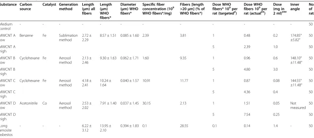
Table 2 shows the mean diameters and mean lengths of our tailor-made MWCNTs A, B, C, D and of the amosite asbestos used.
Characterization of amosite asbestos
The long amosite sample used as reference material was received from Johns Manville Corp. (Littleton, CO, USA) on August 25, 1997.
Dispersion of nanotubes and fibers
For the intraperitoneal test, we performed dispersion of carbon nanotubes and amosite asbestos fibers in“ artifi-cial lung medium” according to Porter and co-workers [17]. Our dispersion medium consisted of Ca2+- and Mg2+-free phosphate-buffered saline, pH 7.4 (Biochrom AG, Berlin, Germany), supplemented with 5.5 mM D-glucose, 0.6 mg/ml bovine serum albumin (Sigma-Aldrich, Seelze, Germany), and 0.01 mg/ml 1,2-dipalmi-toyl-sn-glycero-3-phosphocholine (Sigma-Aldrich, Seelze, Germany). To create the suspensions we used an ultra-sonic homogenizer (Bandelin, Sonoplus HD 2070, VS 70 T) twice for 5 minutes (90% duty cycle, amplitude of 80μm/ss).
Successful homogenization of the suspended nanotubes was proven by SEM. Their length and diameter distribu-tions were measured again (see Figure 8A-E). The SEM images shown were prepared from the suspensions of the respective last pre-test for nanotube dispersion. From each suspension we transferred a very small volume of fluid to the SEM mesh. We cannot exclude that some aggregates may be in the major parts of the suspension that were not
Rittinghausenet al. Particle and Fibre Toxicology2014,11:59 Page 13 of 18
Table 2 Dose groups, substances and dosing Substance Carbon source Catalyst Generation method Length (μm) all fibers
Length (μm) WHO fibers*
Diameter (μm) WHO fibers*
Specific fiber concentration (109 WHO fibers*/mg)
Fibers [length >20μm] (% of WHO fibers*)
Dose WHO fibers* 109per rat (targeted#)
Dose WHO fibers 109per rat (actual##)
Dose (mg in 2 ml)###
Inner angle No. of rats Medium control
- - - 50
MWCNT A low
Benzene Fe Sublimation method
2.72 ± 2.29
8.57 ± 1.51 0.085 ± 1.60 2.39 3.81 1 0.48 0.2 174.85°
±5.82° 50
MWCNT A high
5 2.39 1.0 50
MWCNT B low
Cyclohexane Fe Aerosol method
2.13 ± 2.46
9.30 ± 1.63 0.062 ± 1.71 1.60 9.35 1 0.96 0.6 148.10°
±11.48° 50
MWCNT B high
5 4.80 3.0 50
MWCNT C low
Cyclohexane Fe Aerosol method
4.18 ± 2.41
10.24 ± 1.64
0.040 ± 1.57 10.91 11.77 1 0.87 0.08 144.55°
±11.48° 50
MWCNT C high
5 4.36 0.4 50
MWCNT D low
Acetonitrile Co Aerosol method
2.53 ± 2.02
7.91 ± 1.40 0.037 ± 1.45 30.15 2.13 1 1.51 0.05 Not
measured 50
MWCNT D high
5 7.54 0.25 50
Long amosite asbestos
- - - 6.22 ±
3.12
13.95 ± 2.10
0.394 ± 1.83 0.1 28.55 0.1 0.14 1.4 - 50
Fiber types, lengths and diameters (geometric mean, GMSTD), % of WHO fibers* (length greater than 20μm) and inner angles (to evaluate the degree of circumflexion of the different CNTs, the angle at the concave side [inner angle] was measured between two straight lines, the first from the beginning of the circumflexion to the middle and the second from the middle to the end of the circumflexion).
*WHO fibers: length greater than 5μm, diameter less than 3μm, and a length-to-width ratio (aspect ratio) of at least 3:1.
#
Targeted fiber number dose according to WHO guideline and TRGS 905.
##
Actual fiber number dose; deviations from target values are due to inhomogeneities during successive synthesis or suspension of test items.
###
Total mass dose per rat in mg of substance including also fibers shorter than WHO fibers.
analyzed. The SEM images reflect the same fiber number concentrations as used for intraperitoneal injections.
Dosing scheme
Treatment (see Table 2) consisted in a single intraperi-toneal (i.p.) injection of the test substance in the in-guinal region. We selected the target fiber number concentrations of 1 × 10⁹ and 5 × 10⁹ WHO fibers [1] per animal according to the EU protocol [7] and the German guideline TRGS 905 [62] for carcinogenic, mu-tagenic, and reproduction hazardous materials. WHO fibers are defined as fibers with a length greater than 5 μm and a diameter less than 3 μm and a length-to-width ratio (aspect ratio) of at least 3:1 [7]. As the test items had to be produced successively in aliquots, devi-ations from the initially determined values were found due to inhomogeneities in the final test item bulks.
Thus, the actual doses showed some deviation from the targeted doses. Table 2 shows the actual mass doses per animal. The study duration was scheduled for 24 months after i.p. injection.
The positive control group was treated with amosite asbestos at a ten-fold lower number concentration. We determined this concentration for i.p. administration of amosite asbestos based on published data showing that 1 × 10⁸ WHO amosite asbestos fibers will cause about 50% mesothelioma-bearing rats after two years of experi-mental time [16].
Animal experiment
The study was approved according to the German Animal Welfare Act by the local authority at the LAVES Niedersachsen, Hannover, Germany, No. 33.9-42502-04-11/0507; 33.9-42502-04-11/0743.
Figure 8Scanning electron microscopic photographs of carbon nanotubes and asbestos fibers in suspension.Photographs were taken from liquid formulations of CNT in medium according to Porter (physico-chemical characterization in pre-trials). CNT agglomerates occurred infrequently and are not visible here.A: MWCNT A (left: 10,000×, right: 30,000×).B: MWCNT B (left: 10,000×, right: 30,000×).C: MWCNT C (left: 10,000×, right: 30,000×).D: MWCNT D (left: 10,000×, right: 30,000×).E: Long amosite asbestos (left: 2,000×, right: 7,000×).
Rittinghausenet al. Particle and Fibre Toxicology2014,11:59 Page 15 of 18
For the carcinogenicity test we used 500 male Wistar Han rats Crl:WI(Han), 50 for each group (Charles River Deutschland, Sulzfeld, Germany).
All rats were housed in groups of four rats in polycarbon-ate cages (Macrolon® type IV) on absorbent softwood bed-ding (Lignocell BK 8–15, JRS GmbH & Co KG, Rosenberg, Germany). Tap water (Stadtwerke Hannover, Germany) and pelleted food (R/M-H V1534, Ssniff, Soest, Germany) was offered ad libitum and refreshed at least once weekly. Standard animal housing conditions comprised a temperature of 20 ± 2°C and a 12-hour light/dark cycle.
For unequivocal animal identification, the rats were tattooed at the ears. Animals were checked at least once daily for clinical symptoms, morbidity, or mortality. Body weight recordings were performed at weekly intervals.
Rats were kept for 24 months after i.p. treatment and sacrificed at the end of the study or previously euthanized to avoid unnecessary pain and suffering if they were found morbid and were expected to die soon. All animals which were humanely killed due to a bad clinical condition or which died were included in the mortality curves.
Histology
We performed complete necropsy on all animals that were euthanized either because they were in a bad clinical condition or because a mortality of 80% was reached in the corresponding group, or at the end of the study. Organs and pathologic lesions were fixed in 10% buffered formalin for 24 hours. If subsequent trimming was not possible due to a weekend, the tissue material was trans-ferred to 70% ethanol for further fixation up to 48 hours. All macroscopic lesions, pre-defined parts of diaphragm, abdominal wall, liver, spleen, kidney, mesentery, intestine, lung, and brain were embedded in paraffin. Three-μ m-thick sections were prepared and routinely stained with hematoxylin-eosin.
We used a bright-field microscope for evaluation of the slides. Tumors and pre-neoplastic lesions of the mesothelium were classified according to the INHAND (International Harmonization of Nomenclature and Diag-nostic Criteria for Lesions in Rats and Mice) nomenclature [22]. A second certified veterinary pathologist reviewed tumors and borderline lesions.
Immunohistochemical analysis
From each of the dose groups, we selected ten wax blocks with tumor tissue for immunohistochemistry. A selection factor was the duration of fixation, which we tried to keep as short as possible.
For immunohistochemical detection of the chosen markers for tumor tissue, we cut 3-μm-thick paraffin sections from the histology wax blocks containing the tumor material and mounted them on glass slides. Paraffin
sections were then dewaxed and subject to antigen retrieval methods validated for each of the primary antibodies.
The primary antibodies used comprised monoclonal mouse anti-vimentin (clone V9, isotype IgG1 kappa, Dako Denmark A/S, 2600 Glostrup, Denmark, #M 0725), monoclonal mouse-anti-pan-cytokeratin (clone PCK-26, Sigma-Aldrich Co. LLC, Saint Louis, Missouri 63103, USA, #C-1801), and mouse-anti-podoplanin (clone LF3, Novus Biologicals, Cambridge, UK, #NB110-96423).
Heat-induced antigen retrieval was performed in a citrate-buffered solution. All slides were rinsed with Tris-buffered saline (pH 7.6) plus 0.01% Tween®20 (Merck KGaA, Darmstadt, Germany, 8.22184). We incubated the slides for 20 minutes at 21°C in normal goat serum (Vector Laboratories Inc., Burlingame, CA, USA, S-1000) and then with the primary antibody for 1 hour at 21°C. As secondary antibodies, we applied a biotin-SP-conjugated AffiniPure goat-anti-mouse IgG (Jackson Immunoresearch, West Grove, PA, USA, 111-065-100) for a 30-minute incubation time at 21°C.
Immunostaining was done with a routine method using alkaline phosphatase streptavidin-biotin (Vector Laborato-ries Inc., Burlingame, CA, USA, #S-5100) and Fast Red as chromogen (Fast Red substrate pack, BioGenex, Freemont, CA, USA, #HK182-5 K). The slides were finally counter-stained with Mayer’s hematoxylin (Linaris Biologische Produkte GmbH, Wertheim-Bettingen, Germany, EGH3411). We performed cover slipping using Aquatex® aqueous mounting medium (Merck KGaA, Darmstadt, Germany, #1.08562). Sample permeabilization, antibody concentra-tions, antibody reacconcentra-tions, and staining procedures were optimized for each antibody to get clear and specific immunohistochemical signals.
Statistics
We compared the numbers of rats with and without mesothelioma in the different dose groups with the medium controls by the chi-squared fourfold test, chosing a level of significance of p≤0.05.
Abbreviations
μm:Micrometer; CNT: Carbon nanotubes; i.p: intraperitoneal; MWCNT: Multi-walled carbon nanotube; Nm: Nanometer; SEM: Scanning electron microscopy; SWCNT: Single-walled carbon nanotube; WHO: World Health Organization.
Competing interests
The authors declare that they have no competing interests.
Authors’contributions
SR was involved in planning the pathological investigations and tissue sampling. She performed histopathology and immunohistochemistry of this study and was involved in the interpretation and discussion of results. She wrote major parts of the manuscript and provided the histological photographs. AH performed characterization of the nanotubes in suspension. She evaluated different suspension media to find an optimal method to avoid agglomeration of nanotubes at the time of administration. In addition, she was involved in the dosing of the rats. She was responsible for project management after the death of Bernd Bellmann. OC was involved in the
Rittinghausenet al. Particle and Fibre Toxicology2014,11:59 Page 16 of 18
characterization of the nanotubes and their suspension. He contributed to the chapter“Methods”and was involved in the interpretation and discussion of results. HE reviewed the histopathological slides of tumors. He was involved in the interpretation and discussion of results and review of the manuscript. UH was involved in the design of the study, the interpretation and discussion of results, and the writing of the manuscript. AL generated the nanotubes and performed their characterization. DS was involved in animal necropsy, photographic documentation, discussion of histopathological slides, interpretation and discussion of results, and the writing of the manuscript. All authors read and approved the final manuscript.
Acknowledgments
This project received funding from the German Federal Ministry of Education and Research, grant no: 03X0109A.
The authors are very grateful to Bernd Bellmann (†) for initiating this study. They would like to thank C. Blum, G. Geide, M. Schleiss, A. Schulze, and K. Serwatzki for their excellent technical assistance. The authors wish to thank Karin Schlemminger for language correction.
Author details
1Fraunhofer Institute for Toxicology and Experimental Medicine ITEM, Nikolai-Fuchs-Str. 1, 30625 Hannover, Germany.2Leibniz Institute for Solid State and Materials Research Dresden, PF 270116, 01171 Dresden, Germany.
Received: 9 September 2014 Accepted: 28 October 2014
References
1. World Health Organisation WHO:Reference Methods for Measuring Airborne Man-Made Mineral Fibres (MMMF), WHO/EURO MMMF Reference Scheme, WHO Technical Committee for Monitoring and Evaluating Airborne MMMF. Copenhagen: WHO Regional Office for Europe; 1985.
2. Miller BG, Searl A, Donaldson K, Cullen RT, Bolton RE, Buchanan D, Soutar CA:Influence of fibre length, dissolution and biopersistence on the production of mesothelioma in the rat peritoneal cavity.Ann Occup Hyg 1999,43:155–166.
3. Donaldson K, Poland CA, Murphy FA, MacFarlane M, Chernova T, Schinwald A:Pulmonary toxicity of carbon nanotubes and asbestos - similarities and differences.Adv Drug Deliv Rev2013,65:2078–2086.
4. Mossman B, Light W, Wei E:Asbestos: mechanisms of toxicity and carcinogenicity in the respiratory tract.Ann Rev Pharm Tox1983,23:595–615. 5. Jasani B, Gibbs A:Mesothelioma not associated with asbestos exposure.
Arch Pathol Lab Med2012,136:262–267.
6. Muhle H, Pott F, Bellmann B, Takenaka S, Ziem U:Inhalation and injection experiments in rats to test the carcinogenicity of MMMF.Ann Occup Hyg 1987,31:755–757.
7. JRC EUR 18748 EN–European Commission Joint Research Centre:II3. Carcinogenicity of synthetic mineral fibres after intraperitoneal injection in rats.InMethods for the determination of the hazardous properties for human health of Man Made Mineral Fibres (MMMF). ECB/TM/18(97) rev. Edited by Bernstein DM, Sintes JMR. Ispra, Italy: European Commission Joint Research Centre, Institute for Health and Consumer Protection; 1999. 8. SCOEL:Recommendation from the Scientific Committee on Occupational
Exposure Limits for man-made mineral fibres (MMMF) with no indication for carcinogenicity and not specified elsewhere.SCOEL/SUM/882012,2012:1–17. 9. Pott F, Roller M, Rippe RM, German P-G, Bellmann B:Tumours by the
Intraperitoneal and Intrapleural Routes and Their Significance for the Classification of Mineral Fibres.InMechanisms in Fibre Carcinogenesis. Edited by Brown RC, Hoskins JA, Johnson NF. New York: Plenum Press; 1991:547–565.
10. Hou PX, Xu ST, Ying Z, Yang QH, Liu C, Cheng HM:Hydrogen adsorption/ desorption behavior of multi-walled carbon nanotubes with different diameters.Carbon2003,41:2471–2476.
11. Donaldson K, Aitken R, Tran L, Stone V, Duffin R, Forrest G, Alexander A: Carbon nanotubes: a review of their properties in relation to pulmonary toxicology and workplace safety.Toxicol Sci2006,92:5–22.
12. Poland CA, Duffin R, Kinloch I, Maynard A, Wallace WA, Seaton A, Stone V, Brown S, Macnee W, Donaldson K:Carbon nanotubes introduced into the abdominal cavity of mice show asbestos-like pathogenicity in a pilot study.Nat Nanotechnol2008,3:423–428.
13. Takagi A, Hirose A, Nishimura T, Fukumori N, Ogata A, Ohashi N, Kitajima S, Kanno J:Induction of mesothelioma in p53+/−mouse by intraperitoneal application of multi-wall carbon nanotube.J Toxicol Sci 2008,33:105–116.
14. Muller J, Delos M, Panin N, Rabolli V, Huaux F, Lison D:Absence of carcinogenic response to multiwall carbon nanotubes in a 2-year bioassay in the peritoneal cavity of the rat.Toxicol Sci2009,110:442–448. 15. Nagai H, Okazaki Y, Chew SH, Misawa N, Yamashita Y, Akatsuka S, Ishihara T, Yamashita K, Yoshikawa Y, Yasui H, Jiang L, Ohara H, Takahashi T, Ichihara G, Kostarelos K, Miyata Y, Shinohara H, Toyokuni S:Diameter and rigidity of multiwalled carbon nanotubes are critical factors in mesothelial injury and carcinogenesis.Proc Natl Acad Sci U S A2011,108:E1330–E1338. 16. Davis JM, Bolton RE, Miller BG, Niver K:Mesothelioma dose response
following intraperitoneal injection of mineral fibres.Int J Exp Pathol1991, 72:263–274.
17. Porter D, Sriram K, Wolfarth M, Jefferson A, Schwegler-Berry D, Andrew ME, Castranova V:A biocompatible medium for nanoparticle dispersion. Nanotoxicology2008,2:144–154.
18. Carthew P, Hill RJ, Edwards RE, Lee PN:Intrapleural administration of fibres induces mesothelioma in rats in the same relative order of hazard as occurs in man after exposure.Hum Exp Toxicol1992,11:530–534. 19. Rittinghausen S, Ernst H, Muhle H, Mohr U:Atypical malignant
mesotheliomas with osseous and cartilaginous differentiation after intraperitoneal injection of various types of mineral fibres in rats. Exp Toxicol Pathol1992,44:55–58.
20. Travis WD, Colby TV, Corrin B, Shimosato Y, Brambilla E:Histological typing of lung and pleural tumours.World Health Organization1999,3:25–66. 21. Brambilla E, Travis WD, Colby TV, Corrin B, Shimosato Y:The new world health organization classification of lung tumours.Eur Respir J2001, 18:1059–1068.
22. Greaves P, Chouinard L, Ernst H, Mecklenburg L, Pruimboom-Brees IM, Rinke M, Rittinghausen S, Thibault S, Von Erichsen J, Yoshida T:Proliferative and non-proliferative lesions of the rat and mouse soft tissue, skeletal muscle and mesothelium.J Toxicol Pathol2013,26(3 Suppl):1S–26S.
23. Yaziji H, Battifora H, Barry TS, Hwang HC, Bacchi CE, McIntosh MW, Kussick SJ, Gown AM:Evaluation of 12 antibodies for distinguishing epithelioid mesothelioma from adenocarcinoma: identification of a three-antibody immunohistochemical panel with maximal sensitivity and specificity. Mod Pathol2006,19:514–523.
24. Fassina A, Cappellesso R, Guzzardo V, Dalla Via L, Piccolo S, Ventura L, Fassan M:Epithelial-mesenchymal transition in malignant mesothelioma. Mod Pathol2012,25:86–99. Epub 2011 Oct 7.
25. Marchevsky AM:Application of immunohistochemistry to the diagnosis of malignant mesothelioma.Arch Pathol Lab Med2008,132:397–401. 26. Husain AN, Colby TV, Ordóñez NG, Krausz T, Borczuk A, Cagle PT, Chirieac
LR, Churg A, Galateau-Salle F, Gibbs AR, Gown AM, Hammar SP, Litzky LA, Roggli VL, Travis WD, Wick MR:Guidelines for pathologic diagnosis of malignant mesothelioma: a consensus statement from the international mesothelioma interest group.Arch Pathol Lab Med2009,133:1317–1331. 27. Husain AN, Colby T, Ordóñez N, Krausz T, Attanoos R, Beasley MB, Borczuk AC, Butnor K, Cagle PT, Chirieac LR, Churg A, Dacic S, Fraire A, Galateau-Salle F, Gibbs A, Gown A, Hammar S, Litzky L, Marchevsky AM, Nicholson AG, Roggli V, Travis WD, Wick M:International mesothelioma interest group. Guidelines for pathologic diagnosis of malignant mesothelioma: 2012 update of the consensus statement from the international mesothelioma interest group.Arch Pathol Lab Med2013,137:647–667.
28. Chirieac LR, Pinkus GS, Pinkus JL, Godleski J, Sugarbaker DJ, Corson JM:The immunohistochemical characterization of sarcomatoid malignant mesothelioma of the pleura.Am J Cancer Res2011,1:14–24. Epub 2010 Sep 30. 29. Ordóñez NG:What are the current best immunohistochemical markers
for the diagnosis of epithelioid mesothelioma? A review and update. Hum Pathol2007,38:1–16. Epub 2006 Oct 23.
30. Ordóñez NG:Mesothelioma with signet-ring cell features: report of 23 cases.Mod Pathol2013,26:370–384. Epub 2012 Oct 5.
31. Abe S, Morita Y, Kaneko MK, Hanibuchi M, Tsujimoto Y, Goto H, Kakiuchi S, Aono Y, Huang J, Sato S, Kishuku M, Taniguchi Y, Azuma M, Kawazoe K, Sekido Y, Yano S, Akiyama S, Sone S, Minakuchi K, Kato Y, Nishioka Y: A novel targeting therapy of malignant mesothelioma using anti-podoplanin antibody.J Immunol2013,190:6239–6249.
32. Ohnuma-Koyama A, Yoshida T, Takahashi N, Akema S, Takeuchi-Kashimoto Y, Kuwahara M, Nagaike M, Inui K, Nakashima N, Harada T:Malignant peritoneal
Rittinghausenet al. Particle and Fibre Toxicology2014,11:59 Page 17 of 18