VOL.6 ISSUE 4, 2018; 446 – 450 ; http://ijpda.com; ISSN: 2348-8948
Research Article
Studies On
In Vitro
Anti-Microbial Activity
By Disc Diffusion
Method And FTIR
Analysis OF
Achyranthes
aspera
(L.)
Thafshila Aafrin AM, Anuradha, R*
PG and Research Department of Biochemistry, Sengamala Thayaar Educational Trust Women’s
College, Sundarakkottai, Mannargudi-614 016.
Date Received: 7th March 2018; Date accepted:
26th March 2018; Date Published: 4th April 2018
Abstract
The antimicrobial activity of Achyranthes aspera
against the bacteria and fungi plays a vital role in evaluation of the ethanomedicinal property of plants. FT-IR spectral analysis was carried out in ethanolic extract. The FT-IR analysis has revealed the presence of alcohols, alkanes, terminal al-kyne, alkenes, halogen, ethers, aromatic com-pounds, aliphatic chloro compounds and aryldi-sulphides respectively. This provides and sup-ports the therapeutical information of the plants. The medicinal values of Achyranthes aspera were investigated through agar well disc diffu-sion method. The aqueous and ethanolic extracts of plants to be tested were prepared at a concen-tration of 200µg/mL were dissolved in dimethyl sulphoxide (Dimethyl Sulphoxide (DMSO).The zone of inhibition gives the degree antimicrobial property. It was compared with standard antimi-crobial agents like Gentamycin for bacteria and Amphotericin for fungi. From this study, it can be concluded that Achyranthes aspera exhibits
antimicrobial activity against certain microor-ganisms.
Keywords: Antimicrobial activity, Achyranthes
aspera, FTIR spectral analysis.
Introduction
Medicinal plants can be important source of pre-viously unknown chemical substances with po-tential therapeutic effect. The medicinal use of plants is an ancient tradition, far older than the contemporary sciences of medicine, pharmacolo-gy and chemistry. The World health Organiza-tion (WHO) has estimated that over 75% of the world’s population still relies on plant derived medicines, usually obtained from traditional hea-lers, for its basic health care needs. Herbal medi-cines are in great demand in the developed as well as developing countries for primary health-care because of their wide biological and medi-cinal activities, higher safety margins and lesser costs.
Achyranthes aspera is a species of plant in
the Amaranthaceae family. It is distributed throughout the tropical world. It can be found in many places growing as an introduced spe-cies and a common weed. Achyranthes aspera L. (Latjeera) is an erect or procumbent, annual or perennial herb of about 1-2 meter in height, often with a woody base (Rawat et al., 2008). These phytochemicals are known to possess antioxi-dant (Wong et al., 2009) antibacterial (Nair et al., 2005), antifungal (Khan and Wassilew, 1987), antidiabetic (Singh and Gupta, 2007), anti-inflammatory (Kumar et al., 2008), hypolipidem-ic activity (Durkar et al. 2014) etc and due to these properties they are largely used for medi-cinal purpose.
Infectious diseases
Aafrin TAM & Anuradha, R. ; Int J. Pharm. Drug. Anal, Vol: 6, Issue: 4, 2018; 446-450
Available online at http://ijpda.com
447
to evaluate the antimicrobial activity and FT-IR analysis of Achyranthes aspera extracts against the pathogenic microbes.
MATERIALS AND METHODS
Collection, Identification and Authentication of plant materials
The plant species namely Achyanthes aspera L. plant was collected by in and around Koothanal-lur, Thiruvarur District, Tamil Nadu, India. The plant was identified with the help of the Flora of Presidency of Madras and authenticated by Dr. S. John Britto, RAPINAT Herbarium and Centre for Molecular Systematics, St. Joseph’s college, Tiru-chirappalli (Voucher number of the specimen, AMTA 001) (Gamble, 1997).
Preparation of plant powder
The plant was air dried under shade for 10-15 days. Then the dried material was grinded to fine powder using an electric grinder and stored in air tight bottles. The powder matter was used for further analysis.
Preparation of the aqueous extract
The plant material (Whole plant) was shade dried and coarsely powdered with electrical blender. 200g of Achyranthes aspera was mixed with 1200ml of water. Then it was boiled until it was reduced to one third and filtered. The filtrate was evaporated to dryness. Paste form of the extract obtained was subjected to preclinical screening.
Preparation of the Ethanol extract
Ethanolic extracts was prepared according to the methodology of |Indian pharmacopoeia (Ano-nymous, 1996). The coarse powder material was subjected to Soxhlet extraction separately and successively with 210ml ethanol and 90ml distill-ed water. These extract were concentratdistill-ed to dryness in flash evaporator under reduced pres-sure controlled at a temperature (400C – 500C).
The paste form of the extracts was put in an air tight container stored in refrigerator.
In vitro antimicrobial activity
The antimicrobial activity was performed by disc diffusion method of Beyar etal., 1966..
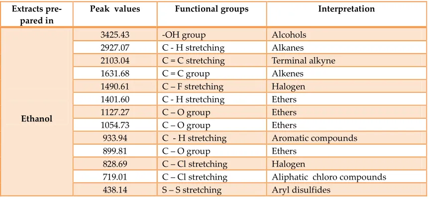
Results And Discussion FTIR analysis
The FT-IR spectrum was used to identify the
functional group of the active components based on the peak value in the region of infra red radia-tion. The ethanolic extracts of Achyranthes aspera
was passed into the FT-IR and functional groups of the components were separated based on its peak ratio. The results showed the presence of alcohols, alkanes, terminal alkyne, alkenes, halo-gen, ethers, aromatic compounds, aliphatic chlo-ro compounds and aryldisulphides respectively
(Table 1 and Figure 1).
Spectral differences are the objective fraction of componential differences. By using, FT-IR spec-trum, we can confirm the functional constituents presence in the given extracts, identify the medi-cinal materials from the adulterate and even eva-luate the qualities (Liu et al., 2005). Many re-searchers applied the FT-IR spectrum as a tool for distinguishing closely associated plants and other organisms (Durates et al., 2008).
Antimicrobial activity
The antimicrobial activities of aqueous and etha-nolic extracts of Achyranthes aspera were studied against two pathogenic bacterial strains (
Escheri-chia coli and Staphylococcus aureus) and two
fun-gal strains (Aspergillus niger and Aspergillus
flavus). Antibacterial and antifungal potential of
aqueous and ethanolic extracts were assessed in terms of zone of inhibition of bacterial and fun-gal growth. The results of the antibacterial and antifungal are presented in Table 2, and Table 3,.
The test organisms used in the study are asso-ciated with various forms of human infections.
E.coli causes septicemias and can infect the gall
bldder, meninges, surgical wounds, skin lesions and the lungs, especially in debliate and the im-munodeficient patients (Dougruz et al., 2008). Whole plant extract of Achyranthes aspera also shows high activity against E.coli with zone of inhibition of 21mm in aqueous extract but poor in ethanolic extract (16mm) as the zone of inhibi-tion 17mm or more considered as high antimi-crobial activity and Staphylococcus aureus with zone of inhibition of 20mm in ethanolic extract but poor in aqueous extract (17mm) as the zone of inhibition 18mm more considered as high an-timicrobial activity (Veeramuthu et al., 2006).
Plate 1 and Plate 2 showed that the antibacterial and antifungal activity of aqueous and ethanolic extract of Achyranthes aspera.
shows the significant activity against most of the tested bacterial and fungal strains. The present study support the traditional usage of plant m terial Achyranthes aspera which possess co pounds with antibacterial and antifungal pote tial that can be used as antimicrobial agents as new drugs for the therapy of infectious diseases caused by pathogens. The results were compared with standard antibiotic drugs gentamycin for bacteria and amphotericin for fungi.
Conclusion
The spectral analysis indicated that the specific functional groups. FT-IR spectroscopy technique
Table 1: FT-IR spectral peak values and functional groups obtained for the whole plant extract in eth Extracts
pre-pared in
Peak values
Ethanol
3425.43 2927.07 2103.04 1631.68 1490.61 1401.60 1127.27 1054.73 933.94 899.81 828.69 719.01 438.14
Figure 1: FT-IR spectral peak values and functional groups obtained for the whole plant extract in eth
ainst most of the tested bacterial and fungal strains. The present study support the traditional usage of plant
ma-which possess com-pounds with antibacterial and antifungal poten-tial that can be used as antimicrobial agents as
drugs for the therapy of infectious diseases caused by pathogens. The results were compared with standard antibiotic drugs gentamycin for
The spectral analysis indicated that the specific IR spectroscopy technique
showed the presence of functional groups which can be identified and further screened for diffe ent kinds of biological activities depending on their therapeutic uses. The antimicrobial activity of aqueous and ethanolic extract of
aspera was evaluated and determined by using
disc diffusion method. The antimicrob
ties of aqueous and ethanolic extract (200µg/ml) were tested against E. coli, S. aureus
A. flavus. The results showed that the
aspera was found to be more effective. On the
basis of the results obtained in the present it is concluded that the whole plant of
ranthes aspera has potent antimicrobial activity.
IR spectral peak values and functional groups obtained for the whole plant extract in eth nol of Achyranthes aspera
Functional groups Interpretation
-OH group Alcohols
C - H stretching Alkanes C = C stretching Terminal alkyne
C = C group Alkenes
C – F stretching Halogen C - H stretching Ethers
C – O group Ethers
C – O group Ethers
C - H stretching Aromatic compounds
C – O group Ethers
C – Cl stretching Halogen
C – Cl stretching Aliphatic chloro compounds S – S stretching Aryl disulfides
IR spectral peak values and functional groups obtained for the whole plant extract in eth nol of Achyranthes aspera
showed the presence of functional groups which further screened for differ-ent kinds of biological activities depending on their therapeutic uses. The antimicrobial activity of aqueous and ethanolic extract of Achyranthes
was evaluated and determined by using disc diffusion method. The antimicrobial activi-ties of aqueous and ethanolic extract (200µg/ml)
E. coli, S. aureus and A. niger,
. The results showed that the Achyranthes
was found to be more effective. On the basis of the results obtained in the present study, it is concluded that the whole plant of
Achy-has potent antimicrobial activity.
IR spectral peak values and functional groups obtained for the whole plant extract in etha-Interpretation
Aliphatic chloro compounds
etha-Aafrin TAM & Anuradha, R. ; Int J. Pharm. Drug. Anal, Vol: 6, Issue: 4, 2018; 446-450
Available online at http://ijpda.com
449
Table 2: In vitro antibacterial activity of aqueous extract of Achyranthes aspera S.No Microorganisms
Zone of inhibition (mm in diameter) Gentamycin
(30µg/ml)
Aqueous extract (200µg/ml)
Ethanolic extract (200µg/ml)
1 Escherinchia coli 23 21 16
2 Staphylococcus aureus 19 17 20
Table 3: In vitro antifungal activity of ethanolic extract of Achyranthes aspera S.No Microorganisms
Zone of inhibition (mm in diameter) Amphotericin
(20µg/ml)
Aqueous extract (200µg/ml)
Ethanolic extract (200µg/ml)
1 Aspergillus niger 15 20 17
2 Aspergillus flavus 13 14 18
Plate 1: Antibacterial activity of aqueous and ethanolic extracts of Achyranthes aspera
Escherichia coli Staphylococcus aureus
Plate 2: Antifungal activity of aqueous and ethanolic extracts of Achyranthes aspera
Acknowledgements
We would like to express my sincere and heart-felt thanks to our beloved correspondent Dr. V. Dhivaharan, M.Sc., D.E.M., Ph.D., Department of Life Sciences, S.T.E.T Women’s College, Sunda-rakkottai, Mannargudi, for encouragement and providing adequate facilities in this research work successfully.
References
Anonymous, 1996.Methodology of Indian
Phar-macopoeia.
Bartlett, Jimmy (2013). Clinical Ocular
Pharmacol-ogy. Elsevier. 214.
Bayer, A.W., Kirby W.M.M., Sherris, J.C., Turck, M. antibiotic susceptibility testing by a standar-dized single disc method. Am. J. Clin. Pathol. 1966; 45: 493-496.
Dogruoz, N., et al., (2008), “Antibacterial Activity of Some Plant Extracts”, IUFS Journal of Biology
Research Article, 67(1):17-21.
Durates, J.M.B., Vogliotti, A., Barbanti, M., 2008. Mazama americana. In: IUCN 2013. IUCN Red List of Threatened Species. Version 2013. 1.
Farnsworth NR, Morris RW. Higher plants--the sleeping giant of drug development. Am J Pharm
Sci Support Public Health. 1976; 148(2):46–52.
Gamble, G. S., Torane, R. C., Mundhe, K. S., Deshpande, N. R. and Salvekar, J. P. J. Chem.
Pharm. Res., 1997, 3(2):465-471.
Griffiths PR, de Haseth JA. Fourier transform infrared spectroscopy. New York: Wiley, 1986.
Hamill, Richard J. (2013. "Amphotericin B Formu-lations: A Comparative Review of Efficacy and Toxici-ty". Drugs. 73 (9): 919–934.
Khan M, Wassilew SW. Natural pesticides from the neem tree and other tropical plants. (Eds)
Kumar A, Ilavarasan R, Jayachandran T, Deeca-raman M, Kumar MR, Aravindan P. Anti in flammatory activity of Syzigium cumini seed. African Journal of Biotechnology, 2008; 7(8): 941-943.
Liu Z, et al. (2005) A novel degron-mediated de-gradation of the RTG pathway regulator, Mks1p, by SCFGrr1. Mol Biol Cell 16(10):4893-904.
Malini A, Deepa E, Gokul B, Prasad S. Non-fermenting gram negative bacilli infections in a tertiary care hospital in Kolar, Karnataka. J Lab
McCann, K.B., Lee, A., Wan, J., Roginski, H. and Coventry, M.J. (2003) The effect of bovine lacto-ferrin and lactoferricin B on the ability of feline calicivirus (a norovirus surrogate) and poliovirus to infect cell cultures. J Appl Microbiol 95, 1026– 1033.
Nair R, Kalariya T, Sumitra C. Antibacterial ac-tivity of some selected Indian medicinal flora. Turkey Journal of Biology, 2005; 29: 41-47. 21.
Rajurkar NS, Nongbri B, Patwardhan AM. Phy-sicochemical and microbial analysis of Umian (Brapani) lake water. Ind J Environ Protec, 2009; 23(6): 633-639. 23.
Rawat P, Kumar M, Sharan K, Chattopadhyay N, Maurya R. Ulmosides A and B: flavonoid 6-C-glycosides from Ulmus wallichiana, stimulating osteoblast differentiation assessed by alkaline phosphatase. Bioorg Med Chem Lett. 2009; 19:4684–4687.
Surewicz WK, Moscarello MA, Mantsch HH (1987). Secondary structure of the hydrophobic myelin protein in a lipid environment as deter-mined by Fourier- transform infrared spectrome-try. J. Biol. Chem. 262:8598-8609.
Veeramuthu D, Muniappan A, Savarimuthu I. Antimicrobial activity of some ethnomedicinal plants used by Paliyar tribe from Tamil Nadu, India. BMC Complementary and Alternative Medicine. 2006;6:35.
Willey J.M., L.M. Sherwood and C.J. Wolverton (2008). Staphylococcae Prescott, Harley and Klein’s Microbiology 7th edition. pp 968-972.
Wong SK, LimYY, Chan EWC. Antioxidant prop-erties of Hibiscus species variation, altitudinal change costal influence and floral colour change. Journal of Tropical Forest Science, 2009; 21: 307-315.