Copyright © 2004, American Society for Microbiology. All Rights Reserved.
Comparison of Blood Smear, Antigen Detection, and Nested-PCR
Methods for Screening Refugees from Regions Where Malaria Is
Endemic after a Malaria Outbreak in Quebec, Canada
Momar Ndao,
1* Etienne Bandyayera,
1Evelyne Kokoskin,
1Theresa W. Gyorkos,
2J. Dick MacLean,
1and Brian J. Ward
1National Reference Centre for Parasitology, McGill University Centre for Tropical Diseases,1and McGill University
Division of Clinical Epidemiology,2Montreal General Hospital, Montreal, Quebec, Canada
Received 11 November 2003/Returned for modification 29 December 2003/Accepted 16 March 2004
The importation of malaria into a region where it is not endemic raises many concerns, including the timely delivery of appropriate care, safety of the blood supply, and the risk of autochthonous transmission. There is presently no consensus on the best way to screen mobile populations for malaria. Between August 2000 and March 2001, 535 refugees arrived in Quebec, Canada, from Tanzanian camps. Within 4 weeks of resettlement of the first group of 224, the McGill University Centre for Tropical Diseases noted an outbreak of malaria across the province (15 cases over a 3-week period). This group (group 1) was traced and screened for malaria between 3 and 4 months after arrival in Canada. Subsequent groups of 106 and 205 refugees were screened immediately upon arrival in Canada (group 2) and immediately prior to their departure from refugee camps (group 3), respectively. A single EDTA-blood sample was obtained from 521 refugees for testing by thick and thin blood smears (groups 1 and 2), antigen detection (ICT Malaria Pf and OptiMAL; group 1 only), and nested PCR (all groups). Overall, 98 of 521 refugees were found to be infected (18.8%). The vast majority of infections (81 of 98) were caused byPlasmodium falciparumalone. Using PCR as the “gold standard,” both microscopy (sensitivity, 50%; specificity, 100%) and antigen detection (ICT sensitivity, 37.5%; ICT specificity, 100%; OptiMAL sensitivity, 29.1%; OptiMAL specificity, 95.6%) performed poorly. None of the PCR-positive subjects were symptomatic at the time of testing, and only two had recently had symptoms compatible with malaria (with or without diagnosis and treatment). Active surveillance of migrants from regions of intense malaria transmission can reduce the risk of morbidity in the migrant population and mitigate against transmission to the host population. Our data demonstrate that PCR is, by far, the most powerful tool for such surveillance.
In the first years of the third millennium, more than 45 million refugees have abandoned their homelands because of oppression, war, famine, and other disasters (47; http://www .refugees.org/world/statistics/wrs02_table3.htm). Malaria is one of the most common causes of serious morbidity and death among refugees and displaced persons (20, 46). Malaria case reports are also increasing in many regions of the world that were previously free of disease (5, 23). Infected refugees mov-ing into areas with little or no disease activity (or immunity) can act as reservoirs and spark full-blown malaria epidemics (21, 22). Refugees and other mobile populations can also take malaria parasites further afield. In recent years, many indus-trialized countries have reported increasing numbers of ma-laria cases in immigrant and refugee communities or in sur-veillance data with high fatality rates (15, 23, 28).
In many developing-world settings, a presumptive diagnosis of malaria is based upon the presence of fever alone. While statistically justifiable in some regions, such an approach inev-itably leads to the overuse of antimalarial drugs. Under ideal circumstances, the clinical suspicion of malaria would be
con-firmed by a laboratory test that is simple to perform, rapid, sensitive, specific, and inexpensive. At the present time, no such test exists. The most common test for malaria diagnosis remains the microscopic examination of Giemsa- or Fields-stained blood smears. However, the examination of blood films requires technical expertise and the availability of a good-quality microscope. Microscopy is also time-consuming and has limited sensitivity when parasitemia is low. During the last decade, several new diagnostic methods for malaria have been developed, including antigen detection (e.g., OptiMAL and ICT) (24a), fluorescence-based assays (e.g., quantitative buffy coat) (26a), and PCR (39a, 42). Each of these tests has strengths and weaknesses in terms of test parameters, cost, and technical complexity.
In August 2000, a charter flight brought 224 refugees from Tanzanian refugee camps to Mirabel airport just north of Montreal, Quebec, Canada. These refugees were immediately dispersed to smaller communities throughout the province to hasten their integration into Quebec society. Within 3 to 4 weeks of their resettlement, the McGill University Centre for Tropical Diseases (TDC), which acts as a reference center for the province, noted a striking increase in requests for confir-matory testing and assistance in treatment. There appeared to be an outbreak of malaria across the province (34a). Malaria parasites were identified in 15 blood smears (11 with
Plasmo-dium falciparum, 2 withPlasmodium ovale, and 2 with mixed
* Corresponding author. Mailing address: National Reference Cen-tre for Parasitology, McGill University CenCen-tre for Tropical Diseases, Montreal General Hospital, Room R3-137, Montreal, Quebec, Can-ada H3G 1A4. Phone: (514) 934-8347. Fax: (514) 934-8347. E-mail: [email protected].
2694
on May 15, 2020 by guest
http://jcm.asm.org/
infection) from African refugees submitted to the TDC during first 6 weeks after their arrival. Parasitemias ranged from 0.3 to 7%. Six children had parasitemias of⬎3%, and one pregnancy was complicated by a diagnosis ofP. falciparum. The arrival of this large group of refugees as well as several subsequent groups prompted us to compare the performances of available malaria tests for the screening of refugees.
MATERIALS AND METHODS
Study subjects.Three groups of refugees (a total of 535 individuals) from Tanzanian camps were screened for malaria. The majority were originally from Burundi (50.9%) and Rwanda (39%), but small numbers originated in Malawi, Tanzania, and the Congo. They were relatively young (mean age, 17 years; range, ⬍1 month to 49 years), there were slightly more females than males (female/male ratio, 244:227), and most had been in refugee camps for at least 1 year (mean, 3.1 ⫾1.6 years; range, 1 to 7 years). Group 1 consisted of 224 refugees who arrived in August 2000 and who were immediately dispersed to communities throughout Quebec. These refugees were recontacted between 3 and 4 months after arrival, and blood was obtained from 210 (94%). At the time of screening, 15 (6.7%) of them had already been diagnosed with malaria during their brief period of residence in Canada (34a). Group 2 consisted of 106 refugees who arrived in October 2000 and were screened immediately upon arrival. All 106 consented to providing a blood sample. Group 3 consisted of 205 refugees scheduled for travel to Canada in June 2001. As part of a treatment trial initiated by Canadian immigration authorities, a blood sample was obtained from these refugees in their camps on the day of departure for the airport. All were subsequently treated with mefloquine (adults, 500 g orally four times a day for 2 days; children, 20 mg/kg orally four times a day for 2 days). None of the subjects in groups 1 to 3 had symptoms compatible with malaria at the time that blood was obtained for screening. All PCR diagnostic tests were performed in a blinded fashion.
Samples.A single venous EDTA-blood sample was collected by venipuncture (for those⬎5 years of age), finger stick (for those⬎1 butⱕ5 years of age), or heel stick (for thoseⱕ1 year of age) from each individual. After thin and thick blood smears were prepared, a few drops (⬃40l/drop) of whole blood were spotted on Whatman no. 4 filter paper and left to air dry. The filter paper samples were stored at⫺20°C in sealed plastic bags containing silica gel until used. The remaining whole blood was frozen at⫺20°C until used for antigen detection. For blood obtained predeparture in Africa (group 3), only filter paper samples were obtained. These were hand carried by immigration personnel to Canada and delivered to the TDC laboratory.
Microscopic examination.Thick and thin smears were prepared for group 1 and 2 refugees. After all of the smears were air dried, the thin smears were fixed in methanol and both thick and thin smears were stained for 45 min in 1:6-diluted
Giemsa stain (pH 7.2) (EM Science, Gibbstown, N.J.). The slides were then rinsed with tap water, air dried, and examined at an oil immersion magnification of⫻100 by TDC technologists. Thick smears were interpreted as negative only after the entire smear was examined (maximum, 30 min). The percent para-sitemia was calculated for the thin smear as total number of infected red blood cells (RBCs) in 100 fields/average number of RBCs per field⫻100 fields. In five cases where parasites were found only in the thick smears, the parasitemia was calculated from the total number of trophozoites per 100 white blood cells, and the percent parasitemia was based on a standard white cell count of 8,000/l and RBCs of 4,000,000 or 5,000,000/l depending on gender.
Antigen detection.The OptiMAL test (Flow Inc., Portland, Oreg.) detects parasite-specific lactate dehydrogenase (LDH) and can be used to detect bothP. falciparumandPlasmodium vivax(36). Each test was carried out with 10l of frozen whole blood and was interpreted according to the manufacturer’s instruc-tions. In the OptiMAL test, there are two diagnostic zones containing different antibodies that recognize eitherP. falciparumalone (lower zone) or bothP. falciparumandP. vivax(upper zone). The ICT Malaria Pf (ICT Diagnostics, Sydney, Australia) detectsP. falciparumonly, using monoclonal antibodies spe-cific for PfHRP-2 antigen (37). This test was also performed according to the manufacturer’s instructions with 10l of frozen whole blood. Although both kits are designed for use with fresh blood samples, the manufacturers’ instructions suggest that they perform equally well with frozen whole blood. Positive and negative control samples were included with each batch tested.
Nested PCR. (i) DNA extraction.Extraction of DNA for PCR was performed as previously described (9). In brief, two 6-mm-diameter filter paper confetti were cut from each blood spot with a chromium-plated paper punch and eluted in 1 ml of double-distilled water for 30 min at room temperature. Between each sampling, the punch was sterilized above a Bunsen burner for⬃3 s and two confetti were cut from a blank filter paper. After centrifugation (7,800⫻gfor 10 min), the supernatant was discarded and 200l of a 2% Chelex 100 (Bio-Rad, Hercules, Calif.) suspension was added to the pellet. The mixture was incubated at 56°C for 30 min and then boiled for 8 min. The sample was then vortexed for 2 min. After a final centrifugation (7,800⫻gfor 5 min), the supernatant was either used immediately for PCR or stored in aliquots at⫺20°C.
[image:2.603.43.544.81.272.2](ii) Amplification and detection.A nested-PCR strategy based upon primers described by Snounou et al. (42) was used, with minor modifications to theP. ovaleprimers. This strategy targets sequences of the 18S ribosomal subunit genes of the four malaria parasites for amplification. The primer sequences and reac-tion condireac-tions are outlined in Table 1. The first amplificareac-tion reacreac-tion used 10 l of the extracted DNA as the template for each PCR (final reaction volume, 50 l). The second amplification was accomplished by using 2l of the first product as the DNA template for each PCR (final reaction volume, 20l). Genomic DNA samples prepared from healthy individuals with no history of malaria were included as negative controls in all PCR assay runs. In order to prevent cross-contamination, different sets of pipettes and work areas were used for template TABLE 1. Characteristics of primer pairsadescribed by Snounou et al.
Primer
name Nucleotide sequence (5⬘33⬘) Parasite targeted
Annealing temp (°C)b
Amplicon length
(bp)
rPLU1 TCAAAGATTAAGCCATGCAAGTGA Plasmodiumsp. (first amplification) 55 ⬃640
rPLU5 CCTGTTGTTGCCTTAAACTCC
rPLU3 TTTTTATAAGGATAACTACGGAAAAGCTGT Plasmodiumsp. (second amplification) 62 240 rPLU4 TACCCGTCATAGCCATGTTAGGCCAATACC
rFAL1 TTAAACTGGTTTGGGAAAACCAAATATATT P. falciparum 58 205
rFAL2 ACACAATGAACTCAATCATGACTACCCGTC
rMAL1 ATAACATAGTTGTACGTTAAGAATAACCGC P. malariae 58 144
rMAL2 AAAATTCCCATGCATAAAAAATTATACAAA
rVIV1 CGCTTCTAGCTTAATCCACATAACTGATAC P. vivax 58 117
rVIV2 ACTTCCAAGCCGAAGCAAAGAAAGTCCTTA
rOVA3 CGGGGAAATTTCTTAGATTGC P. ovale 58 456
rOVA4 GAGAAACAGCATGAATTGCG
aAll primers except those forP. ovalewere described by Snounou et al. (42). bIn all cases the annealing time was 60 s.
on May 15, 2020 by guest
http://jcm.asm.org/
preparation, preparation of master mix for PCR, addition of template to first and second “nests,” and PCR assays. Movement between PCR areas was unidirec-tional on any given work day. Detection of the amplified DNA was accomplished by electrophoresis of 10l of the second PCR product on 2% agarose gels stained with 1 mg of ethidium bromide per liter. Fluorescent bands were visu-alized by using UV illumination and photographed with Polaroid film. All PCRs were conducted using a programmable thermal cycler (PTC 200; MJC Research, Watertown, Mass.).
Calculations.Sensitivity was calculated as TP/(TP⫹FN)⫻100%, specificity was calculated as TN/(TN⫹FP)⫻100%, positive predictive value (PPV) was calculated as TP/(TP⫹FP)⫻100%, and negative predictive value (NPV) was calculated as TN/(TN⫹FN)⫻100%, where TP is the number of true-positive results, TN is the number of true-negative results, FP is the number of false-positive results, and FN is the number of false-negative results. The efficiency was determined as described by Weigle et al. (49) by using the formula 1⫺(FN⫹ FP/Total)⫻100.
RESULTS
Blood smear.Overall, a malaria infection was identified in 13.9% (44 of 316) of the refugees in groups 1 and 2 for whom smears were available. In group 1, malaria parasites were found in 24 of 210 smears (11.4%) (Table 2).P. falciparumwas identified in the large majority of cases (21 of 24 [87.5%]), whilePlasmodium malariaeandP. ovalewere identified in 2 of 24 (8.3%) and 1 of 24 (4.2%) cases, respectively. No mixed infections were detected. In general, the level ofP. falciparum parasitemia was low (mean, 0.023%; range,⬍0.001 to 0.1%). In group 2, malaria parasites were identified in 20 of 106 smears (18.8%). In these cases, onlyP. falciparumandP. vivax were identified in 19 of 20 (95%) and 1 of 20 (5%) subjects, respectively. Again, the level of parasitemia was quite low (mean, 0.005%; range, ⬍0.001 to 0.04%). Six of these cases were identified with gametocytes only or very low numbers of
trophozoites. As noted above, no blood smears were available for group 3.
Antigen detection.Antigen detection (group 1 samples only) identified a malaria infection in 9.3% of the subjects. Only 21 of 210 (10%) were positive by OptiMAL, and 18 of 210 (8.5%) were positive by ICT. If the PCR assay is considered the “gold standard,” all of the infections identified by antigen detection were caused byP. falciparum.
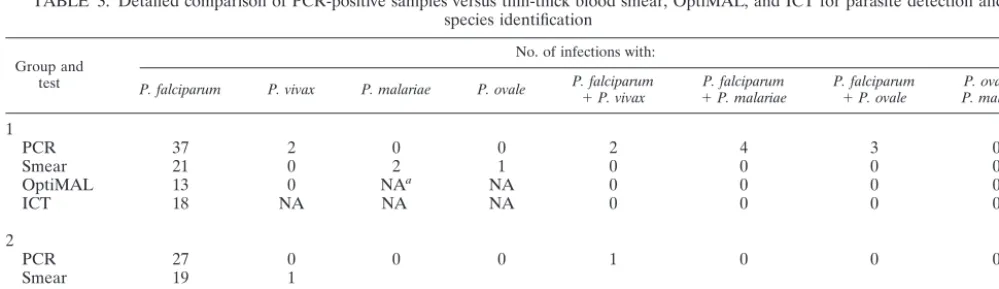
PCR.Overall, PCR identified a malaria infection in 98 of 521 (18.8%) of the refugees screened. The large majority of these infections were caused byP. falciparum(81 withP.
fal-ciparumalone, 2 withP. vivaxalone, 3 withP. ovalealone, and
12 mixed infections with P. falciparumand eitherP. vivax[3 infections]),P. ovale[4], orP. malariae[5]). The PCR preva-lence rates in the three groups ranged from 10.7 to 26.4%. A total of 48 of 210 (22.8%) were infected in group 1 (37 withP.
falciparumalone, 2 withP. vivaxalone, and 9 mixed infections
withP. falciparumwithP. vivax[2],P. ovale[3], orP. malariae
[4]). In groups 2 and 3, 28 of 106 (26.4%) and 22 of 205 (10.7%) were found to harbor a malaria infection, respectively. Again the majority of infections wereP. falciparumalone (44 of 50) in groups 2 and 3, but 3P. ovaleinfections and 3 mixed infections were also identified (Table 3).
Comparison of the screening tests.Comparisons between assays were based only on samples from subjects withP.
falci-paruminfection in group 1 for whom all four test results were
available. All specimens that were positive by either smear or ICT antigen detection were also positive by PCR. As noted above, the OptiMAL test was positive for seven samples that were negative by all other assays. The PCR assay identified a substantial number of malaria infections that were missed ei-ther by smear (32 of 76), by both of the antigen detection tests (27 of 48), or by all three assays (24 samples).
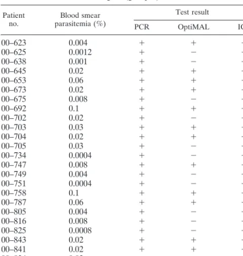
[image:3.603.41.285.99.168.2]There were also significant discrepancies between micros-copy and antigen detection and between the two antigen detection kits. In particular, there was a surprisingly large number of smear-positive (mean parasitemia, 0.012%) but an-tigen-negative samples for both the OptiMAL (n ⫽ 11) and ICT (n⫽10) tests (Table 4). There was also a single case of an apparently false-positive smear (smear positive, PCR nega-tive). Upon exhaustive review of the original smears, this ap-parent discrepancy was resolved in favor of the PCR result.
TABLE 2. Comparison of results for blood smears and OptiMAL and ICT tests for detection ofP. falciparuminfection in group 1
samples with the PCR test as the gold standard
PCR result (n)
No. with the following test result:
Blood smear OptiMAL ICT
Positive Negative Positive Negative Positive Negative
Positive (48) 24 24 14 34 18 30
Negative (162) 0 162 7 155 0 162
TABLE 3. Detailed comparison of PCR-positive samples versus thin-thick blood smear, OptiMAL, and ICT for parasite detection and species identification
Group and test
No. of infections with:
P. falciparum P. vivax P. malariae P. ovale P. falciparum⫹P. vivax ⫹P. falciparumP. malariae P. falciparum⫹P. ovale P. malariaeP. ovale⫹
1
PCR 37 2 0 0 2 4 3 0
Smear 21 0 2 1 0 0 0 0
OptiMAL 13 0 NAa NA 0 0 0 0
ICT 18 NA NA NA 0 0 0 0
2
PCR 27 0 0 0 1 0 0 0
Smear 19 1
3, PCR 17 0 0 3 0 1 1 0
aNA, not applicable.
on May 15, 2020 by guest
http://jcm.asm.org/
[image:3.603.42.542.572.716.2]With PCR as the gold standard assay, the sensitivities and specificities as well as the PPVs and NPVs of the other tests were calculated (Table 5). Although the specificities of the tests were quite good (range, 95.6 to 100%), they all had unacceptably low sensitivities (range, 29.1 to 37.5%).
DISCUSSION
In Canada, the United States, and other developed coun-tries, the importation of malaria by travelers, immigrants, and refugees is a significant and growing health problem (3, 5, 54). In Canada, 6,670 cases of malaria were reported between 1985 and 1997 (3). In 1996 alone, more than 400 cases of malaria were reported in the province of Ontario (13), and country-wide reporting rates reached very high levels in 1997 (1,029 cases), coincident with increased P. vivaxdisease activity in South Asia (26). Slightly more than 1,000 malaria cases are recorded each year in the United States, and 118 malaria-related deaths were reported between 1979 and 1998 (average
of 6 deaths/year) (44). The reasons for the almost threefold-higher per capita rate of imported malaria reported in Canada compared with the United States are not yet understood (3). Many other developed countries report similar experiences with imported malaria (33), and this trend in malaria impor-tation has important implications for clinical care, blood safety, and the possibility of autochthonous transmission of disease (33, 34). At present, few developed countries have established protocols for screening travelers, immigrants, or refugees from regions where malaria is endemic, and most jurisdictions rely exclusively on questionnaire-based exclusion criteria to pre-vent transfusion-associated malaria. The screening of refugees from regions where malaria is endemic regions may be partic-ularly important since high levels of transmission can occur in the suboptimal living conditions that these individuals fre-quently experience (43). There is presently no consensus on the optimal protocol for screening such refugees, in part due to the absence of an “ideal” test.
Microscopy has traditionally been considered the gold stan-dard test for malaria diagnosis. Under optimum conditions, microscopy can detect 20 to 50 parasites per l of blood (0.0004 to 0.001% parasitemia) (11), but such sensitivity is rarely achieved under routine laboratory conditions. This is particularly true in the case of imported malaria, since the expertise of microscopists in countries where the disease is not endemic has been revealed as a major problem (7, 12). The interpretation of a blood smear, particularly at low levels of parasitemia, requires considerable skill (48). Milne et al. found that more than 10% of the blood films submitted to the Lon-don-UK Malaria Reference Laboratory had been read as false negative (31). More recently, it was found that 10 to 15% of laboratories in Quebec routinely misdiagnose quality assur-ance smears with low parasitemia, despite a 3-year, intensive effort to improve the diagnosis of malaria (E. Kokoskin, per-sonal communication). Misdiagnoses can lead to inappropriate therapy or delays in diagnosis and treatment that have been implicated in malaria-associated deaths in developed countries (19). Since none of the refugees in our study were symptomatic at the time that blood was obtained and almost all had lived for many years in refugee camps (i.e., areas with active malaria transmission), it was not surprising that microscopy detected
⬍50% of those infected. The level of parasitemia was low in the large majority of the refugees tested (mean, 0.014%), and single trophozoites or gametocytes were the basis for the mi-croscopic diagnosis for many subjects. Indeed, it is likely that the 15- to 30-min microscopic examination performed by the skilled technologists of the TDC in the present study actually overestimates the sensitivity of this technique when performed in most laboratories.
Low parasitemia has long been recognized as the Achille’s heel of the commercial antigen detection kits as well. Initially reported to be⬃90% sensitive for the diagnosis of falciparum malaria compared with microscopy (8, 39), subsequent studies suggested that sensitivities at low (⬍0.002%) parasitemia could drop to as low as 11 to 40% (17, 40, 51). It was therefore understandable that neither the OptiMAL (sensitivity,
[image:4.603.44.284.107.360.2]⬃29.1%) nor the ICT (sensitivity,⬃37.5%) kit performed par-ticularly well as screening tools for the refugees in our study. Although the product monographs for both kits suggest that either fresh or frozen whole blood can be used, we do not know
TABLE 4. Detailed comparison of PCR and antigen detection (OptiMAL and ICT) in samples with defined levels of parasitemia
by blood smear for diagnosis of malaria in East African refugees (group 1)
Patient
no. parasitemia (%)Blood smear
Test result
PCR OptiMAL ICT
00–623 0.004 ⫹ ⫹ ⫺
00–625 0.0012 ⫹ ⫺ ⫺
00–638 0.001 ⫹ ⫺ ⫺
00–645 0.02 ⫹ ⫹ ⫺
00–653 0.06 ⫹ ⫹ ⫺
00–673 0.02 ⫹ ⫹ ⫹
00–675 0.008 ⫹ ⫺ ⫹
00–692 0.1 ⫹ ⫹ ⫹
00–702 0.02 ⫹ ⫺ ⫹
00–703 0.03 ⫹ ⫹ ⫹
00–704 0.02 ⫹ ⫹ ⫹
00–705 0.03 ⫹ ⫺ ⫹
00–734 0.0004 ⫹ ⫺ ⫹
00–747 0.008 ⫹ ⫹ ⫺
00–749 0.004 ⫹ ⫺ ⫺
00–751 0.0004 ⫹ ⫺ ⫺
00–758 0.1 ⫹ ⫹ ⫹
00–787 0.06 ⫹ ⫹ ⫹
00–805 0.004 ⫹ ⫺ ⫹
00–816 0.008 ⫹ ⫺ ⫺
00–825 0.0008 ⫹ ⫺ ⫺
00–843 0.02 ⫹ ⫹ ⫹
00–841 0.02 ⫹ ⫹ ⫹
00–834 0.02 ⫹ ⫹ ⫹
TABLE 5. Comparison of sensitivities, specificities, efficiencies, PPVs, and NPVs of blood smears and OptiMAL and ICT tests for
detection ofP. falciparuminfection in group 1 samples with the PCR test as the gold standarda
Test Sensitivity(%) Specificity(%) PPV (%) NPV (%) Efficiency(%)
Blood smear 50 100 100 87 89
OptiMAL 29.1 95.6 66.6 82 81
ICT 37.5 100 100 89 86
aSee Table 2.
on May 15, 2020 by guest
http://jcm.asm.org/
[image:4.603.42.283.659.716.2]if improved sensitivity would have been achieved by testing fresh blood specimens in this study. These kits also have major limitations in their abilities to identify nonfalciparum malaria. Since HRP2 (the target antigen in the ICT kit) is expressed only byP. falciparum, this test could be expected to give neg-ative results for P. vivax, P. ovale, orP. malariae infections; many cases of nonfalciparum malaria may therefore be misdi-agnosed as malaria negative. Finally, there is evidence that
some P. falciparumstrains also lack the HRP2 gene and will
therefore never give a positive result with this test (38). With the PCR assay as the gold standard test, there were no false-positive results with the ICT test in our hands. Although the OptiMAL kit is based upon monocloncal antibodies with spec-ificities for bothP. falciparumandP. vivaxLDH enzymes, this test also performed poorly in our study population. First, very
fewP. vivaxinfections were identified in the refugees (n⫽5),
and second, the OptiMAL kit yielded a positive (P. falciparum) signal in seven subjects who were negative by all other tests. Similar (presumed) false-positive results have been reported by Iqbal et al. (18). The OptiMAL kit also missed two mixedP.
falciparum-P. vivax infections, identifying only theP.
falcipa-rum component. Although the sensitivity of OptiMAL is known to decline sharply with the initiation of the treatment (32, 35), almost all of the refugees in the present study denied recent antimalarial use. The poor performance of the antigen detection kit in our hands may reflect regional variations in the genetic determinants of parasite-specific LDH or quality as-surance problems with these kits (29). Most disturbing, there were important discrepancies between the smear and the an-tigen detection tests. The ICT and OptiMAL tests failed to detect malaria antigens in 10 and 11 smear-positive and PCR-positive cases, respectively.
The PCR-based method was used as the reference standard due to its established sensitivity and specificity and its advan-tages over microscopy, particularly in cases with low-level par-asitemia (17, 41). It has been estimated that PCR can detect malaria infections with parasitemias as low as 5 parasites/l (0.0001% parasitemia) (52). The capacity to establish a spe-cies-specific diagnosis and recognize mixed infections makes PCR a very attractive screening tool (16, 41, 45). The nested-PCR approach used in the present study proved to be simple and highly reproducible. We did not compare PCR with fresh blood versus blood spotted on filter paper. However, the dried blood spot technique is far more practical: it is inexpensive, it is technically simple, and, once dried, the nucleic acids are stable over a wide range of temperatures and over time (4, 30). Although the use of dried whole-blood spots on filter paper may result in a minor loss of sensitivity (K. Kain, personal communication), we feel that the advantages in collection, transport, and storage outweigh any slight loss in sensitivity. The ability to “project” this PCR-based test into the refugee camps was amply demonstrated by group 3. Although none of the PCR-positive refugees in our study were symptomatic at the time of the testing, all those in whom malaria parasites were identified were treated (i.e., received immediate therapy as well as primaquine forP. vivaxandP. ovaleinfections after testing). While the treatment of nonfalciparum infections would be considered “standard of care” because of possible relapse, there is very little evidence on which to base a decision to treat (or not to treat) asymptomatic and presumably
par-tially immune persons with low P. falciparum parasitemias. Whether or not such persons are likely to develop symptomatic disease at some point in the future is presently unknown.
While there is no evidence-based rationale to treatP.
falci-paruminfections in these asymptomatic persons for their own
benefit, there are several parasitologic and ecologic factors that lead to public health arguments in favor of treatment. The most important parasitologic factor is the fact that some
Plas-modiumspecies can either persist (P. malariae) or recrudesce
(P. ovale and P. vivax) over prolonged periods of time. The
degree to which P. falciparumpersists in persons with some degree of immunity is presently unknown. Most blood banks in developed countries prevent immigrants or travelers from do-nating blood for 2 to 3 years after they leave an area where malaria is endemic. Such exclusions depend critically upon the truthfulness of the donor. Transfusion-associated cases of ma-laria have occurred in recent years in the European Commu-nity, Canada, and the United States (39, 50, 53). The time period between the reported exposure to malaria and the do-nation of blood products that transmitted the infection varies from one report to another. Mungai et al. reported intervals of 44 years for P. malariae, 7 years forP. ovale, 5 years for P.
falciparum, and 2.5 years forP. vivaxinfection (34). The
prin-cipal ecologic factor that favors treating asymptomatic subjects
withP. falciparumis the fact that many regions of the world
that are presently free of malaria nonetheless have a wide range of vector-competent mosquito species (e.g., Anopheles
freeborni,Anopheles quadrimaculatus,Anopheles punctipennis,
andAnopheles earleiin eastern and southern North America)
(6, 25, 50, 53; http://www.nehc.med.navy.mil/downloads /nepmu2/canadaMFP-vrap.pdf). Although indigenous trans-mission of malaria has not recently been reported in Canada, malaria was endemic in most of southern Canada until early in the 20th century, and local spread following importation has repeatedly been reported in both the United States (2, 6, 24, 25, 27) and Europe (14, 23, 33).
In this study, we confirmed that some refugee populations can have a very high prevalence of asymptomatic malaria in-fections (1, 10). The risks associated with such inin-fections apply primarily to the refugees themselves (e.g., possible develop-ment of disease) and, to an undefined extent, to their host populations (e.g., transfusion-associated malaria and autoch-thonous transmission). Standard microscopy and antigen de-tection performed poorly in our study, most likely because our partially immune subjects had very low parasite densities. These data raise important questions with regard to refugee and immigrant populations from regions where malaria is en-demic. (i) Should health authorities screen and treat or treat without screening? (ii) Under what circumstances would mass treatment be acceptable? (iii) What screening tests should be used, and when should testing be performed relative to depar-ture for the host country? Finally, if PCR is used to screen these populations, the health implications of a positiveP.
fal-ciparumPCR in an otherwise asymptomatic subject need to be
established. While the answers to some of these questions will require carefully designed studies, our research demonstrates that PCR can be a practical and effective surveillance tool for imported malaria.
on May 15, 2020 by guest
http://jcm.asm.org/
ACKNOWLEDGMENTS
We gratefully acknowledge Sylvie Aubin for help in the follow-up of subjects in group 1 and Lyne Cedilotte, Irene McArthur, and Ewa Rydzik for technical assistance.
This study was supported by grants from Health Canada (HT070-010033/01/SS, 4500013744, and 222000-02201-20HG-254000).
This study was approved by the Research Ethics Committee of the McGill University Health Centre at the Montreal General Hospital.
REFERENCES
1. Babiker, H. A., A. M. Abdel-Muhsin, L. C. Ranford-Cartwright, G. Satti, and D. Walliker.1998. Characteristics of Plasmodium falciparum parasites that survive the lengthy dry season in eastern Sudan where malaria transmission is markedly seasonal. Am. J. Trop. Med. Hyg.59:582–590.
2. Brook, J. H., C. A. Genese, P. B. Bloland, J. R. Zucker, and K. C. Spitalny.
1994. Malaria probably locally acquired in New Jersey. N. Engl. J. Med.
331:22–23.
3. Canadian Communicable Diseases Report.2000. Canadian recommenda-tions for the prevention and treatment of malaria among international trav-ellers. Can. Commun. Dis. Rep.26(Suppl. 2):i-vi, 1–42.
4. Carducci, C., L. Ellul, I. Antonozzi, and A. Pontecorvi.1992. DNA elution and amplification by polymerase chain reaction from dried blood spots. BioTechniques13:735–737.
5. Causer, L. M., R. D. Newman, J. M. Roberts, G. Stennies, P. B. Bloland, M. E. Parise, and R. W. Steketee.2002. Malaria surveillance—United States, 2000. Morb. Mortal. Wkly. Rep. CDC Surveill. Summ.51:9–21.
6. Centers for Disease Control and Prevention.1995. Local transmission of Plasmodium vivax malaria—Houston, Texas. Morb. Mortal. Wkly. Rep.44:
295–303.
7. Chiodini, P. L.1998. Non-microscopic methods for diagnosis of malaria. Lancet351:80–81.
8. Cooke, A. H., P. L. Chiodini, T. Doherty, A. H. Moody, J. Ries, and M. Pinder.1999. Comparison of a parasite lactate dehydrogenase-based immu-nochromatographic antigen detection assay (OptiMAL) with microscopy for the detection of malaria parasites in human blood samples. Am. J. Trop. Med. Hyg.60:173–176.
9. De Almeida, P. J. L. P., M. Ndao, N. Meirvenne, and S. Geerts.1998. Diagnostic evaluation of PCR on dried blood samples from goats experi-mentally infected with Trypanosoma brucei brucei. Acta Trop.70:269–276. 10. Franks, S., K. A. Koram, G. E. Wagner, K. Tetteh, D. McGuinness, J. G. Wheeler, F. Nkrumah, L. Ranford-Cartwright, and E. M. Riley.2001. Fre-quent and persistent, asymptomatic Plasmodium falciparum infections in African infants, characterized by multilocus genotyping. J. Infect. Dis.183:
796–804.
11. Guerin, P. J., P. Olliaro, F. Nosten, P. Druilhe, R. Laxminarayan, F. Binka, W. L. Kilama, N. Ford, and N. J. White.2002. Malaria: current status of control, diagnosis, treatment, and a proposed agenda for research and de-velopment. Lancet Infect. Dis.2:564–573.
12. Hanscheid, T.1999. Diagnosis of malaria: a review of alternatives to con-ventional microscopy. Clin. Lab. Haematol.21:235–245.
13. Health Canada.1996. Fatal falciparum malaria in Canadian travellers. Can. Commun. Dis. Rep.20–22:165–168.
14. Huerga, H., and R. Lopez-Velez.2001. Imported malaria in immigrant and travelling children in Madrid. Eur. J. Clin. Microbiol. Infect. Dis.20:591–593. 15. Huerga, H., and R. Lopez-Velez.2002. Infectious diseases in sub-Saharan African immigrant children in Madrid, Spain. Pediatr. Infect. Dis. J.21:830– 834.
16. Humar, A., M. A. Harrington, and K. C. Kain.1997. Evaluation of a non-isotopic polymerase chain reaction-based assay to detect and predict treat-ment failure of Plasmodium vivax malaria in travellers. Trans. R. Soc. Trop. Med. Hyg.91:406–409.
17. Humar, A., C. Ohrt, M. A. Harrington, D. Pillai, and K. C. Kain.1997. Parasight F test compared with the polymerase chain reaction and micros-copy for the diagnosis of Plasmodium falciparum malaria in travelers. Am. J. Trop. Med. Hyg.56:44–48.
18. Iqbal, J., A. Sher, P. R. Hira, and R. Al Owaish.1999. Comparison of the OptiMAL test with PCR for diagnosis of malaria in immigrants. J. Clin. Microbiol.37:3644–3646.
19. Kain, K. C., M. A. Harrington, S. Tennyson, and J. S. Keystone.1998. Imported malaria: prospective analysis of problems in diagnosis and man-agement. Clin. Infect. Dis.27:142–149.
20. Kamel, W. W.1997. Health dilemmas at the borders—a global challenge. World Health Forum18:9–16.
21. Kanji, N., and T. Harpham.1992. From chronic emergency to development: an analysis of the health of the urban poor in Luanda, Angola. Int. J. Health Serv.22:349–363.
22. Kazmi, J. H., and K. Pandit.2001. Disease and dislocation: the impact of refugee movements on the geography of malaria in NWFP, Pakistan. Soc. Sci. Med.52:1043–1055.
23. Kockaerts, Y., S. Vanhees, D. C. Knockaert, J. Verhaegen, M. Lontie, and
W. E. Peetermans.2001. Imported malaria in the 1990s: a review of 101 patients. Eur. J. Emerg. Med.8:287–290.
24. Layton, M., M. E. Parise, C. C. Campbell, R. Advani, J. D. Sexton, E. M. Bosler, and J. R. Zucker.1995. Mosquito-transmitted malaria in New York City, 1993. Lancet346:729–731.
24a.Lee, S. H., U. A. Kara, E. Koay, M. A. Lee, S. Lam, and D. Teo.2002. New strategies for the diagnosis and screening of malaria. Int. J. Hematol.
76(Suppl. 1):291–293.
25. MacArthur, J. R., T. H. Holtz, J. Jenkins, J. P. Newell, J. E. Koehler, M. E. Parise, and S. P. Kachur.2001. Probable locally acquired mosquito-trans-mitted malaria in Georgia, 1999. Clin. Infect. Dis.32:E124–E128. 26. MacLean, D. J., A. M. Demers, M. Ndao, E. Kokoskin, B. J. Ward, and T. W.
Gyorkos.Twenty years of malaria surveillance in Canada; epidemics missed, lessons learned. Emerg. Infect. Dis., in press.
26a.Makler, M. T., C. J. Palmer, and A. L. Ager.1998. A review of practical techniques for the diagnosis of malaria. Ann. Trop. Med. Parasitol.92:419– 433.
27. Maldonado, Y. A., B. L. Nahlen, R. R. Roberto, M. Ginsberg, E. Orellana, M. Mizrahi, K. McBarron, H. O. Lobel, and C. C. Campbell.1990. Transmission of Plasmodium vivax malaria in San Diego County, California, 1986. Am. J. Trop. Med. Hyg.42:3–9.
28. Martens, P., and L. Hall.2000. Malaria on the move: human population movement and malaria transmission. Emerg. Infect. Dis.6:103–109. 29. Mason, D. P., F. Kawamoto, K. Lin, A. Laoboonchai, and C.
Wongsrichana-lai.2002. A comparison of two rapid field immunochromatographic tests to expert microscopy in the diagnosis of malaria. Acta Trop.82:51–59. 30. McCabe, E. R. B., S. Z. Huang, W. K. Seltzer, and M. L. Law.1987. DNA
microextraction from dried blood spots on filter paper blotters: potential applications to newborn screening. Hum. Genet.75:213–216.
31. Milne, L. M., M. S. Kyi, P. L. Chiodini, and D. C. Warhurst.1994. Accuracy of routine laboratory diagnosis of malaria in the United Kingdom. J. Clin. Pathol.47:740–742.
32. Moody, A., A. Hunt-Cooke, E. Gabbett, and P. Chiodini.2000. Performance of the OptiMAL malaria antigen capture dipstick for malaria diagnosis and treatment monitoring at the Hospital for Tropical Diseases, London. Br. J. Haematol.109:891–894.
33. Muentener, P., P. Schlagenhauf, and R. Steffen.1999. Imported malaria (1985–95): trends and perspectives. Bull. W. H. O.77:560–566.
34. Mungai, M., G. Tegtmeier, M. Chamberland, and M. Parise.2001. Trans-fusion-transmitted malaria in the United States from 1963 through 1999. N. Engl. J. Med.344:1973–1978.
34a.Ndao, M., E. Bandyayera, E. Kokoskin, T. Gyorkos, D. Diemerk, J. D. MacLean, R. St-John, and B. J. Ward.Malaria ‘epidemic’ in Quebec: diag-nosis and response to imported malaria. Can. Med. Assoc. J., in press. 35. Oduola, A. M., G. O. Omitowoju, A. Sowunmi, M. T. Makler, C. O. Falade,
D. E. Kyle, F. A. Fehintola, O. A. Ogundahunsi, R. C. Piper, B. G. Schuster, and W. K. Milhous.1997. Plasmodium falciparum: evaluation of lactate dehydrogenase in monitoring therapeutic responses to standard antimalarial drugs in Nigeria. Exp. Parasitol.87:283–289.
36. Palmer, C. J., J. F. Lindo, W. I. Klaskala, J. A. Quesada, R. Kaminsky, M. K. Baum, and A. L. Ager.1998. Evaluation of the OptiMAL test for rapid diagnosis ofPlasmodium vivaxandPlasmodium falciparummalaria. J. Clin. Microbiol.36:203–206.
37. Parra, M. E., C. B. Evans, and D. W. Taylor.1991. Identification of Plas-modium falciparumhistidine-rich protein 2 in the plasma of humans with malaria. J. Clin. Microbiol.29:1629–1634.
38. Pieroni, P., C. D. Mills, C. Ohrt, M. A. Harrington, and K. C. Kain.1998. Comparison of the ParaSight-F test and the ICT Malaria Pf test with the polymerase chain reaction for the diagnosis of Plasmodium falciparum ma-laria in travellers. Trans. R. Soc. Trop. Med. Hyg.92:166–169.
39. Proux, S., L. Hkirijareon, C. Ngamngonkiri, S. McConnell, and F. Nosten.
2001. Paracheck-Pf: a new, inexpensive and reliable rapid test for P. falci-parum malaria. Trop. Med. Int. Health6:99–101.
39a.Rubio, J. M., A. Benito, P. J. Berzosa, J. Roche, S. Puente, M. Subirats, R. Lopez-Velez, L. Garcia, and J. Alvar.1999. Usefulness of seminested mul-tiplex PCR in surveillance of imported malaria in Spain. J. Clin. Microbiol.
37:3260–3264.
40. Singh, N., N. Valecha, and V. P. Sharma.1997. Malaria diagnosis by field workers using an immunochromatographic test. Trans. R. Soc. Trop. Med. Hyg.91:396–397.
41. Snounou, G., S. Viriyakosol, W. Jarra, S. Thaithong, and K. N. Brown.1993. Identification of the four human malaria parasite species in field samples by the polymerase chain reaction and detection of a high prevalence of mixed infections. Mol. Biochem. Parasitol.58:283–292.
42. Snounou, G., S. Viriyakosol, X. P. Zhu, W. Jarra, L. Pinheiro, V. E. do Rosario, S. Thaithong, and K. N. Brown.1993. High sensitivity of detection of human malaria parasites by the use of nested polymerase chain reaction. Mol. Biochem. Parasitol.61:315–320.
43. Stauffer, W. M., D. Kamat, and P. F. Walker.2002. Screening of interna-tional immigrants, refugees, and adoptees. Primary Care29:879–905. 44. Stoppacher, R., and Adams, S. P.2003. Malaria deaths in the United States:
case report and review of deaths, 1979–1998. J. Forensic Sci.48:404–408.
on May 15, 2020 by guest
http://jcm.asm.org/
45. Tirasophon, W., P. Rajkulchai, M. Ponglikitmongkol, P. Wilairat, V. Boon-saeng, and S. Panyim.1994. A highly sensitive, rapid, and simple polymerase chain reaction-based method to detect human malaria (Plasmodium falci-parum and Plasmodium vivax) in blood samples. Am. J. Trop. Med. Hyg.
51:308–313.
46. Toole, M. J., and R. J. Waldman.1990. Prevention of excess mortality in refugee and displaced populations in developing countries. JAMA263:3296–3302. 47. Toole, M. J., and R. J. Waldman.1997. The public health aspects of complex
emergencies and refugee situations. Annu. Rev. Public Health18:283–312. 48. Warhurst, D. C., and J. E. Williams.1996. Laboratory diagnosis of malaria.
J. Clin. Pathol.49:533–538.
49. Weigle, K. A., M. Escobar, A. L. Arias, F. Martinez, and C. Rojas.1993. A clinical prediction rule for American cutaneous leishmaniasis in Colombia. Int. J. Parasitol.21:548–558.
50. Wilton, P.1998. Malaria may be on move to “tropical” Canada. Can. Med. Assoc. J.158:160.
51. World Health Organization.1996. A rapid dipstick antigen capture assay for the diagnosis of falciparum malaria. W. H. O. Informal Consultation on Recent Advances in Diagnostic Techniques and Vaccines for Malaria. Bull. W. H. O.74:47–54.
52. Zalis, M. G., M. F. Ferreira-da-Cruz, H. C. Balthazar-Guedes, D. M. Banic, W. Alecrim, J. M. Souza, P. Druilhe, and C. T. Daniel-Ribeiro.1996. Malaria diagnosis: standardization of a polymerase chain reaction for the detection of Plasmodium falciparum parasites in individuals with low-grade para-sitemia. Parasitol. Res.82:612–616.
53. Zucker, J. R.1996. Changing patterns of autochthonous malaria transmis-sion in the United States: a review of current outbreaks. Emerg. Infect. Dis.
2:2–37.
54. Zucker, J. R.1996. Changing patterns of autochthonous malaria transmis-sion in the United States: a review of recent outbreaks. Emerg. Infect. Dis.
2:37–43.