Copyright © 2002, American Society for Microbiology. All Rights Reserved.
Detection in
Escherichia coli
of the Genes Encoding the Major
Virulence Factors, the Genes Defining the O157:H7 Serotype,
and Components of the Type 2 Shiga Toxin Family
by Multiplex PCR
Gehua Wang,* Clifford G. Clark, and Frank G. Rodgers†
National Laboratory for Enteric Pathogens, National Microbiology Laboratory, Winnipeg, Canada Received 6 May 2002/Accepted 8 July 2002
Strains of Shiga toxin-producingEscherichia coli(STEC) have been associated with outbreaks of diarrhea, hemorrhagic colitis, and hemolytic-uremic syndrome in humans. Most clinical signs of disease arise as a consequence of the production of Shiga toxin 1 (Stx1), Stx2 or combinations of these toxins. Other major virulence factors include enterohemorrhagicE. colihemolysin (EHEChlyA), and intimin, the product of the
eaeA gene that is involved in the attaching and effacing adherence phenotype. In this study, a series of multiplex-PCR assays were developed to detect the eight most-importantE. coligenes associated with viru-lence, two that define the serotype and therefore the identity of the organism, and a built-in gene detection control. Those genes detected werestx1,stx2,stx2c,stx2d,stx2e,stx2f, EHEChlyA, andeaeA, as well asrfbE, which
encodes theE. coliO157 serotype;fliC, which encodes theE. coli flagellum H7 serotype; and theE. coli16S rRNA, which was included as an internal control. A total of 129E. colistrains, including 81 that were O157:H7, 10 that were O157:non-H7, and 38 that were non-O157 isolates, were investigated. Among the 129 samples, 101 (78.3%) werestxpositive, while 28 (21.7%) were lackedstx. Of these 129 isolates, 92 (71.3%) were EHEChlyA
positive and 96 (74.4%) wereeaeApositive. All STEC strains were identified by this procedure. In addition, all Stx2 subtypes, which had been initially identified by PCR-restriction fragment length polymorphism, were identified by this method. A particular strength of the assay was the identification of these 11 genes without the need to use restriction enzyme digestion. The proposed method is a simple, reliable, and rapid procedure that can detect the major virulence factors ofE. coliwhile differentiating O157:H7 from non-O157 isolates.
Many strains ofEscherichia coliproduce a variety of potent toxins, including Shiga-like toxins (Stx). Indeed it is often by virtue of these toxins that Shiga toxin producingE. coli(STEC) isolates express virulence for humans (10, 12). These strains are often referred to as verotoxin-producingE. colidue to the effect these toxins have on Vero cells in culture (12, 15). Such toxigenic isolates have been identified as a worldwide cause of serious human gastrointestinal disease, often with severe com-plicating problems that include bloody diarrhea, hemorrhagic colitis (HC), and the life-threatening condition hemolytic-ure-mic syndrome (HUS) (10).
Using in vitro and animal model studies, several groups have reported that a number of factors account for the virulence of STEC isolates, and prominent among these is the Stx group of toxins (4, 12, 15). Based on serological methods and DNA sequence analysis, these Stx toxins have been divided in to two major subclasses, Stx1 and Stx2 (16; S. C. Head, M. A. Karmali, M. E. Roscoe, M. Petric, N. A. Strockbine, and I. K. Wachs-muth, Letter, Lancetii:751, 1988). Since there is no currently available specific treatment for HUS, there is an urgent need for effective preventive measures based on a detailed
under-standing of the epidemiology of STEC infections. Such mea-sures will also be dependent on the availability of rapid, sen-sitive, simple, and reproducible procedures for the detection of these pathogens and for the characterization of their toxins both in samples from human specimens and those of nonhu-man origin such as food and water.
Although Stx1 is relatively homogeneous, five subtypes of Stx2 have been identified, and these include Stx2, Stx2c (15, 20), Stx2d (27, 28, 31), Stx2e (11, 21), and Stx2f (7, 35). Indeed, based on both restriction fragment length polymorphisms (RFLP) of the B-subunit encoding the DNA fragments ob-tained by PCR and digoxigenin oligonucleotide labeling, DNA probes specific forstx2B-subunit genes encoding Stx2, Stx2c,
and Stx2d have been further confirmed (4, 6, 8, 9, 14, 15, 24–26, 31, 37, 38). Other major virulence factors ascribed to the pathogen include a plasmid-encoded enterohemolysin from STEC (enterohemorrhagicE. coli[EHEC]hlyA) that is often associated with severe clinical disease in humans (33, 34) as well as intimin, the product of theeaeAgene involved in the bacterial attaching and effacing adherence phenomenon and clustered in a pathogenicity island termed the locus for entero-cyte effacement (22, 23). In addition, the presence of theehl1
gene, which encodes an enterohemolysin that is unrelated to the EHEChlyA genotype, has been associated with a severe outbreak ofE. colidisease among neonates (2).
For detection purposes, a number of multiplex-PCR assays have been developed for the various virulence genes associated withE. colistrains. Such assays are usually aimed at detecting
* Corresponding author. Mailing address: Special Projects Unit, Na-tional Laboratory for Enteric Pathogens, NaNa-tional Microbiology Lab-oratory, 1015 Arlington St., Winnipeg, Manitoba R3E 3R2, Canada. Phone: (204) 789-6077. Fax: (204) 789-2018. E-mail: Gehua_Wang @hc-sc.gc.ca.
† Present address: Department of Microbiology, Rudman Hall, Uni-versity of New Hampshire, Durham, NH 03824.
3613
on May 15, 2020 by guest
http://jcm.asm.org/
the genes stx1; stx2;stx2e;rfbEO157, which encodes theE. coli
somatic antigen O157;fliCH7, which encodes theE. coli
struc-tural flagella antigen H7; uidA, which encodes  -glucuroni-dase; EHEChlyA and eaeA; and the genes for the cytotoxic necrotizing factors, heat-labile toxin; heat-stable toxin; entero-invasive toxin, and the enteroaggregative protein (4, 6, 8, 9, 14, 24, 26, 39).
In this study, a multiplex-PCR assay is described that uses three primer sets to detect the genes for 10E. coliO157:H7 genes simultaneously. The genes detected were the eight vir-ulence genesstx1,stx2,stx2c,stx2d,stx2e,stx2f, EHEChlyA, and
eaeAand the two genesrfbEO157and fliCH7to provide
geno-typic identification of the O157:H7 serotype most commonly associated with disease. As an internal positive control for each reaction, primers were also designated to amplify theE. coli
16S rRNA. Validation of the multiplex-PCR primers was per-formed and interpreted using individual primers by PCR-RFLP analysis (31, 38). In addition, a total of 129 isolates ofE. coliO157 and non-O157 were characterized for the presence of the various virulence genes.
MATERIALS AND METHODS
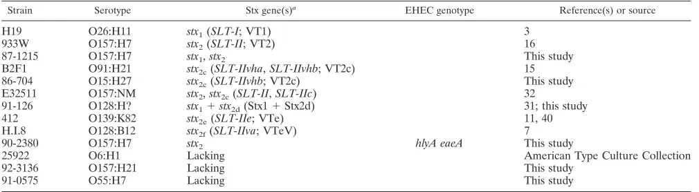
Bacterial strains and culture media.A total of 129E. coliisolates derived from the culture collection of the National Laboratory for Enteric Pathogens were used in this study, and these included 79E. coliO157:H7 isolates, five O157:NM (nonmotile) isolates, seven O157:non-H7 isolates (one each of O157: H10, H19, H21, H43, and H45 and two of H16), 12 non-O157:H7 isolates (two isolates of O27:H7, three of O18:H7, five of O55:H7, and one each of O156:H7 and O83:H7), six non-O157:NM isolates (one each of O1:NM, O7:NM, O91: NM, and O rough:NM and two isolates of O111:NM), 14 non-O157:non-H7 isolates (one isolate of O6:H1; two of O103:H2; one each of O146:H21, O26: H11, O70:H11, O91:H21, O139:K82, O128:B12, and O15:H27; two of O128:H untypeable, 1 of O113:H21, and 1 of O rough:H21), three isolates of O untype-able:H7, 1 of O untypable:H8, and 2 of O untypeable:H untypeable. Of these, 101 strains were STEC and 28 were negative for Stx. The control strains had been previously defined in terms of virulence factors and toxigenicity with respect to the genesstx1, stx2, stx2c,stx2-stx2c,stx2d,stx2e,stx2f, EHEChlyA, andeaeA(Table
1).
DNA isolation.Total DNA was isolated from 0.5 ml of brain heart infusion broth culture grown overnight for all the bacterial strains used in the study. The procedure used for DNA isolation was as described previously (38). DNA sam-ples were dissolved in Tris-EDTA buffer (10 mM Tris, 1 mM EDTA at pH 8.0), and the DNA concentration was determined in micrograms per milliliter at an optical density reading ofA260. The template DNA concentration used was 2
g/ml.
Primer design.Oligonucleotides ranging from 19- to 25-mers were selected from the published DNA sequences ofE. coliusing Oligo software (version 3.4).
Synthesis of oligonucleotides was carried out at the DNA Core Facility at the National Microbiology Laboratory, Winnipeg, Canada. For multiplex PCR, three primers sets were prepared. Set A was designed to amplifystx1,stx2,stx2f, and 16S
rRNA; set B was designed to amplifystx2c,stx2e,eaeA, and 16S rRNA; and set C
was designed to amplifystx2d, EHEChlyA,rfbEO157, andfliCH7as well as 16S
rRNA. The primer sequences used in the multiplex PCR are outlined in Table 2.
Multiplex-PCR conditions.Three sets of primer mixtures were prepared with slight modification according to the instructions supplied with the AmpliTaq Gold kit (Applied Biosystems, Foster City, Calif). In general, all of the multiplex primer sets contained 200M deoxynucleoside triphosphates, 2.5l of 10⫻
reaction buffer (100 mM Tris-HCl at pH 8.3, 500 mM KCl), 1.5 mM MgCl2, and
a 0.1M concentration of theE. coli16S rRNA (E16S) primers. In addition to these, set A contained a 0.5M concentration of each of the primers Stx1, Stx2, and Stx2f together with 2.5 U ofTaqDNA polymerase (AmpliTaq Gold; Applied Biosystems) and 5 ng of template DNA. The volume of this mix was adjusted to 25l with sterile water. In addition to the common constituents, the multiplex primer set B included primers at the following concentrations: 1.5M Stx2c, 0.4
M Stx2e, and 0.75M EAE. Multiplex primer set C comprised common components plus primers at the following concentrations: 1.5M Stx2d, 1.0M (each) HlyA and RfbE, and 0.4M FliC (Table 2). DNA amplification was carried out in a Perkin-Elmer thermocycler 2400 using an initial denaturation step at 95°C for 8 min, followed by 30 cycles of amplification with denaturation at 95°C for 30 s, annealing at 58°C for 30 s, and extension at 72°C for 30 s, ending with a final extension at 72°C for 7 min.
RESULTS
Multiplex PCR for the detection of E. colivirulence genes and O157:H7 serotype genes.The reaction conditions for the multiplex-PCR assay were optimized to ensure that all of the target gene sequences were satisfactorily amplified. Initially, equimolar primer concentrations of 0.5M each were used in the multiplex PCR, but there was uneven amplification, and some of the products were barely visible even after the reaction was optimized for the cycling conditions. Overcoming this problem required changing the proportions of the various primers in the reaction mixture to give an increase in the concentration of primers for the “weak” loci and a decrease in the primer concentration for the “strong” loci. The final con-centration of the primers (0.1 to 1.5M) varied considerably among the loci and was established empirically.
The primers were designed to target the coding regions of the genes, and care was taken to avoid areas of homology within the structural genes encoding the Stx2 family of toxins. The primers used in each set had identical annealing temper-atures, which reduced the possibility of the occurrence of un-wanted bands originating from nonspecific amplification.
Fig-TABLE 1. Characteristics of reference strains used in this study
Strain Serotype Stx gene(s)a EHEC genotype Reference(s) or source
H19 O26:H11 stx1(SLT-I; VT1) 3
933W O157:H7 stx2(SLT-II; VT2) 16
87-1215 O157:H7 stx1,stx2 This study
B2F1 O91:H21 stx2c(SLT-IIvha,SLT-IIvhb; VT2c) 15
86-704 O15:H27 stx2c(SLT-IIvhb; VT2c) This study
E32511 O157:NM stx2,stx2c(SLT-II,SLT-IIc) 32
91-126 O128:H? stx1⫹stx2d(Stx1⫹Stx2d) 31; this study
412 O139:K82 stx2e(SLT-IIe; VTe) 11, 40
H.I.8 O128:B12 stx2f(SLT-IIva; VTeV) 7
90-2380 O157:H7 stx2 hlyA eaeA This study
25922 O6:H1 Lacking American Type Culture Collection
92-3136 O157:H21 Lacking This study
91-0575 O55:H7 Lacking This study
aData in parentheses are Shiga-like toxin genes, Stx toxins, and/or verotoxins.
on May 15, 2020 by guest
http://jcm.asm.org/
[image:2.587.42.547.84.223.2]ure 1 shows the presence of the amplified product profiles after agarose gel electrophoresis, when DNA extracted from a ref-erenceE. colistrain (positive control) was used as the template in the PCR using the multiplex primer sets. The four bands in set A, stx1, stx2, stx2f, and 16S rRNA, were amplified
consis-tently even when mixtures of DNA derived from the same strains were tested (Fig. 1A). Similarly, four bands were ob-tained in set B when a mixture of DNA extracts from the corresponding strains that carried the genesstx2c,stx2e,eaeA,
and 16S rRNA was tested (Fig. 1B). For set C, which contained primers to amplify the genes stx2d, EHEC hlyA, rfbEO157,
fliCH7, and 16S rRNA, a total of five bands was obtained from
the positive-control DNA (Fig. 1C). The various control strains corresponded to the predicted sizes (Table 2). As a negative control, all sets were tested withE. colistrain ATCC 25922, and only the 16S rRNA band was observed (lanes 12 in Fig. 1A and B; lane 11 in Fig. 1C). Genomic DNA fromAeromonas hydrophilaandCampylobacter jejuniwas also tested using these three primer sets, and none gave PCR amplification bands (data not shown).
To substantiate the multiplex-PCR technique, 129 strains of
E. colithat were tested by multiplex PCR were also screened for the presence of individual toxin genes by using the methods described previously (14, 17, 18, 24, 30).stx2subtype,stx2c, and
stx2d genes were confirmed by PCR-RFLP analysis (31, 38).
The serotype identity of theE. coliO157:H7 isolates and the non-O157:H7 strains was determined at the National Labora-tory for Enteric Pathogens. Agreement between the toxigenic profile and O157:H7 serotype results and the multiplex-PCR data was observed (Table 3). However, 3 of 11 phenotypically NM strains showed gene-positive results for H7 by the PCR assay, and one of these was the reference stain E32511, previ-ously shown to be genotypically H7. An internal control ofE. coli16S rRNA was present in all of theE. colisamples, thus confirming the presence and the quality ofE. coliDNA am-plification as well as validating the PCR conditions.
Primary validation of amplicons.The sizes of the amplicons obtained from the multiplex primer sets were identical to those predicted from the design of the primers (Table 2). The am-plicons from the control strains were subjected to further con-firmation and characterization by digestion with restriction endonucleases with cleavage sites within each of the ampli-cons. The restriction enzymes used and the predicted product sizes are given in Table 4. Enzyme fragments with the antici-pated sizes were obtained in all cases (data not shown).
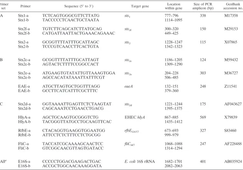
[image:3.587.47.541.83.429.2]Analysis of results. Among the 129 strains tested, 101 (78.3%) were positive for Stx toxins. All of O157:H7 strains wereeaeAand EHEChlyApositive. A total of 96 (74.4%)E. TABLE 2. Primers used in this study
Primer
set Primer Sequence (5⬘to 3⬘) Target gene within geneLocation amplicon (bp)Size of PCR accession no.GenBank
A Stx1-a TCTCAGTGGGCGTTCTTATG stx1 777–796 338 M17358
Stx1-b TACCCCCTCAACTGCTAATA 1114–1095
Stx2f-a TGTCTTCAGCATCTTATGCAG stx2f 300–320 150 M29153
Stx2f-b CATGATTAATTACTGAAACAGAAAC 449–425
Stx2-a GCGGTTTTATTTGCATTAGC stx2 1228–1247 115 X07865
Stx2-b TCCCGTCAACCTTCACTGTA 1342–1323
B Stx2c-a GCGGTTTTATTTGCATTAGT stx2c 1186–1205 124 M59432
Stx2c-b AGTACTCTTTTCCGGCCACT 1309–1290
Stx2e-a ATGAAGTGTATATTGTTAAAGTGGA stx2e 204–228 303 M36727
Stx2e-b AGCCACATATAAATTATTTCGT 506–485
EAE-a ATGCTTAGTGCTGGTTTAGG eaeA 132–151 248 Z11541
EAE-b GCCTTCATCATTTCGCTTTC 379–360
C Stx2d-a GGTAAAATTGAGTTCTCTAAGTAT stx2d 1221–1244 175 AF043627
Stx2d-b CAGCAAATCCTGAACCTGACG 1395–1375
HlyA-a AGCTGCAAGTGCGGGTCTG EHEChlyA 867–885 569 X79839
HlyA-b TACGGGTTATGCCTGCAAGTTCAC 1435–1412
RfbE-a CTACAGGTGAAGGTGGAATGG rfbEO157 673–693 327 S83460
RfbE-b ATTCCTCTCTTTCCTCTGCGG 999–979
FliC-a TACCATCGCAAAAGCAACTCC fliCH7 1068–1088 247 AF228488
FliC-b GTCGGCAACGTTAGTGATACC 1314–1294
Alla E16S-a CCCCCTGGACGAAGACTGAC E. coli16S rRNA 1682–1701 401 AB035924
E16S-b ACCGCTGGCAACAAAGGATA 2082–2063
aUsed in all sets as the internal control.
on May 15, 2020 by guest
http://jcm.asm.org/
coliisolates were positive for theeaeAgene, while seveneaeA
positives were detected among strains otherwise lacking stx. The ability of the C set of primers to identify O157:H7 from otherE. colistrains was determined by analyzing 79 O157:H7, 5 O157:NM, 7 O157:non-H7, and 38 non-O157E. coliisolates. Two of the five O157:NM strains and one of six non-O157:NM strains werefliCH7gene positive, indicating that these isolates
were genetically H7 with flagellum antigens that were either not expressed or not detectable in serotyping tests (Table 3) (5). All of the 129 samples tested contained theE. coli 16S rRNA gene.
DISCUSSION
STEC strains have been associated with outbreaks of disease that included cases of HC and HUS in humans. The two major categories of E. coli Stx toxins are Stx1 and Stx2. Stx1 is a relatively homogeneous family of toxins that show identity with the Shiga toxins ofShigella dysenteriae. Stx2 toxins, however, are a more heterogeneous group that are serologically distinct from Stx1. Within the Stx2 toxin family, Stx2c was formerly subdivided into Stx2-Va and Stx2-Vb (15, 38). These are only partially neutralized by antiserum to Stx2 (14; Head et al., letter). Stx2d shows a low cytotoxicity in Vero cells (27, 28, 31), while Stx2e is cytotoxic only in Vero cells and has been asso-ciated with porcine edema disease (11, 21). Stx2f (also called VTeV) has low-level cytotoxicity for Vero cells and is readily neutralized by antisera that are raised against Stx2 and Stx2e (7, 35).
Among STEC isolates, certain strains appear to have a greater degree of virulence for humans, while some data sug-gest that toxin type is important in determining the probability of developing HUS. Indeed, it has been shown epidemiologi-cally that Stx2 is more critical than Stx1 for the development of HUS, in that strains producing Stx2 were more frequently associated with cases of HUS than were those isolates express-ing Stx1 only (10). In addition to serological differences, the Stx2 group of toxins may differ in terms of their in vitro or in vivo properties. Experiments with clones carrying chimeric
FIG. 1. Shown are multiplex-PCR amplification profiles from ref-erenceE. colistrains with primer set A (A), B (B), and C (C). (A) Lane 1, 100-bp DNA ladder (Bethesda Research Laboratories Inc., Gaith-ersburg, Md.); lane 2,stx1and 16S rRNA (E. colistrain H19); lane 3,
stx2and 16S rRNA (strain 90-2380); lane 4,stx1,stx2, and 16S rRNA (strain 87-1215); lane 5, 16S rRNA (formerlystx2Va, strain 91-2245); lane 6, 16S rRNA (formerlystx2Vb, strain 86-704); lane 7,stx2(stx2-stx2c,
strain E32511); lane 8,stx1and 16S rRNA (stx1-stx2d, strain 91-126); lane 9, 16S rRNA (stx2e, strain 412); lane 10,stx2fand 16S rRNA (stx2f, strain H.I.8); lane 11, stx1, stx2, stx2f, and 16S rRNA; lane 12, stx negative control (ATCC 25922). (B) Lane 1, 100-bp DNA ladder (Bethesda Research Laboratories Inc.); lane 2, amplification products ofeaeAand 16S rRNA (stx1, strain H19); lane 3,eaeAand 16S rR-NA(stx2, strain 90-2380); lane 4,eaeAand 16S rRNA(stx1-stx2, strain 87-1215); lane 5,eaeA,stx2c, and 16S rRNA (formerlystx2Va, strain 91-2245); lane 6,stx2cand 16S rRNA (formerlystx2Vb, strain 86-704); lane 7,eaeA,stx2cand 16S rRNA (stx2-stx2c, strain E32511); lane 8, 16S rRNA(stx1-stx2d, strain 91-126); lane 9, stx2e and 16S rRNA (stx2e, strain 412); lane 10,eaeAand 16S rRNA (stx2f, strain H.I.8); lane 11,
eaeA,stx2c,stx2e, and 16S rRNA; lane 12,stxnegative control (ATCC 25922). (C) Lanes 1 and 12, 100-bp DNA ladder (Bethesda Research Laboratories Inc.); lane 2,hlyAand 16S rRNA (strain H19); lane 3,
hlyA,rfbEO157,fliCH7, and 16S rRNA(strain 90-2380, O157:H7); lane 4,
hlyA,rfbEO157,fliCH7, and 16S rRNA (strain E32511, O157: NM); lane 5,rfbEO157and 16S rRNA (O157: H21); lane 6,fliCH7and 16S rRNA (O55: H7); lane 7,hlyA,stx2d, and 16S rRNA (stx1-stx2d, strain 91-126); lane 8, 16S rRNA (strain 412); lane 9, 16S rRNA (strain H.I.8); lane 10, positive control ofhlyA,rfbEO157,fliCH7,stx2d, and 16S rRNA; lane 11,stxnegative control (ATCC 25922).
on May 15, 2020 by guest
http://jcm.asm.org/
O48/OX3bstx2operons indicated that the increased virulence
was a function of the A subunit ofstx2/OX3b. This differs in the
A-subunit structure from that ofstx2/O48 by only two amino
acids (Met-43Thr and Gly-1023Asp, respectively). These findings raise the possibility that naturally occurring Stx2 se-quence variations may directly impact the capacity of a given Stx-producingE. colistrain to cause severe disease (29).
The use of multiplex PCR or PCR-RFLP analysis to char-acterize the various subtypes of thestx2genes has been well
[image:5.587.52.539.84.505.2]documented (4, 6, 8, 9, 14, 24–26, 31, 38). Lin et al. (19) introduced common primers for PCR-RFLP analysis in order to detect the genes for various Stx toxins. However, all the PCR-related methods require restriction digestion to achieve identification. A multiplex PCR-based diagnostic protocol is described for the detection of those genes encoding the various
TABLE 3. Major virulence genes detected by the three primer sets in the multiplex PCR analysis
Toxin(s) confirmed by PCR or PCR-RFLP
analysis (n)a Serotype (n) b
No. of genes detected by PCR with primer set:
A B C
stx1 stx2 stx2f stx2c stx2e eaeA stx2d hlyA rfbEO157 fliCH7
Stx1 (13) Non-O157:non-H7 (5) 5 —c — — — 4 — 5 — —
O111:NM (1) 1 — — — — 1 — 1 — —
O91:NM (1) 1 — — — — — — 1 — —
O157:H7 (5) 5 — — — — 5 — 5 5 5
O157:NM (1) 1 — — — — 1 — 1 1 1
Stx1⫹Stx2 (27) O UT:H UTd(1) 1 1 — — — — — — — —
O157:H7 (26) 26 26 — — — 26 — 26 26 26
Stx1⫹Stx2c (2) O157:H7 (1) 1 — — 1 — 1 — 1 1 1
O111:NM (1) 1 — — 1 — 1 — — — —
Stx1⫹Stx2d (3) O128:H? (2) 2 — — — — — 2 2 — —
O rough:NM (1) 1 — — — — — 1 — — —
Stx2 (16) O157: H7 (16) — 16 — — — 16 — 16 16 16
Stx2⫹Stx2c (27) O157:NM (1) — 1 — 1 — 1 — 1 1 1
O157:H7 (26) — 26 — 26 — 26 — 26 26 26
Stx2c (10) O91:H21 (1) — — — 1 — — — 1 — —
Non-O157:non-H7 (3) — — — 3 — — — 1 — —
O157:H7 (5) — — — 5 — 5 — 5 5 5
O55:H7 (1) — — — 1 — 1 — — — 1
Stx2d (1) O UT:H8 (1) — — — — — — 1 — — —
Stx2e (1) O139:K82 (1) — — — — 1 — — — — —
Stx2f (1) O128:B12 (1) — — 1 — — 1 — — — —
Stx negative (28) O157:non-H7 (7) — — — — — 6 — — 7 —
O157:NM (3) — — — — — — — — 3 —
Non-O157:H7 (11) — — — — — 1 — — — 11
O UT:H7 (3) — — — — — — — — — 3
Non-O157:NM (2) — — — — — — — — — 1
Non-O157:non-H7 (1) — — — — — — — — — —
O UT:H UT (1) — — — — — — — — — —
Total 101/129 45 70 1 39 1 96 4 92 91 106
aAs outlined in references 17, 18, 31, and 38.
bNumbers in parentheses are numbers of serotypes tested. c—, tested negative for the gene.
dUT⫽untypable.
TABLE 4. Predicted sizes of restriction fragments and enzymes used for RFLP analysis of amplified products of multiplex PCR
Genes ampliconPCR size (bp)
Multiplex primer
set(s) Enzyme
Expected sizes of restriction fragments (bp)
stx1 338 A BglI 136, 202
stx2 115 A BsrDI 37, 78
stx2f 150 A AluI 54, 96
stx2e 303 B TaqI 51, 87, 165
stx2c 124 B HhaI 48, 76
eaeA 248 B AluI 109, 139
stx2d 175 C RsaI 66, 109
EHEChlyA 569 C ApaI 299, 270
rfbEO157 327 C AluI 80, 93, 154
fliCH7 247 C AluI 40, 207
E. coli16S rRNA 401 A, B, and C RsaI 156, 245
on May 15, 2020 by guest
http://jcm.asm.org/
[image:5.587.300.541.591.728.2]Stx toxins, including Stx1, Stx2, Stx2c, Stx2d, Stx2e, and Stx2f, EHEChlyA, andeaeA, together with the genes governing the serotype O157 (rfbE) and H7 (fliC) in the absence of restric-tion enzyme digesrestric-tion. Compared to the individual primers and PCR-RFLP analysis results, the multiplex-PCR primer sets proved to be highly specific, they gave consistent results, and they were effective in detecting all 11 genes, including the internal control gene. All primers were gene specific, as dem-onstrated by restriction fragment lengths obtained after spe-cific restriction endonuclease digestion of the amplicons.
In this study, the toxin genotype and O157:H7 serotype of a range ofE. coli strains was demonstrated (Table 3). Of 81 STEC O157:H7 clinical isolates (including the 2 that were serotypically O157:NM but were PCR positive forfliCH7), all
were positive for the EHEChlyAandeaeAgenes. These find-ing were in agreement with previous reports (1, 33). Interest-ingly, among 20 of the non-O157 STEC isolates, 11 (55%) were EHEChlyApositive and 8 (40%) wereeaeApositive. Three of 10 strains positive for the stx2c gene only were negative for
EHEChlyA, while the referencestx2cgene-positive strain
(se-rotype O91:H21, an isolate from a patient with HUS) was EHEChlyApositive and lackedeaeA, suggesting that theeaeA
may not be an essential major virulence factor for the acqui-sition of HUS. All four strains that possessed thestx2d
geno-type lackedeaeA. Of these four strains, three possessed both the stx1 and stx2d genotype, while one was positive for stx2d
alone. Two of these werehlyA positive. This suggested that STEC strains without EHEChlyAmay possess reduced patho-genicity or may even be nonpathogenic in humans (36). Fur-thermore, among the 28 non-STEC isolates (Stx negative), 7 wereeaeApositive and none possessed the EHEChlyAgene. This suggested that EHEChlyA may be a more critical viru-lence factor for disease thaneaeA(Table 3). Further studies are required on EHEChlyA, eaeA, and the various Stx com-ponents in order to elucidate the role that these toxins play in STEC disease in general and in HUS and HC in particular.
In total 11 phenotypic nonmotileE. coli isolates were ana-lyzed. Of these, three werefliCH7positive. Two of these were
O157:NM strains (including reference strain E32511), and one was an O1:NM strain. All three were confirmed as H7 positive using primer FLICh7-F and FLICh7-R (14). AnE. colifliCH7
sequence comparison (GenBank accession no. AF228487 for O157:H7, AF228495 for O19ab:H7, AF228496 for O53:H7, AF228489 for O55:H7, and U47614 for O157:NM) also con-firmed that fliC is highly conserved among different sero-groups. Therefore, it would appear that some ofE. colistrains that are serologically NM are genetically H7 (5). The factors that governfliCexpression are unclear; however, environmen-tal factors or other related genes may play key roles.
From these data we conclude that the multiplex primer sets described are specific and give highly consistent results. The use of this three-tube assay should allow simultaneous detec-tion of the major virulence factors associated withE. coliO157 and non-O157 strains and avoids the need for endonuclease digestion steps.
ACKNOWLEDGMENTS
We thank David Woodward, Richard Caldeira, Jennifer Campbell, David Spreitzer, Kelly Robinson, Kevin Hill, and Julie Walsh for valuable technical assistance.
REFERENCES
1.Boerlin, P., S. A. McEwen, B. P. Franziska, J. B. Wilson, R. P. Johnson, and C. L. Gyles.1999. Associations between virulence factors of Shiga toxin-producingEscherichia coliand disease in humans. J. Clin. Microbiol.37:497– 503.
2.Chinen, I., M. Rivas, V. Soriano, E. Miliwebsky, G. F. Galvez, G. Chillemi, A. Baschkier, G. Wang, R. Caldeira, D. L. Woodward, and F. G. Rodgers.
2002.Escherichia coli ehl1gene-positive serotype O18ac:H31 associated with an outbreak of diarrhea in a neonatal nursery in Neuquen City, Argentina. J. Clin. Microbiol.40:1225–1229.
3.De Grandis, S., J. Ginsberg, M. Toone, S. Climie, J. Friesen, and J. Brunton.
1987. Nucleotide sequence and promoter mapping of theEscherichia coli
Shiga-like toxin operon of bacteriophage H-19B. J. Bacteriol.169:4313– 4319.
4.Feng, P., and S. R. Monday.2000. Multiplex PCR for detection of trait and virulence factors in enterohemorrhagicEscherichia coliserotypes. Mol. Cell. Probes14:333–337.
5.Fields, P. I., K. Blom, H. J. Hughes, L. O. Helsel, P. Feng, and B. Swami-nathan.1997. Molecular characterization of the gene encoding H antigen in
Escherichia coliand development of a PCR-restriction fragment length poly-morphism test for identification ofE. coliO157:H7 and O157:NM. J. Clin. Microbiol.35:1066–1070.
6.Fratamico, P. M., L. K. Bagi, and T. Pepe.2000. A multiplex polymerase chain reaction assay for rapid detection and identification ofEscherichia coli
O157:H7 in foods and bovine feces. J. Food Prot.63:1032–1037. 7.Gannon, V. P., C. Teerling, S. A. Masri, and C. L. Gyles.1990. Molecular
cloning and nucleotide sequence of another variant of theEscherichia coli
Shiga-like toxin II family. J. Gen. Microbiol.136:1125–1135.
8.Gannon, V. P., S. D’Souza, T. Graham, R. K. King, K. Rahn, and S. Read.
1997. Use of the flagella H7 gene as a target in multiplex PCR assays and improved specificity in identification of enterohemorrhagicEscherichia coli
strains. J. Clin. Microbiol.35:656–662.
9.Gannon, V. P., S. D’Souza, T. Graham, and R. K. King.1997. Specific identification ofEscherichia coliO157:H7 using a multiplex PCR assay. Adv. Exp. Med. Biol.412:81–82.
10.Griffin, P. M., and R. V. Tauxe.1991. The epidemiology of infections caused byEscherichia coliO157:H7, other enterohemorrhagicE. coli, and the asso-ciated hemolytic uremic syndrome. Epidemiol. Rev.13:60–98.
11.Gyles, C. L., S. A. De Grandis, C. MacKenzie, and J. L. Brunton.1988. Cloning and nucleotide sequence analysis of the genes determining verocy-totoxin production in a porcine edema disease isolate ofEscherichia coli. Microb. Pathog.5:419–426.
12.Gyles, C. L.1992.Escherichia colicytotoxins and enterotoxins. Can. J. Mi-crobiol.38:734–746.
13.Hii, J. H., C. Gyles, T. Morooka, M. A. Karmali, R. Clarke, S. DeGrandis, and J. L. Brunton.1991. Development of verotoxin 2- and verotoxin 2 variant (VT2v)-specific oligonucleotide probes on the basis of the nucleotide sequence of the B cistron of VT2v fromEscherichia coliE32511 and B2F1. J. Clin. Microbiol.29:2704–2709.
14.Hu, Y., Q. Zhang, and J. C. Meitzler.1999. Rapid and sensitive detection of
Escherichia coliO157:H7 in bovine faeces by a multiplex PCR. J. Appl. Microbiol.87:867–876.
15.Ito, H., A. Terai, H. Kurazono, Y. Takeda, and M. Nishibuchi.1990. Cloning and nucleotide sequencing of Vero toxin 2 variant genes fromEscherichia coliO91:H21 isolated from a patient with the hemolytic uremic syndrome. Microb. Pathog.8:47–60.
16.Jackson, M. P., R. J. Neill, A. D. O’Brien, R. K. Holmes, and J. W. Newland.
1987. Nucleotide sequence analysis and comparison of the structural genes for Shiga-like toxin, I., and Shiga-like toxin II encoded by bacteriophages fromEscherichia coli.FEMS Microbiol. Lett.44:109–114.
17.Johnson, W. M., D. R. Pollard, H. Lior, S. D. Tyler, and K. R. Rozee.1990. Differentiation of genes coding for Escherichia coli verotoxin 2 and the verotoxin associated with porcine edema disease (VTe) by the polymerase chain reaction. J. Clin. Microbiol.28:2351–2353.
18.Johnson, W. M., S. D. Tyler, G. Wang, and H. Lior.1991. Amplification by the polymerase chain reaction of a specific target sequence in the gene coding forEscherichia coliverotoxin (VTe variant). FEMS Microbiol Lett.
68:227–230.
19.Lin, Z., H. Kurazono, S. Yamasaki, and Y. Takeda.1993. Detection of various variant verotoxin genes inEscherichia coliby polymerase chain re-action. Microbiol. Immunol.37:543–548.
20.Lindgren, S. W., J. E. Samuel, C. K. Schmitt, and A. D. O’Brien.1994. The specific activities of Shiga-like toxin type II (SLT-II) and SLT-II-related toxins of enterohemorrhagicEscherichia colidiffer when measured by Vero cell cytotoxicity but not by mouse lethality. Infect. Immun.62:623–631. 21.Marques, L. R. M., J. S. M. Peiris, S. J. Cryz, and A. D. O’Brien.1987.
Escherichia colistrains isolated from pigs produce a variant of Shiga-like toxin II. FEMS Microbiol. Lett.44:33–38.
22.McDaniel, T. K., K. G. Jarvis, M. S. Donnenberg, and J. B. Kaper.1995. A genetic locus of enterocyte effacement conserved among diverse enterobac-terial pathogens. Proc. Natl. Acad. Sci. USA92:1664–1668.
on May 15, 2020 by guest
http://jcm.asm.org/
23.McDaniel, T. K., and J. B. Kaper.1997. A cloned pathogenicity island from enteropathogenicEscherichia coliconfers the attaching and effacing pheno-type onE. coliK12. Mol. Microbiol.2:399–407.
24.Meng, J., S. Zhao, M. P. Doyle, S. E. Mitchell, and S. Kresovich.1997. A multiplex PCR for identifying Shiga-like toxin-producingEscherichia coli
O157:H7. Lett. Appl. Microbiol.24:172–176.
25.Meng, J., S. Zhao, and M. P. Doyle.1998. Virulence genes of Shiga toxin-producingEscherichia coliisolated from food, animals and humans. Int. J. Food Microbiol.45:229–235.
26.Pass, M. A., R. Odedra, and R. M. Batt.2000. Multiplex PCRs for identifi-cation ofEscherichia colivirulence genes. J. Clin. Microbiol.38:2001–2004. 27.Paton, A. W., J. C. Paton, M. W. Heuzenroeder, P. N. Goldwater, and P. A. Manning.1992. Cloning and nucleotide sequence of a variant Shiga-like toxin II gene fromEscherichia coliOX3:H21 isolated from a case of sudden infant death syndrome. Microb. Pathog.13:225–236.
28.Paton, A. W., J. C. Paton, P. N. Goldwater, M. W. Heuzenroeder, and P. A. Manning.1993. Sequence of a variant Shiga-like toxin type-I operon of
Escherichia coliO111:H-. Gene129:87–92.
29.Paton, A. W., A. J. Bourne, P. A., Manning, and J. C. Paton.1995. Compar-ative toxicity and virulence ofEscherichia coliclones expressing variant and chimeric Shiga-like toxin type II operons. Infect. Immun.63:2450–2458. 30.Paton, A. W., and J. C. Paton.1997. Detection and characterization of Shiga
toxigenicEscherichia coliby using multiplex PCR assays forstx1,stx2,eaeA, enterohemorrhagicE. coli hlyA,rfb0111, andrfb0157. J. Clin. Microbiol.
36:598–602.
31.Pierard, D., G. Muyldermans, L. Moriau, D. Stevens, and S. Lauwers.1998. Identification of new verocytotoxin type 2 variant B-subunit genes in human and animal. Escherichia coli isolates. J. Clin. Microbiol.36:3317–3322. 32.Schmitt, C. K., M. L. McKee, and A. D. O’Brien.1991. Two copies of
Shiga-like toxin II-related genes common in enterohemorrhagicEscherichia
colistrains are responsible for the antigenic heterogeneity of the O157:H⫺
strain E32511. Infect. Immun.59:1065–1073.
33.Schmidt, H., L. Beutin, and H. Karch.1995. Molecular analysis of the plasmid-encoded hemolysin ofEscherichia coliO157:H7 strain EDL 933. Infect. Immun.63:1055–1061.
34.Schmidt, H., and H. Karch.1996. Enterohemolytic phenotypes and geno-types of Shiga-toxin-producingEscherichia coliO111 strains from patients with diarrhea and hemolytic-uremic syndrome. J. Clin. Microbiol.34:2364– 2367.
35.Schmidt, H., J. Scheef, S. Morabito, A. Caprioli, L. H. Wieler, and H. Karch.
2000. A new Shiga toxin 2 variant (Stx-2f) fromEscherichia coliisolated from pigeons. Appl. Environ. Microbiol.66:1205–1208.
36.Stephan, R., and L. E. Hoelzle.2000. Characterization of shiga toxin type 2 variant B-subunit inEscherichia colistrains from asymptomatic human car-riers by PCR-RFLP. Lett. Appl. Microbiol.31:139–142.
37.Thomas, A., H. R. Smith, and B. J. Rowe.1993. Use of digoxigenin-labelled oligonucleotide DNA probes for VT2 and VT2 human variant genes to differentiate Vero cytotoxin-producingEscherichia colistrains of serogroup O157. J. Clin. Microbiol.31:1700–1703.
38.Tyler, S. D., W. M. Johnson, H. Lior, G. Wang, and K. R. Rozee.1991. Identification of verotoxin type 2 variant B-subunit genes inEscherichia coli
by the polymerase chain reaction and restriction fragment length polymor-phism analysis. J. Clin. Microbiol.29:1339–1343.
39.Wang, L., D. Rothemund, H. Curd, and P. R. Reeves.2000. Sequence diver-sity of theEscherichia coliH7fliCgenes: implication for a DNA-based typing scheme forE. coliO157:H7. J. Clin. Microbiol.38:1786–1790.
40.Weinstein, D. L., M. P. Jackson, J. E. Samuel, R. K. Holmes, and A. D. O’Brien.1988. Cloning and sequencing of a Shiga-like toxin type II variant fromEscherichia colistrain responsible for edema disease of swine. J. Bac-teriol.170:4223–4230.