0095-1137/06/$08.00
⫹
0
doi:10.1128/JCM.00299-06
Copyright © 2006, American Society for Microbiology. All Rights Reserved.
Use of the Phoenix Automated System for Identification of
Streptococcus
and
Enterococcus
spp.
Gioconda Brigante,
1Francesco Luzzaro,
1Alessia Bettaccini,
1Gianluigi Lombardi,
1Francesca Meacci,
2Beatrice Pini,
1Stefania Stefani,
3and Antonio Toniolo
1*
Laboratory of Medical Microbiology, Ospedale di Circolo and University of Insubria, Varese,
1Department of Molecular Biology,
University of Siena, Siena,
2and Department of Microbiological Sciences, University of Catania, Catania,
3Italy
Received 10 February 2006/Returned for modification 3 April 2006/Accepted 10 July 2006
The Phoenix system (Becton Dickinson Diagnostic Systems, Sparks, MD) was evaluated for identification (ID) to
the species level of streptococci and enterococci. Two hundred clinical isolates were investigated: beta-hemolytic
streptococci (
n
ⴝ
50),
Streptococcus pneumoniae
organisms (
n
ⴝ
46), viridans group streptococci (
n
ⴝ
31),
Entero-coccus faecium
(
n
ⴝ
36),
Enterococcus faecalis
(
n
ⴝ
25), and other catalase-negative cocci (
n
ⴝ
12). The API system
(bioMe
´rieux, Marcy l’E
´toile, France) was used as a comparator. Molecular methods (sequencing of 16S rRNA and
zwf
and
gki
genes and
ddl
gene amplification) were used to investigate discordant results. Upon resolution of
discrepancies, correct species ID was achieved by the Phoenix system for 121/129 (93.8%) streptococci and 63/70
(90.0%) enterococci. Excellent results were obtained for
S. pneumoniae
(45/45) and beta-hemolytic streptococci
(49/50). With regard to viridans streptococci, the accuracy of the Phoenix system was 83.9%. Among the latter
organisms, the best performance was obtained with isolates of the
Streptococcus sanguinis
group and
Streptococcus
anginosus
group; problems were instead encountered with the
Streptococcus mitis
group. Four
E. faecium
and three
E. faecalis
isolates were misidentified as
Enterococcus casseliflavus/Enterococcus gallinarum
or
Enterococcus durans
.
Thus, these isolates were identified only at the genus level. Compared with commercially available systems, the
Phoenix system appears a reliable diagnostic tool for identifying clinically relevant streptococci and enterococci. The
SMIC/ID-2 panel proved particularly effective for beta-hemolytic streptococci and pneumococci.
Catalase-negative, gram-positive cocci are a heterogeneous
group of 17 genera that include streptococci, enterococci, and
nonstreptococcal, nonenterococcal species (9, 10). Over 70
streptococcal and enterococcal species have been implicated in
human disease (10, 27). Of these, only a few are known to
cause important infections (e.g.,
Streptococcus pneumoniae
,
Streptococcus pyogenes
,
Streptococcus agalactiae
,
Enterococcus
faecalis
,
Enterococcus faecium
, and viridans group
strepto-cocci).
A number of manual, semiautomated, and automated
sys-tems are reported to produce acceptable identification (ID)
results for
S. pneumoniae
, beta-hemolytic streptococci, and
enterococcal species (26, 27). These systems, however, were
shown not to be sufficiently accurate in identifying streptococci
of the viridans group (13, 20), organisms of complex taxonomy
(2, 10, 26). The performance of some automated systems has
been evaluated with regard to catalase-negative, gram-positive
cocci (7, 13, 14, 17, 24). Reproducibility and accuracy of
re-sults, turnaround time, availability of data for epidemiological
monitoring, and cost-effectiveness constitute the main reasons
supporting the choice of automated systems.
Becton Dickinson (BD Diagnostic Systems, Sparks, MD)
has introduced the Phoenix automated microbiology system
for ID and antimicrobial susceptibility testing (AST) of human
pathogenic bacteria, including enterobacteria, nonfermenting
gram-negative bacteria, staphylococci, and enterococci (5, 8,
11, 25). Recently, the SMIC/ID-2 panel, dedicated to ID and
AST of streptococcal species, was launched (15, 18). This study
was designed to evaluate the performance of the Phoenix
sys-tem for identification of streptococcal and enterococcal
iso-lates at the species level.
MATERIALS AND METHODS
Clinical isolates.Clinical isolates were obtained from routine clinical speci-mens at the Microbiology Laboratory of the Ospedale di Circolo, Varese, Italy. A total of 200 nonduplicated isolates of gram-positive, catalase-negative cocci were studied. The following strains were investigated:S. pneumoniae(n⫽46),S. pyogenes(n⫽ 15),S. agalactiae (n⫽15), Streptococcus dysgalactiaesubsp.
equisimilis(n⫽20), viridans group streptococci (n⫽31),E. faecium(n⫽36),
E. faecalis(n⫽25), other enterococcal species (n⫽9), and other catalase-negative, gram-positive cocci (n⫽3). Isolates were stored at⫺70°C in Todd-Hewitt broth containing 20% glycerol. Before ID assays were performed, all strains were passed twice on Mueller-Hinton agar containing 5% sheep blood (Oxoid SpA, Milan, Italy) to get them to an active-growth stage following met-abolic inactivity while frozen.
Phoenix system procedures. The Phoenix system uses different panels for gram-positive cocci. The SMIC/ID-2 panel is dedicated to streptococci and the PMIC/ID-14 panel to enterococci and staphylococci. All panels include two separate sections: wells on the left contain ID substrates, and wells on the right side are dedicated to AST. Panel inoculation was performed according to the manufacturer’s instructions. Both panel sections were inoculated, but only ID results have been taken into consideration for this study. After overnight culture, bacteria were suspended in the ID broth. Turbidity was adjusted to a 0.5 Mc-Farland standard by using the CrystalSpec Nephelometer (Becton Dickinson). Panels were then sealed, logged, loaded into the instrument, and incubated at 35°C. Kinetic, colorimetric, and fluorescent signals were automatically collected by the instrument every 20 min until results were completed.
Comparator biochemical ID method.Two different API ID systems (bio-Me´rieux, Marcy l’E´ toile, France) were used to identify streptococcal and entero-coccal isolates at the species level. The API 20 Strep system was used for beta-hemolytic streptococci. The rapid ID 32 Strep system was used for
entero-* Corresponding author. Mailing address: Laboratory of Medical
Mi-crobiology, University of Insubria and Ospedale di Circolo e Fondazione
Macchi, Viale Borri 57, 21100, Varese, Italy. Phone: 39-0332-278.309. Fax:
39-0332-260.517. E-mail: [email protected].
3263
on May 16, 2020 by guest
http://jcm.asm.org/
cocci and non-beta-hemolytic streptococci. Inoculation, reading, and interpreta-tion of panels were performed according to the manufacturer’s instrucinterpreta-tions.
Data analysis and resolution of discrepancies.Isolates that were equally identified at the species level by both the API and the Phoenix systems were considered to be correctly identified and included in the “concordant ID” cate-gory. Due to the inability of the Phoenix system to discriminate between Entero-coccus casseliflavusandEnterococcus gallinarum, the classification of an isolate as
E.casseliflavus/E. gallinarumby the Phoenix system was considered concordant when the isolate was identified by the API system as eitherE.casseliflavusorE.
gallinarum. Isolates with discordant species ID (i.e., an ID produced by the Phoenix system that differed from that obtained with the API system) were retested using both the Phoenix and the API systems. When discrepant results persisted, bacterial ID was investigated by molecular methods.
Amplification and sequencing of the 16S rRNA gene.Bacterial DNA was extracted from pure cultures by using a QIAamp DNA mini kit (QIAGEN, Basel, Switzerland). DNA eluted in Tris-EDTA was stored at⫺80°C. ABI 2400 thermal cyclers and PCR reagents were from Applied Biosystems (Foster City, CA). AmpliTaq Gold polymerase, PCR buffer II, standard deoxynucleoside triphosphate mixture, and the universal 16S rRNA gene primers 8f (5⬘-GAGA GTTTGATCCTGGCTCAG-3⬘) and 1492r (5⬘-TACGGCTACCTTGTTACGA CT-3⬘) were used to produce a 1,498-bp amplicon (23). Amplification products were purified using a Mini Elut PCR purification kit (QIAGEN) and directly sequenced using an ABI 310 genetic analyzer (Applied Biosystems). Sequences for both DNA strands were determined, each by using the product of a different PCR as a template. Analysis and comparison of sequence data were carried out at the BLAST interface (http://www.ncbi.nlm.nih.gov/BLAST/) and ClustalW interface (http://www.ebi.ac.uk/clustalw/) websites.
Amplification and sequencing of housekeeping genes of viridans group strep-tococci.Samples were amplified with degenerate primers specific for the internal fragments of thezwf(encoding glucose-6-phosphate dehydrogenase) andgki
(encoding glucose kinase) streptococcal genes (21). Two primer pairs were used: 5⬘-CCG(T/G)ATCGACCATTA(T/C)CTTGG(T/C)AAGG-3⬘ and 5⬘-TC(A/T) GTCAG(T/A)CGTTTACCTGT(A/G)CGGA-3⬘for thezwfgene and 5⬘-GGC ATTGGAATGGGATCACCAGG-3⬘and 5⬘-CCGATAA(C/T)TCCAGCGTCA TTTCC-3⬘for thegkigene. Amplicons of 453 bp (zwf) and 624 bp (gki) were directly sequenced.
Amplification of housekeeping genes in enterococci.The ID ofE. faeciumand
E. faecalisisolates was confirmed by amplification of a fragment internal to the
ddlgene encoding aD-Ala-D-Ala ligase (4, 6). The reaction mixture contained 250 ng of DNA as a template, 50 pmol of each primer, 200 pmol per liter of each deoxynucleoside triphosphate (dATP, dCTP, dGTP, and dTTP), 10 mM Tris-HCl (pH 8.8), 1.5 mM MgCl2, 50 mM KCl, and 2 U of AmpliTaq Gold. Upon electrophoresis on a 2% agarose gel, ethidium bromide-stained DNA fragments were visualized under UV light with a Kodak CF440 camera (NEN Life Science Products, Boston, MA).
Quality controls.The following strains were included in each run:S. pneu-moniaeATCC 49619,S. agalactiaeATCC 13813, andE. faecalisATCC 29212. The identification results obtained with the above-mentioned reference strains were consistently satisfactory.
RESULTS
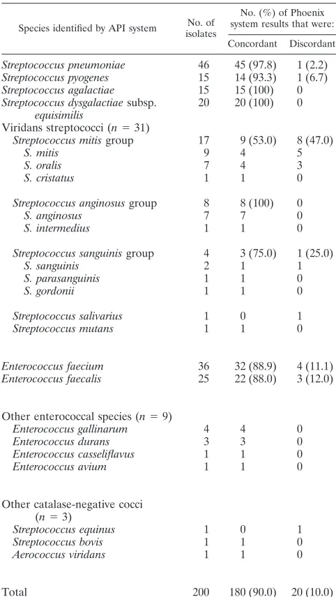
Biochemical identification of clinical isolates.
Compared
with the API system, the Phoenix system correctly identified
180/200 (90.0%) test organisms at the species level: 116/129
(89.9%) streptococci, 63/70 (90%) enterococci, and 1/1
Aero-coccus viridans
isolate. Results are summarized in Table 1.
Concordant ID for
S. pneumoniae
was obtained in 45/46
cases, since one isolate (identified as
S
.
pneumoniae
by the API
system) was classified as a “not identified organism” by the
Phoenix system. After resolution of discrepancies, however,
this isolate was ultimately defined as
Streptococcus mitis
by
sequencing the 16S rRNA gene. Fourteen out of 15 (93.3%)
S.
pyogenes
and 21/31 (67.7%) viridans streptococci isolates were
correctly identified by the Phoenix system. Among the latter,
8/8 members of the
Streptococcus anginosus
group, one
Strep-tococcus mutans
isolate, 3/4 members of the
Streptococcus
san-guinis
group, and 9/17 members of
S. mitis
group were correctly
identified. Correct species ID was obtained for 90% of the
enterococci. Four
E. faecium
and three
E. faecalis
isolates were
misidentified as
E. casseliflavus/E. gallinarum
or
Enterococcus
durans
. Thus, these isolates were identified only at the genus
level. Overall, the Phoenix system correctly identified all
iso-lates of the following species:
S. pneumoniae
,
S. agalactiae
,
S.
dysgalactiae
subsp.
equisimilis
, and
Streptococcus bovis
.
Analysis of discrepancies.
Species ID of 20 discordant
iso-lates was investigated by molecular methods. As shown in
Table 2, sequencing of the 16S rRNA gene assessed that one
S.
mitis
isolate was not identified by either the API or the Phoenix
system. One
S. pyogenes
isolate (not identified by the Phoenix
system) was correctly identified by the API system. Five
iso-TABLE 1. Results of biochemical identification of streptococci and
enterococci by the Phoenix system, with the API system
used as a comparator
aSpecies identified by API system No. of isolates
No. (%) of Phoenix system results that were:
Concordant Discordant
Streptococcus pneumoniae
46
45 (97.8)
1 (2.2)
Streptococcus pyogenes
15
14 (93.3)
1 (6.7)
Streptococcus agalactiae
15
15 (100)
0
Streptococcus dysgalactiae
subsp.
equisimilis
20
20 (100)
0
Viridans streptococci (
n
⫽
31)
Streptococcus mitis
group
17
9 (53.0)
8 (47.0)
S. mitis
9
4
5
S. oralis
7
4
3
S. cristatus
1
1
0
Streptococcus anginosus
group
8
8 (100)
0
S. anginosus
7
7
0
S. intermedius
1
1
0
Streptococcus sanguinis
group
4
3 (75.0)
1 (25.0)
S. sanguinis
2
1
1
S. parasanguinis
1
1
0
S. gordonii
1
1
0
Streptococcus salivarius
1
0
1
Streptococcus mutans
1
1
0
Enterococcus faecium
36
32 (88.9)
4 (11.1)
Enterococcus faecalis
25
22 (88.0)
3 (12.0)
Other enterococcal species (
n
⫽
9)
Enterococcus gallinarum
4
4
0
Enterococcus durans
3
3
0
Enterococcus casseliflavus
1
1
0
Enterococcus avium
1
1
0
Other catalase-negative cocci
(
n
⫽
3)
Streptococcus equinus
1
0
1
Streptococcus bovis
1
1
0
Aerococcus viridans
1
1
0
Total
200
180 (90.0)
20 (10.0)
aResults refer to the biochemical identification obtained by the API system.
Results obtained with the Phoenix system are shown as concordant or discordant with API results, without taking into account the resolution of discrepancies by the molecular methods reported in Table 2.
on May 16, 2020 by guest
http://jcm.asm.org/
[image:2.585.300.541.97.525.2]lates (which had been assigned to the
S. mitis
or
Streptococcus
salivarius
group by the API system) did belong to the
S.
san-guinis
group (
Streptococcus parasanguinis
,
n
⫽
2;
S. sanguinis
,
n
⫽
2; and
Streptococcus gordonii
,
n
⫽
1). The
above-men-tioned five isolates were correctly classified at the species level
by the Phoenix system, not by the API system. One discordant
isolate (resolved by 16S rRNA gene sequencing as
S. gordonii
)
was not identified by the Phoenix system and was identified
only at the group level by the API. Sequencing of the 16S
rRNA and the housekeeping
zwf
and
gki
genes failed to resolve
five additional isolates at the species level. The most probable
molecular ID for those five isolates appeared to be
S. mitis
,
Streptococcus oralis
, or
S. pneumoniae
(species belonging to the
S. mitis
group) (26).
Concerning seven enterococci identified by the Phoenix
sys-tem as
E. casseliflavus/E. gallinarum
(
n
⫽
5) or
Enterococcus
durans
(
n
⫽
2), PCR analysis of the
ddl
gene confirmed the IDs
given by the API system (four
E. faecium
and three
E. faecalis
isolates).
The performance of the Phoenix and API systems with
re-gard to discordant isolates is summarized in Table 3. Of 13
streptococci, 9 were correctly identified at the species or group
level by the Phoenix system and 5 by the API system. Three
isolates could not be identified by either system. The correct
IDs for seven discordant enterococci were given by the API
system, not by the Phoenix system. Of the streptococci that
could not be resolved at the species level by molecular
meth-ods, 4/5 were identified at the group level by both the API and
the Phoenix systems. Overall, taking into consideration species
IDs given by molecular methods, the accuracy of the Phoenix
system in identifying streptococci rose from 89.9% to 93.8%.
For enterococci, accuracy of ID at the species level remained
at 90%.
DISCUSSION
Automated systems may have significant diagnostic impact
on diseases caused by streptococci and enterococci, especially
with regard to aggressive infections and drug-resistant
iso-lates (26).
[image:3.585.45.542.82.287.2]The Phoenix automated system did agree with the API system
for 89.9% of streptococcal IDs. Upon resolution of discrepancies,
accuracy for streptococci rose to 93.8%. The performance of the
new SMIC/ID-2 panel dedicated to streptococci was excellent
for beta-hemolytic streptococci (49/50) and
S
.
pneumoniae
(45/
46). Only one
S
.
pyogenes
isolate and one
S
.
pneumoniae
isolate
were not identified. It should be noted that the latter isolate
(reported by the API system as
S. pneumoniae
) was ultimately
identified as
S
.
mitis
by molecular methods. This brings the
accuracy for
S. pneumoniae
to 100% and underlines difficulties
that may be encountered in the biochemical identification of
streptococcal isolates by commercial methods (3, 16, 22).
TABLE 2. Analysis of 20 discordant isolates
aAPI system result (no. of isolates)
Discrepant result provided by the Phoenix system (no. of isolates)
Species (no. of isolates) identified by molecular method
16S rRNA gene sequencing zwfandgkigene sequencing ddlgene amplification
ID by molecular analysis
S. pneumoniae
(1)
NI
S. mitis
—
—
S. mitis
S. pyogenes
(1)
NI
S. pyogenes
—
—
S. pyogenes
S. mitis
(5)
S. parasanguinis
S. parasanguinis
—
—
S. parasanguinis
S. parasanguinis
S. parasanguinis
—
—
S. parasanguinis
S. oralis
S. mitis
,
S. oralis
,
S. pneumoniae
S. mitis
,
S. pneumoniae
—
Unresolved
S. sanguinis
S. sanguinis
—
—
S. sanguinis
NI
S. mitis
,
S. oralis
,
S. pneumoniae
S. mitis
,
S. oralis
,
S. pneumoniae
—
Unresolved
S. oralis
(3)
S. sanguinis
S. sanguinis
—
—
S. sanguinis
S. cristatus
S. mitis
,
S. oralis
,
S. pneumoniae
S. mitis
,
S. oralis
,
S. pneumoniae
—
Unresolved
S. pneumoniae
S. mitis
,
S. oralis
,
S. pneumoniae
S. mitis
,
S. oralis
—
Unresolved
S. salivarius
(1)
S. gordonii
S. gordonii
—
—
S. gordonii
S. sanguinis
(1)
NI
S. gordonii
—
—
S. gordonii
S. equinus
(1)
S. mitis
S. mitis
,
S. oralis
,
S. pneumoniae
S. mitis
,
S. pneumoniae
—
Unresolved
E. faecium
(4)
E. casseliflavus/
E. gallinarum
(2)
—
—
E. faecium
(4)
E. faecium
(4)
E. durans
(2)
—
—
E. faecalis
(3)
E. casseliflavus/
gallinarum
(3)
—
—
E. faecalis
(3)
E. faecalis
(3)
aMolecular methods were used in an attempt to resolve discrepant identification results provided by the two biochemical methods, i.e., the API system and the
Phoenix system.zwf, glucose 6-phosphate dehydrogenase gene;gki, glucose kinase gene;ddl,D-Ala-d-Ala ligase gene ofE. faeciumandE. faecalis; NI, not identified; —, not done.
TABLE 3. Analysis of 20 discordant isolates
Molecular identification at the species or group level (no. of isolates)
No. of isolates for which biochemical identification matched identification by:
API system Phoenix system Neither
Species ID
S. pyogenes
(1)
1
S. sanguinis
(2)
2
S. parasanguinis
(2)
2
S. gordonii
(2)
1
1
S. mitis
(1)
1
E. faecium
(4)
4
E. faecalis
(3)
3
Group ID
S. mitis
group (5)
4
a4
a1
aIdentification by the API and the Phoenix systems gave results that were
concordant at the group level but discordant at the species level.
on May 16, 2020 by guest
http://jcm.asm.org/
[image:3.585.43.281.565.708.2]Taken together, the results confirm the documented ability
of automated systems in identifying beta-hemolytic
strepto-cocci and
S. pneumoniae
(18, 24). Kanemitsu et al. (18)
re-ported that the Phoenix system performed satisfactorily with
regard to beta-hemolytic streptococci (
⬎
90% concordance
with a manual biochemical test supplemented by hemolysis
data and serological grouping) and behaved less brilliantly with
S. pneumoniae
(85.9% concordance). Better performances
with the Phoenix system were reported by Hirakata et al. (15);
concordance with the comparator (phenotypic tests and
sero-logical grouping) was
⬎
90% for
S. pneumoniae
and
⬎
95% for
beta-hemolytic streptococci.
With regard to viridans group streptococci, the performance
of automated systems has been reported as problematic; only
55% (6/11) of
S. bovis
isolates and 40% (6/15) of viridans group
streptococci were correctly identified at the species level by the
VITEK 2 system (13). On the other hand, the cited Japanese
studies on Phoenix panels evaluated IDs of viridans
strepto-cocci only at the group level (
S. anginosus
group or
S. mitis
group) (15). The performance of the Phoenix SMIC/ID-2
panel for species ID of viridans streptococci was evaluated for
the first time by this study. The results for the Phoenix system
were in agreement either with the API system or with
molec-ular methods for 26/31 viridans streptococci (83.9%).
Discrep-ancies between the Phoenix and the API systems were
encoun-tered especially within the
S
.
mitis
group, possibly due to close
genetic relations among members of this group (19). Four of
eight discordant isolates belonging to the
S. mitis
group were
not resolved by molecular methods. The remaining four
iso-lates were correctly identified exclusively by the Phoenix
sys-tem (
S. parasanguinis
,
n
⫽
2;
S. sanguinis
,
n
⫽
2). Thus, the
Phoenix system appeared to correctly identify 8/9 members of
the
S
.
sanguinis
group.
Among enterococci, correct IDs were achieved in 90% of
cases by the Phoenix system. Discrepancies were limited to
E.
faecalis
and
E. faecium
. Problems in identifying enterococci
with automated systems have already been reported. For
in-stance, the VITEK 2 system failed to identify substantial
num-bers (9% to 37%) of
E. faecium
and
E. faecalis
isolates (1, 7,
12). The latter isolates were most frequently identified as
E.
casseliflavus/E. gallinarum
. Similarly, the Phoenix system has
been reported to misidentify
E. faecium
and
E. faecalis
as
E.
casseliflavus/E. gallinarum
(5, 11). The present results show
that automated ID of enterococci remains a problem. In a
clinical laboratory, however, the simple motility test usually
allows for discrimination of
E. casseliflavus
and
E. gallinarum
from other enterococci (27), thus improving ID accuracy.
In conclusion, the Phoenix system appears a reliable tool for
identification of clinically relevant streptococcal and enterococcal
species. The new SMIC/ID-2 panel proved particularly effective
for beta-hemolytic streptococci and pneumococci. Though not
perfect, ID performance with viridans group streptococci
ap-peared to be superior to those of currently available systems.
ACKNOWLEDGMENTS
The excellent technical contribution of Vito Elia, Mirta Broggi,
Clara De Bortoli, Pasquale Abbate, Riccardo Fusar-Imperatore,
Ser-gio Gallazzi, Paola Caputo, Rosalia Bonta
`, Francesco Tucci, and
Nun-zia Vocino is gratefully acknowledged.
This work was supported by grants from the Italian Ministry
Edu-cation, University and Scientific Research (MIUR, Rome, Italy), and
the Italian Ministry of Health (ISS, Rome, Italy).
REFERENCES
1.Abele-Horn, M., L. Hommers, R. Trabold, and M. Frosch.2006. Validation of VITEK 2 4.01 software for detection, identification, and classification of glycopeptide-resistant enterococci. J. Clin. Microbiol.44:71–76.
2.Arbique, J. C., C. Poyart, P. Trieu-Cuot, G. Quesne, M. da Glo´ria S. Car-valho, A. G. Steigerwalt, R. E. Morey, D. Jackson, R. J. Davidson, and R. R. Facklam.2004. Accuracy of phenotypic and genotypic testing for identifica-tion ofStreptococcus pneumoniaeand description ofStreptococcus pseudo-pneumoniaesp. nov. J. Clin. Microbiol.42:4686–4696.
3.Bosshard, P. P., S. Abels, M. Altwegg, E. C. Bo¨ttger, and R. Zbinden.2004. Comparison of conventional and molecular methods for identification of aerobic catalase-negative gram-positive cocci in the clinical laboratory. J. Clin. Microbiol.42:2065–2073.
4.Depardieu, F., B. Perichon, and P. Courvalin.2004. Detection of the van alphabet and identification of enterococci and staphylococci at the species level by multiplex PCR. J. Clin. Microbiol.42:5857–5860.
5.Donay, J. L., D. Mathieu, P. Fernandes, C. Pre´germain, P. Bruel, A. Warg-nier, I. Casin, F. X. Weill, P. H. Lagrange, and J. L. Herrmann.2004. Evaluation of the automated Phoenix system for potential routine use in the clinical microbiology laboratory. J. Clin. Microbiol.42:1542–1546. 6.Dutka-Malen, S., S. Evers, and P. Courvalin.1995. Detection of
glycopep-tide resistance genotypes and identification to the species level of clinically relevant enterococci by PCR. J. Clin. Microbiol.33:24–27.
7.Eisner, A., G. Gorkiewicz, G. Feierl, E. Leitner, J. Ko¨fer, H. H. Kessler, and E. Marth. 2005. Identification of glycopeptide-resistant enterococci by VITEK 2 system and conventional and real-time polymerase chain reaction. Diagn. Microbiol. Infect. Dis.53:17–21.
8.Endimiani, A., F. Luzzaro, A. Tamborini, G. Lombardi, V. Elia, R. Belloni, and A. Toniolo.2002. Identification and antimicrobial susceptibility testing of clinical isolates of nonfermenting gram-negative bacteria by the Phoenix automated microbiology system. New Microbiol.25:323–329.
9.Facklam, R., and J. A. Elliott.1995. Identification, classification and clinical relevance of catalase-negative, gram-positive cocci, excluding the strepto-cocci and enterostrepto-cocci. Clin. Microbiol. Rev.8:479–495.
10.Facklam, R.2002. What happened to the streptococci: overview of taxo-nomic and nomenclature changes. Clin. Microbiol. Rev.15:613–630. 11.Fahr, A. M., U. Eigner, M. Armbrust, A. Caganic, G. Dettori, C. Chezzi, L.
Bertoncini, M. Benecchi, and M. G. Menozzi.2003. Two-center collaborative evaluation of the performance of the BD Phoenix automated microbiology system for identification and antimicrobial susceptibility testing of Entero-coccusspp. andStaphylococcusspp. J. Clin. Microbiol.41:1135–1142. 12.Garcia-Garrote, F., E. Cercenado, and E. Bouza.2000. Evaluation of a new
system, VITEK 2, for identification and antimicrobial susceptibility testing of enterococci. J. Clin. Microbiol.38:2108–2111.
13.Gavin, P. J., J. R. Warren, A. A. Obias, S. M. Collins, and L. R. Peterson.
2002. Evaluation of the Vitek 2 system for rapid identification of clinical isolates of gram-negative bacilli and members of the familyStreptococcaceae. Eur. J. Clin. Microbiol. Infect. Dis.21:869–874.
14.Guthrie, L. L., S. Banks, W. Setiawan, and K. B. Waites.1999. Comparison of MicroScan MICroSTREP, PASCO, and Sensititre MIC panels for deter-mining antimicrobial susceptibilities ofStreptococcus pneumoniae. Diagn. Microbiol. Infect. Dis.33:267–273.
15.Hirakata, Y., J. Matsuda, M. Nakano, T. Hayashi, S. Tozaka, T. Takezawa, H. Takahashi, Y. Higashiyama, Y. Miyazaki, S. Kamihira, and S. Kohno.
2005. Evaluation of the BD Phoenix automated microbiology system SMIC/ID panel for identification and antimicrobial susceptibility testing of
Streptococcusspp. Diagn. Microbiol. Infect. Dis.53:169–173.
16.Jensen, T. G., H. B. Konradsen, and B. Bruun.1999. Evaluation of the rapid ID 32 Strep system. Clin. Microbiol. Infect.5:417–423.
17.Jorgensen, J. H., M. L. McElmeel, and S. A. Crawford.1998. Evaluation of the Dade MicroScan MICroSTREP antimicrobial susceptibility testing panel with selectedStreptococcus pneumoniaechallenge strains and recent clinical isolates. J. Clin. Microbiol.36:788–791.
18.Kanemitsu, K., H. Kunishima, K. Inden, M. Hatta, H. Harigae, K. Ishizawa, and M. Kaku.2005. Evaluation of the BD Phoenix SMIC/ID, a new strep-tococci identification and antimicrobial susceptibility panel, for potential routine use in a university-based clinical microbiology laboratory. Diagn. Microbiol. Infect. Dis.53:101–105.
19.Kawamura, Y., X. G. Hou, F. Sultana, H. Miura, and T. Ezaki.1995. De-termination of 16S rRNA sequences ofStreptococcus gordoniiand phyloge-netic relationships among members of the genusStreptococcus. Int. J. Syst. Bacteriol.45:406–408.
20.Kikuki, K., T. Enari, K. Totsuka, and K. Shimizu.1995. Comparison of phenotypic characteristics, DNA-DNA hybridization results, and results with a commercial rapid biochemical and enzymatic reaction system for identifi-cation of viridans group streptococci. J. Clin. Microbiol.33:1215–1222.
on May 16, 2020 by guest
http://jcm.asm.org/
21.Kiratisin, P., L. Li, P. R. Murray, and S. H. Fischer.2005. Use of house-keeping gene sequencing for species identification of viridans streptococci. Diagn. Microbiol. Infect. Dis.51:297–301.
22.Kirschner, C., K. Maquelin, P. Pina, N. A. Ngo Thi, L.-P. Choo-Smith, G. D. Sockalingum, C. Sandt, D. Ami, F. Orsini, S. M. Doglia, P. Allouch, M. Mainfait, G. J. Puppels, and D. Naumann.2001. Classification and identi-fication of enterococci: a comparative phenotypic, genotypic, and vibrational spectroscopic study. J. Clin. Microbiol.39:1763–1770.
23.Lane, D. J.1991. 16S/23S rRNA sequencing, p. 115–148.InE. Stackebrandt and M. Goodfellow (ed.), Nucleic acid techniques in bacterial systematics. Wiley, Chichester, United Kingdom.
24.Ligozzi, M., C. Bernini, M. G. Bonora, M. de Fatima, J. Zuliani, and R. Fontana.2002. Evaluation of the VITEK 2 system for identification and
antimicrobial susceptibility testing of medically relevant gram-positive cocci. J. Clin. Microbiol.40:1681–1686.
25.O’Hara, C. M.2005. Manual and automated instrumentation for identifica-tion of Enterobacteriaceaeand other aerobic gram-negative bacilli. Clin. Microbiol. Rev.18:147–162.
26.Ruoff, K., L. R. A. Whiley, and D. Beighton.2003.Streptococcus, p. 405–421.
InP. R. Murray, E. J. Barron, J. H. Jorgensen, M. A. Pfaller, and R. H. Yolken (ed.), Manual of clinical microbiology, 8th ed. American Society for Microbiology, Washington, D.C.
27.Teixeira, L. M., and R. R. Facklam.2003.Enterococcus, p. 422–433.InP. R. Murray, E. J. Barron, J. H. Jorgensen, M. A. Pfaller, and R. H. Yolken (ed.), Manual of clinical microbiology, 8th ed. American Society for Microbiology, Washington, D.C.