0095-1137/96/$04.0010
Copyrightq1996, American Society for Microbiology
Application of Long PCR Method to Identification of
Variations in Nucleotide Sequences among
Varicella-Zoster Virus Isolates
MICHIKO TAKAYAMA,1* NAOHIDE TAKAYAMA,2NAOKI INOUE,1† ANDYOSUKE KAMEOKA3
Department of Virology I1and Gene Bank,3National Institute of Health, Tokyo 162, and
Tokyo Metropolitan Komagome Hospital, Tokyo 113,2Japan
Received 3 June 1996/Returned for modification 9 August 1996/Accepted 3 September 1996
Restriction fragment length polymorphism (RFLP) analysis of whole viral DNA of varicella-zoster virus (VZV) requires the time-consuming and laborious preparation of a large amount of purified viral DNA. RFLP analysis of small DNA fragments amplified by PCR was developed as an alternative method. However, its use has been limited because of the small number of variations in VZV. To overcome these drawbacks and to identify variations in VZV, we developed an RFLP analysis method combined with the long PCR method which has recently been developed for the amplification of DNA fragments between 5 and 35 kb in length. We amplified three DNA regions ranging from 6.8 to 11.4 kb and demonstrated that RFLP analyses of these regions allowed for the classification of 40 VZV isolates in Japan into 17 groups. One-fourth of the isolates
contained a nucleotide difference of C versus T, which abolished theStyI site at position 76530; this alteration
was linked to the reportedPstI site polymorphism at position 69349 (nucleotide positions are based on those
of strain Dumas). Nucleotide sequence variation in the examined regions among VZV isolates in Japan was estimated at roughly less than 0.05%, confirming the previously proposed idea that VZV is genetically stable and not highly diversified. Our method will be useful for studies of the molecular epidemiology of VZV.
Restriction fragment length polymorphism (RFLP) analysis of whole viral DNA has been widely used for molecular epi-demiological research on herpesviruses (3, 9, 10, 14, 18, 20, 23). However, the method has two drawbacks. First, it requires a large amount of purified viral DNA. Preparation of viral DNA, especially that from cell-associated herpesviruses such as vari-cella-zoster virus (VZV), is time-consuming and laborious. In addition, passage of virus-infected cells in vitro to obtain suf-ficient viral DNA may increase the chance of selecting variant viruses with altered nucleotide sequences, although herpesvi-ruses (in this case, VZV) are in general genetically stable (10, 22, 24). Second, detection of polymorphisms with restriction enzymes that yield large numbers of viral DNA fragments must be done in conjunction with Southern blotting, which results in complicated procedures. Recently, RFLP analysis of small DNA fragments amplified by PCR has been developed as an alternative method for determining the molecular epidemiol-ogy of herpesviruses (for example, see references 4 and 7). The method can be performed in less time and with smaller amounts of material. However, its application to VZV has been limited, because the lengths of the DNA fragments am-plified by conventional PCR are too short to reveal the limited genetic variations among VZV isolates. Reported RFLP anal-yses of PCR products of VZV are mainly dependent on vari-ations in the copy numbers of repetitive sequences such as R2 and R5 (12, 21). Thus far, only substitutions in aPstI cleavage site and in aBglI site were reported as markers for the classi-fication of VZV isolates by the PCR method (1, 13, 16, 21).
More recently, a long PCR method has been developed for
the amplification of DNA covering nucleotide sequences as long as 35 kb (2, 5, 6, 11). In this study, we applied the method to RFLP analyses of VZV DNA and demonstrated its ease and utility for the identification of nucleotide sequence variations among VZV isolates.
MATERIALS AND METHODS
VZV isolates.Forty VZV isolates from vesicular fluids of Japanese patients with varicella or herpes zoster obtained from 1976 to 1994 in our laboratory were used (see Table 3). The isolates were propagated in human embryonic lung cells, and their genomic DNAs were extracted from nucleocapsid preparations as described previously (22).
DNA primers.The nucleotide sequence of the VZV Dumas strain (GenBank accession number X04370) (8) was used to design the primers listed in Table 1. The three fragments amplified with these primers cover the regions spanning genes 12 to 16, 38 to 43, and 54 to 60, respectively. These regions contain the coding sequences for thymidylate synthetase (gene 13), glycoprotein V (gpV; gene 14), DNA polymerase accessory protein (gene 16), major capsid protein
(gene 40), helicase-primase (gene 55), thePstI site polymorphism (13), and R2
and R5 variable regions (8).
PCR conditions.Forty microliters of a reaction mixture containing 2.5 mM
MgCl2, 50 to 200 ng of template viral DNA, 0.2mM (each) primer, 400mM
(each) deoxynucleoside triphosphate (dNTP), and 2 U of LATaqDNA
poly-merase (LA PCR kit, version 2; Takara Shuzo Biomedicals, Co. Ltd., Shiga,
Japan) were subjected to PCR. PCR conditions were 1 min at 948C for 1 cycle,
25 s at 988C and 15 min at 688C for 30 cycles, and 10 min at 728C for 1 cycle
(Quick Thermo Personal QTP-1; Nippon Genetics, Co. Ltd., Tokyo, Japan). Reactions were done under hot-start conditions by using AmpliWax PCR Gem 50 beads (Perkin-Elmer, Branchburg, N.J.). One microliter of the amplified product was separated by 0.8% agarose gel electrophoresis and was visualized by ethidium bromide staining.
RFLP analyses.The PCR products were dissolved in 30ml of 10 mM Tris-HCl (pH 8.0)–0.1 mM EDTA after ethanol precipitation. One to three microliters of the solution was used for digestion with the restriction enzymes listed in Table 1. Digestion with these enzymes yielded adequate numbers of fragments for com-parison of cleavage patterns.
Nucleotide sequence analysis.DNA fragments of 163 bp containing theStyI cleavage site variation described later were amplified from PCR products of the
gene 38 to 43 region with primers 59-ATCGGATGCGGCTATTCGCA-39
(po-sitions 76434 to 76453) and 59-CGTCTTGAATCCGCGGCATT-39(positions
76636 to 76617) and were purified after separation by agarose gel electrophore-sis. Their nucleotide sequences were determined directly by the dideoxy-chain
* Corresponding author. Mailing address: Department of Virology I, National Institute of Health, 1-23-1 Toyama, Shinjuku-ku, Tokyo 162, Japan. Fax: 81-3-5285-1150.
† Present address: Department of Medicine, Markey Molecular Medicine Center, University of Washington, Seattle, WA 98195-7720.
2869
on May 15, 2020 by guest
http://jcm.asm.org/
termination method with aTaqDyedeoxy Terminator Cycle Sequencing Kit (Applied Biosystems, Inc., Foster City, Calif.).
RESULTS
Conditions for amplification of long DNA sequences.
Con-ditions for PCR amplification of the 6.8- to 11.4-kb regions of VZV DNA were determined under the conditions suggested by the manufacturer’s instruction. First, the concentrations of MgCl2and dNTP, which are the most critical parameters of long PCR, were determined. In the presence of 0.3 to 0.4 mM dNTPs, reaction mixtures containing 1.0, 1.5, 2.0 or 2.5 mM MgCl2were compared for their efficiencies in PCR. The reac-tion mixture containing 2.0 or 2.5 mM MgCl2was required to yield PCR products, and PCR with the mixture containing 1.0 or 1.5 mM MgCl2did not yield any product. Second, 0.1 or 0.2
mM DNA primers were sufficient for PCR, and a high concen-tration of primers (1.0 mM) resulted in the production of additional shorter bands. Third, a 13- to 15-min extension period at 688C per cycle was sufficient for amplification of the 6.8- to 11.4-kb sequences. Fourth, in addition to VZV DNA prepared by the method described previously (22), that pre-pared from the supernatant of a sonicated lysate of infected cells after a low-speed centrifugation was competent as the template for amplification. Finally, hot-start reaction condi-tions effectively reduced the number of background bands. Figure 1 shows the amplification products obtained from two to three isolates by long PCR under the optimized conditions. VZV DNA was prepared from approximately 105 virus-infected cells. The amount of DNA extracted was sufficient for more than 20 long PCRs, and each reaction yielded sufficient product to allow RFLP analysis with more than 20 different restriction enzymes. Whole VZV DNA prepared from about 106 infected cells was required for RFLP analysis with 10 different enzymes by the conventional methodology (data not shown). Therefore, the long PCR method required less than 1% of the amount of whole viral DNA required by conven-tional RFLP analysis.
RFLP analysis of the gene 12 to 16 region.RFLPs of the
gene 12 to 16 region among 40 isolates were analyzed by digestion of the long PCR products with BamHI, BanIII,
[image:2.612.351.523.69.211.2]EcoRI,StyI,HinfI,AluI,CfoI,HaeIII,MspI, orRsaI. Repre-sentative results of the RFLP analyses are shown in Fig. 2. In comparison with strain Dumas, the BanIII site at position 18877 and the HinfI site at position 18874 were both absent
FIG. 1. Long PCR products from DNAs of VZV isolates. The gene 12 to 16 region (lanes 1 to 3), the gene 38 to 43 region (lanes 4 and 5), and the gene 54 to 60 region (lanes 6 and 7) were amplified from different VZV isolates under the optimized conditions described in the Materials and Methods section and were
separated in a 0.8% agarose gel. Lane M, bacteriophagelDNA digested with
HindIII. TABLE 1. Primers for long PCR and restriction enzymes used for RFLP analyses Region Sequence of primers Position Size of
amplified product (kbp)
Enzyme(s) 6b p 5b p 4b p Genes 12 to 16 5 9 -TTATGGTTCTTGGACGTTGTGGACGCCAGGGTA -3 9 16970–17002 6.8 Bam HI, Ban III, Eco RI, Sty I Hin fI Alu I, Cfo I, Hae III, Msp I, Rsa I 5 9 -TGGATTTGAGGTCGCGTACAGACGATGCTTTGGAC -3 9 23793–23759 Genes 38 to 43 5 9 -AGAGCGCCTAAATATGCTATATAACGCCTCCCAGC -3 9 68726–68760 11.4 Bam HI, Bgl I, Dra I, Kpn I, Pst I, Sty I Hin fI Cfo I, Hae III, Mbo I, Rsa I, Taq I 5 9 -ATAGGTCCGATTAACGATGCAGGTAGTGCTGCCTG -3 9 80157–80123 Genes 54 to 60 5 9 -ATTCACATAACCCTCGAGCATGCCGTTGATACTG -3 9 94596–94629 7.7 Bgl I, Dra I, Mlu I, Pst I Hin fI, Mva I Alu I, Hae III, Msp I, Rsa I 5 9 -AAGCACGCTGCCAAGTACGCACTAAGAATGCTTAG -3 9 102280–102246
on May 15, 2020 by guest
http://jcm.asm.org/
[image:2.612.124.241.84.726.2]from 4 of the 40 isolates examined (Table 2; Fig. 2). Because the recognition sequences of the two enzymes partially overlap, it is likely that the four isolates contained a single nucleotide alteration of either a T at position 18877 or a C at position 18878. An additionalStyI,RsaI, orMspI site and the absence of anRsaI orHaeIII site were detected as minor variations (Table 2; Fig. 2). Although theCfoI site at position 19118 described in the Dumas strain was not observed in any of the isolates, the digestion patterns obtained withBamHI,EcoRI,AluI, orCfoI were identical among the 40 isolates.
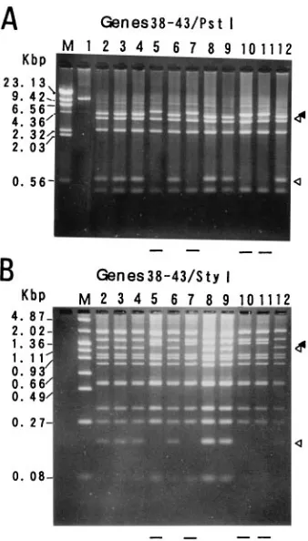
RFLP analysis of the gene 38 to 43 region.In the gene 38 to
43 region, a base substitution from A to G at nucleotide 69349 in aPstI site was previously identified as a marker for grouping VZV isolates (13). Lack of thePstI site was observed in 11 of the 40 isolates. Figure 3A shows thePstI digestion patterns for some of the isolates that contained this alteration (lanes 5, 7, 10, and 11). The absence of theStyI site at position 76530 was observed in the same isolates that lacked thePstI site at posi-tion 69349 (Fig. 3B, lanes 5, 7, 10, and 11). DNA fragments of 163 bp containing theStyI site alteration were amplified from 9 of the 11 mutants, and the nucleotide sequences of these
fragments were determined. The nucleotide sequences of the nine mutants were identical and they contained a one-base substitution, C to T, at position 76530 in theStyI site compared with that of the Dumas strain. RFLPs of the gene 38 to 43 region among the isolates were observed in digests withCfoI and HinfI, but not in those with BamHI, BglI, DraI, KpnI,
HaeIII,MboI,RsaI, andTaqI (Table 2).
RFLP analysis of the gene 54 to 60 region.It was reported
that although the Oka vaccine strain had oneBglI site in the gene 54 to 60 region, most of isolates in the United States lacked the BglI site (1, 16). All of the 40 Japanese isolates examined contained theBglI site identical to that in the Oka vaccine strain (data not shown).
PstI digestion of this region yielded two fragments. The shorter digest was heterogeneous in length (Fig. 4). TheHinfI,
MluI, andMvaI fragments that overlap the shortPstI fragment also varied in length (data not shown). Taking account of the differences of fragment length, the variations seem to be de-rived from variations of the copy number of the R5 repeat sequence. DraI, MluI, HinfI, MvaI, AluI, HaeIII, and MspI digests did not reveal any polymorphisms except for those caused by R5 copy number variation.
RFLP analysis of the Oka vaccine strain.In addition to the
40 isolates, the three regions of the Oka vaccine strain were amplified by long PCR. The PCR products were digested with all restriction enzymes listed in Table 1 exceptMluI. ThePstI site at position 69349 and the StyI site at 76530 were absent from the Oka vaccine strain, and the R5 copy number was two. The other restriction sites examined were the same as those in the Dumas strain. As a result, the Oka vaccine strain was classified into group 8-b (Table 2).
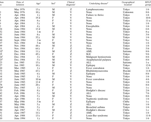
Epidemiological and RFLP analyses of 40 VZV isolates. Clinical data for patients from whom 40 VZV strains were isolated are presented in Table 3. Isolates 202, 224, and 225 were obtained from patients who were hospitalized in the same ward during the same period. Because the isolates from the three patients were classified into the same RFLP group, it is likely that the patients were infected with the same virus in the hospital, as reported previously (22). The other isolates were found to have no epidemiological relationship to one another. Isolates 75, 124, and 367 were obtained from neonates within 19 days after birth. Two of three neonates (infected with iso-lates 124 and 367) were clinically suspected of being infected in utero. No individual who received the Oka varicella vaccine was included in our study except the individual infected with isolate 429. Isolate 429 was grouped into group 1-c, which is different from that of the vaccine strain. Seven isolates (isolates 10, 202, 224, 225, 302, 378, and 435) were grouped into group 8-b which is the same as that of the Oka vaccine strain. How-ever, these isolates had not originated from the Oka strain, because they were isolated before 1987, when the Oka vaccine became commercially available in Japan, and none of the seven patients had received test varicella vaccines. No correlation was found among the year of virus isolation, primary or sec-ondary infection, the clinical severity of the infection, and RFLP groups. Because all except one of the isolates used in this study were obtained from patients who lived in or around Tokyo, there is no geographical bias to the RFLP grouping.
DISCUSSION
Progress in molecular epidemiology of VZV infections has been hampered by two factors: the difficulty in obtaining a large quantity of viral DNA because of the highly cell-associ-ated nature of the virus and the high degree of conservation from isolate to isolate. In this study, we established the
condi-FIG. 2. RFLP analyses of the gene 12 to 16 regions of various VZV isolates.
The enzymes used wereBanIII (A) andHaeIII andHinfI (B). Samples were run
on 0.8% (A) and 4% (B) agarose gels. The isolates in lanes 1 to 10 are inde-pendent and different among the three photographs. The restriction enzyme site which separates the band indicated with a closed arrowhead into the two bands indicated with two open arrowheads was absent from the isolates marked with
minus signs at the bottom of each panel. In panel B, theHinfI digests indicated
with a closed arrowhead contain a single band (257 bp) in lanes 1 to 5 and an additional band (259 bp) in lanes 2, 3, and 5. The digests indicated with the lower open arrowhead contain a single band (77 bp) in lanes 2, 3, and 5 and two bands
similar in length (76 and 77 bp) in lanes 1 and 4. Lane M, bacteriophagelDNA
digested withHindIII.
on May 15, 2020 by guest
http://jcm.asm.org/
tions for PCR amplification of VZV genomic segments longer than 6 kb and demonstrated the ease and utility of using the long PCR products for discriminating VZV isolates. The long PCR method enabled us to shorten the time and reduce the amount of labor required to prepare the materials needed for RFLP analysis. The three regions amplified in this study cover about 20% of the VZV genome. In addition to the three regions, we also amplified a different 15-kb sequence under the same conditions established in the study. However, RFLP analyses of this region of some isolates failed to distinguish them from each other (data not shown). Considering the ca-pacity of the long PCR method, fewer than 10 sets of primers may be sufficient to span the entire VZV genome. It is neces-sary to analyze the remaining regions, which may give more information for classifying VZV isolates.
[image:4.612.57.555.81.286.2]During preliminary experiments, some difficulty was encoun-tered in amplification of the region containing the R4 repeat sequence (data not shown). It is possible that R4 was unstable
FIG. 3. RFLP analyses of the gene 38 to 43 region digested withPstI (A) and
StyI (B). The isolates in lanes 2 through 12 are identical between the two panels.
The digests were analyzed in 1.4% (A) and 4.0% (B) agarose gels. The
undi-gested amplified product is in lane 1 of panel A. BacteriophagelDNA digested
withHindIII and pHY300PLK DNA digested withHindIII andHaeIII (Takara
[image:4.612.94.263.370.667.2]Shuzo, Co., Ltd.) were used as size standards in lane M of panels A and B, respectively. Symbols are as described in the legend to Fig. 2.
FIG. 4. Analysis of R5 copy number.PstI digests of DNA fragments
ampli-fied from the gene 54 to 60 region were separated in a 1.4% agarose gel. Bands marked with a bracket contain R5 and differ in length by increments of the R5 length (112 bp). The copy numbers of R5 of the isolates are one (lane 2), two (lanes 1, 3 to 6, and 9 to 11), and three (lanes 7 and 8). Lane M, bacteriophage
lDNA digested withHindIII.
TABLE 2. RFLPs found in 40 VZV isolates
Variation group
RFLPa
No. of isolates
Genes 12 to 16 Genes 38 to 43 Genes 54 to 60
BanIII/HinfI HaeIII MspI RsaI StyI PstI StyI CfoI HinfI RsaI R5b
1-a 1 1 1 1 1 1 1 1 1 1 1 1
1-b 1 1 1 1 1 1 1 1 1 1 2 15
1-c 1 1 1 1 1 1 1 1 1 1 3 3
2-b 1 2 1 1 1 1 1 1 1 2 2 1
3-b 1 1 11 1 1 1 1 1 1 1 2 1
4-b 1 1 1 2 1 1 1 1 1 1 2 1
5-b 1 1 1 11 1 1 1 1 1 1 2 1
6-b 1 1 1 1 1 1 1 1 11 1 2 1
7-b 1 1 1 1 1 1 1 1 2 1 2 1
8-a 1 1 1 1 1 2 2 1 1 1 1 1
8-b 1 1 1 1 1 2 2 1 1 1 2 7
9-b 1 1 1 1 1 2 2 2 1 1 2 1
9-c 1 1 1 1 1 2 2 2 1 1 3 1
10-b 1 1 1 1 1 2 2 1 1 11 2 1
11-a 2 1 1 1 1 1 1 1 1 1 1 1
11-b 2 1 1 1 1 1 1 1 1 1 2 2
12-b 2 1 1 1 11 1 1 1 1 1 2 1
a1
, restriction site identical to that of the Dumas strain;11, gain of an additional restriction site;2, loss of a restriction site.
b
Copy number of R5 on the basis of length variations ofPstI,HinfI,MluI, andMvaI digests.
on May 15, 2020 by guest
http://jcm.asm.org/
[image:4.612.346.526.533.674.2]during passage of the virus, as suggested by the others (10, 21). Similarly, difficulty in cloning R3 and the instability of R3 have been reported (8). Although a difference in R2 copy number among isolates was reported (15, 21), we found no obvious difference in R2 copy number among the isolates that we analyzed. Although the overall G1C content of VZV DNA is 46%, those of R1, R2, R3, R4, and R5 are 71, 63, 86, 82, and 44%, respectively. Because the amplification of repeat se-quences with high G1C contents has been reported to be difficult (17), the results regarding the R2 copy number should be confirmed by another method. However, because RFLP analyses combined with long PCR do not necessarily depend on variations in the repeat sequences to distinguish isolates, it could be possible to design regions for amplification that do not contain repeat sequences with high G1C contents, such as R1 to R4.
RFLP analysis of the long PCR products identified 12
vari-able loci among 40 isolates on the basis of the presence or absence of restriction enzyme cleavage sites. Combination of these loci with the R5 copy number allowed classification of the 40 isolates into 17 groups (Table 2). One-fourth of the isolates contained a nucleotide alteration of C to T at position 76530, and that alteration was linked to the reportedPstI site polymorphism at position 69349. The StyI site-less alteration will be a useful marker for molecular epidemiology. We nei-ther observed any difference in in vitro growth property among the groups nor found any relationship between the group(s) and the clinical presentations of the patients from whom the isolates were obtained.
On the basis of our observations, we were able to calculate and approximate the rate of nucleotide sequence variation among Japanese VZV isolates. The calculation was performed as follows: (number of restriction enzyme cleavage sites as identified from electrophoresis patterns)3(number of nucle-TABLE 3. Clinical data and RFLP variation groups for 40 VZV isolates
Isolate no.
Date of
isolation Age
a Sexb Clinical
diagnosisc Underlying diseased
Geographical
locatione
RFLP group
9 May 1976 13 y M Z Lymphosarcoma Tokyo 1-b
10 May 1976 10 y M V None Unknown 8-b
57 Apr. 1984 1 y M V Failure to thrive Tokyo 1-b
75 Apr. 1984 19 d F V None Tokyo 10-b
78 Apr. 1984 8 y F V None Tokyo 11-a
79 Apr. 1984 5 y F V None Tokyo 1-b
80 Apr. 1984 4 y M V Encephalitis Tokyo 1-b
124 June 1984 13 d M V None Hyogo 6-b
132 June 1984 1 m F V None Tokyo 8-a
135 June 1984 8 y M V None Tokyo 3-b
136 June 1984 35 y M V None Tokyo 1-b
160 Sept. 1984 2 m F V None Tokyo 9-c
197 Nov. 1984 5 m M V None Tokyo 1-b
199 Nov. 1984 48 y M Z ALL Tokyo 1-b
201 Nov. 1984 64 y F Z None Tokyo 5-b
202f Dec. 1984 11 y M Z AML Saitama 8-b
205 Dec. 1984 49 y F Z SLE Tokyo 9-b
224f Dec. 1984 3 y M V Malignant hystiocytosis Tokyo 8-b
225f Dec. 1984 5 y M V Anaphylactoid purpura Tokyo 8-b
223 Jan. 1985 13 y M Z ALL Saitama 1-a
227 Jan. 1985 10 y M V Epilepsy Tokyo 12-b
260 Mar. 1985 1 y F V Fever convulsion Tokyo 1-b
273 May 1985 43 y F Z Rhabdomyosarcoma Ibaraki 11-b
302 June 1985 6 y M V Epilepsy Tokyo 8-b
364 June 1985 1 y F V None Tokyo 1-b
365 June 1985 10 m M V Fever convulsion Tokyo 4-b
367 June 1985 12 d F V None Tokyo 1-b
378 June 1985 20 y F V None Tokyo 8-b
429g Dec. 1985 1 y M V None Tokyo 1-c
433 Feb. 1986 8 y F Z Hodgkin’s disease Tokyo 2-b
442 Feb. 1986 5 y M Z ALL Tokyo 7-b
435 Apr. 1986 5 y M V None Tokyo 8-b
436 Apr. 1986 4 y F V Nephrotic syndrome Tokyo 1-b
437 May 1986 5 m F V Epilepsy Chiba 1-c
438 May 1986 5 y M Z ALL Tokyo 1-b
439 July 1986 5 y F V Bronchial asthma Tokyo 1-b
430 Apr. 1987 5 y M Z Hodgkin’s disease Chiba 1-c
431 June 1987 14 y F Z AML Tokyo 1-b
292 Apr. 1988 13 y F Z Louis-Bar syndrome Tokyo 11-b
810 June 1994 1 y F V None Tokyo 1-b
a
y, year; m, month; d, day.
b
M, male; F, female.
c
Z, zoster; V, varicella.
d
ALL, acute lymphocytic leukemia; AML, acute myelogenous leukemia; SLE, systemic lupus erythematosus.
e
Chiba, Saitama, and Ibaraki are situated around Tokyo, and Hyogo is in the west of Japan, far from Tokyo.
f
Isolates 202, 224, and 225 were taken from the patients who were hospitalized in the same ward at the same period.
g
The patient was inoculated with the Oka varicella vaccine 9 days before the onset of varicella.
on May 15, 2020 by guest
http://jcm.asm.org/
[image:5.612.67.556.82.475.2]otide residues recognized by the enzyme) for each enzyme. The values for each of the enzymes used were summed. The sum was multiplied by the number of epidemiologically inde-pendent isolates, which was 38. The result, 65,000, is the total number of nucleotide residues analyzed. The total number of variations in all of the isolates examined, 28, was divided by the total number of residues analyzed (65,000). The result is the frequency of nucleotide sequence variations. Although this formula is based on a simple assumption that each RFLP was caused by one base alteration and does not account for varia-tions in repeated sequence copy number, we think that the result provides a close approximation of reality. The extent of variations was calculated to be 0.043%; in other words, one base per 2.3 kb of sequence would be expected to differ from isolate to isolate, confirming the genetic stability and limited diversity of VZV isolates (9, 24). The nucleotide sequence diversity of herpes simplex virus type 1 isolates in Japan was estimated to be 0.37% (19).
Our method will be helpful for investigating variations in VZV DNA that are based on geographical distribution, differ-ences in clinical presentations, and consequdiffer-ences of antiviral drug treatment. Application of the method to molecular epi-demiology based on geographical distribution and to distin-guishing patients with VZV reactivation from those with VZV reinfection is under way. The Oka vaccine is now used not only in Japan but also in several countries, including the United States. Therefore, identification of the genetic markers specific for the vaccine strain should be necessary for quality control and monitoring of vaccination programs. Although some RFLP loci for distinguishing the Oka vaccine and its parent strains from other field isolates were previously proposed on the basis of analyses of whole viral DNA (9, 10, 21), genetic markers unique to the Oka vaccine strain have not yet been conclusively established. In this study, we demonstrated that the Oka vaccine strain is classified in group 8-b (Table 2). To discriminate the vaccine strain from any isolates, it will be essential to examine RFLPs with different restriction enzymes and to analyze the other regions of the VZV genome.
Finally, because field isolates of human herpesviruses 6A, 6B, and 7 and human cytomegalovirus are highly cell associ-ated and are difficult to grow, the long PCR RFLP method will also be useful in studies with these viruses.
ACKNOWLEDGMENTS
We thank Philip E. Pellett and Masato Tashiro for critical reading of the manuscript and A. Oya and T. Kitamura for their support.
REFERENCES
1.Adams, S. G., D. E. Dohner, and L. D. Gelb.1989. Restriction fragment differences between the genomes of the Oka varicella vaccine virus and
American wild-type varicella-zoster virus. J. Med. Virol.29:38–45.
2.Barnes, W. M.1994. PCR amplification of up to 35-kb DNA with high
fidelity and high yield fromlbacteriophage templates. Proc. Natl. Acad. Sci.
USA91:2216–2220.
3.Buchman, T. G., B. Roizman, G. Adams, and B. H. Stover.1978. Restriction endonuclease fingerprinting of herpes simplex virus DNA: a novel
epidemi-ological tool applied to a nosocomial outbreak. J. Infect. Dis.138:488–498.
4.Cen, H., M. C. Breinig, R. W. Atchison, M. Ho, and J. L. C. McKnight.1991. Epstein-Barr virus transmission via the donor organs in solid organ trans-plantation: polymerase chain reaction and restriction fragment length
poly-morphism analysis of IR2, IR3, and IR4. J. Virol.65:976–980.
5.Cheng, S., S.-Y. Chang, P. Gravitt, and R. Respess.1994. Long PCR. Nature
(London)369:684–685.
6.Cheng, S., C. Fockler, W. M. Barnes, and R. Higuchi.1994. Effective am-plification of long targets from cloned inserts and human genomic DNA.
Proc. Natl. Acad. Sci. USA91:5695–5699.
7.Chou, S.1990. Differentiation of cytomegalovirus strains by restriction anal-ysis of DNA sequences amplified from clinical specimens. J. Infect. Dis.
162:738–742.
8.Davison, A. J., and J. E. Scott.1986. The complete DNA sequence of
varicella-zoster virus. J. Gen. Virol.67:1759–1816.
9.Hayakawa, Y., S. Torigoe, K. Shiraki, K. Yamanishi, and M. Takahashi.
1984. Biologic and biophysical markers of a live varicella vaccine strain (Oka): identification of clinical isolates from vaccine recipients. J. Infect.
Dis.149:956–963.
10. Hayakawa, Y., T. Yamamoto, K. Yamanishi, and M. Takahashi.1986. Anal-ysis of varicella-zoster virus DNAs of clinical isolates by endonuclease
HpaI. J. Gen. Virol.67:1817–1829.
11. Her, C., and R. M. Weinshilboum.1995. Rapid restriction mapping by use of
long PCR. BioTechniques19:530–532.
12. Hondo, R., and Y. Yogo.1988. Strain variation of R5 direct repeats in the right-hand portion of the long unique segment of varicella-zoster virus
DNA. J. Virol.62:2916–2921.
13. Hondo, R., Y. Yogo, M. Yoshida, A. Fujima, and S. Itoh.1989. Distribution of varicella-zoster virus strains carrying a PstI-site-less mutation in Japan and
DNA change responsible for the mutation. Jpn. J. Exp. Med.59:233–237.
14. Kilpatrick, B. A., E.-S. Huang, and J. S. Pagano.1976. Analysis of cytomeg-alovirus genomes with restriction endonucleases HinD III and EcoR-1. J.
Vi-rol.18:1095–1105.
15. Kinchington, P. R., J. Remenick, J. M. Ostrove, S. E. Straus, W. T. Ruyechan, and J. Hay.1986. Putative glycoprotein gene of varicella-zoster virus with variable copy numbers of a 42-base-pair repeat sequence has
homology to herpes simplex virus glycoprotein C. J. Virol.59:660–668.
16. LaRussa, P., O. Lungu, I. Hardy, A. Gershon, S. P. Steinberg, and S. Silverstein.1992. Restriction fragment length polymorphism of polymerase chain reaction products from vaccine and wild-type varicella-zoster virus
isolates. J. Virol.66:1016–1020.
17. McConlogue, L., M. A. D. Brow, and M. A. Innis.1988.
Structure-indepen-dent DNA amplification by PCR using 7-deaza-29-deoxyguanosine. Nucleic
Acids Res.16:9869.
18. Pellett, P. E., G. J. Lindquester, P. Feorino, and C. Lopez.1990. Genomic
heterogeneity of human herpesvirus 6 isolates. Adv. Exp. Med. Biol.278:9–
18.
19. Sakaoka, H., K. Kurita, Y. Iida, S. Takada, K. Umene, Y. T. Kim, C. S. Ren, and A. J. Nahmias.1994. Quantitative analysis of genomic polymorphism of herpes simplex virus type 1 strains from six countries: studies of molecular
evolution and molecular epidemiology of the virus. J. Gen. Virol.75:513–
527.
20. Straus, S. E., J. Hay, H. Smith, and J. Owens.1983. Genome differences
among varicella-zoster virus isolates. J. Gen. Virol.64:1031–1041.
21. Takada, M., T. Suzutani, I. Yoshida, M. Matoba, and M. Azuma.1995. Identification of varicella-zoster virus strains by PCR analysis of three repeat
elements and aPstI-site-less region. J. Clin. Microbiol.33:658–660.
22. Takayama, M., N. Takayama, Y. Kameoka, K. Hachimori, K. Kaneda, and M. Minamitani.1989. Comparative restriction endonuclease analysis of
vari-cella-zoster virus clinical isolates. Med. Microbiol. Immunol.178:61–67.
23. Wyatt, L. S., and N. Frenkel.1992. Human herpesvirus 7 is a constitutive
inhabitant of adult human saliva. J. Virol.66:3206–3209.
24. Zweerink, H. J., D. H. Morton, L. W. Stanton, and B. J. Neff.1981. Restric-tion endonuclease analysis of the DNA from varicella-zoster virus: stability
of the DNA after passage in vitro. J. Gen. Virol.55:207–211.