mouse model of chronic asthma
Mang Yu, … , Mindy Tsai, Stephen J. Galli
J Clin Invest.
2011;
121(8)
:3133-3143.
https://doi.org/10.1172/JCI43598
.
Asthma is considered a Th2 cell–associated disorder. Despite this, both the Th1 cell–
associated cytokine IFN-
g
and airway neutrophilia have been implicated in severe asthma.
To investigate the relative contributions of different immune system components to the
pathogenesis of asthma, we previously developed a model that exhibits several features of
severe asthma in humans, including airway neutrophilia and increased lung IFN-
g
. In the
present studies, we tested the hypothesis that IFN-
g
regulates mast cell function in our
model of chronic asthma. Engraftment of mast cell–deficient
Kit
W-sh/W-shmice, which
develop markedly attenuated features of disease, with wild-type mast cells restored disease
pathology in this model of chronic asthma. However, disease pathology was not fully
restored by engraftment with either IFN-
g
receptor 1–null (
Ifngr1
–/–) or Fc
e
receptor 1
g
–null
(
Fcer1g
–/–) mast cells. Additional analysis, including gene array studies, showed that mast
cell expression of IFN-
g
R contributed to the development of many Fc
e
RI
g
-dependent and
some Fc
e
RI
g
-independent features of disease in our model, including airway
hyperresponsiveness, neutrophilic and eosinophilic inflammation, airway remodeling, and
lung expression of several cytokines, chemokines, and markers of an alternatively activated
macrophage response. These findings identify a previously unsuspected IFN-
g
/mast cell
axis in the pathology of chronic allergic inflammation of the airways in mice.
Research Article
Immunology
Find the latest version:
Identification of an IFN-
γ
/mast cell axis
in a mouse model of chronic asthma
Mang Yu,1 Michael R. Eckart,2 Alexander A. Morgan,3 Kaori Mukai,1 Atul J. Butte,3Mindy Tsai,1 and Stephen J. Galli1,4
1Department of Pathology, 2Department of Protein and Nucleic Acid Biotechnology Facility, 3Department of Pediatrics and Center for Pediatric Bioinformatics,
and 4Department of Microbiology and Immunology, Stanford University School of Medicine, Stanford, California, USA.
Asthma is considered a Th2 cell–associated disorder. Despite this, both the Th1 cell–associated cytokine IFN-
γ
and airway neutrophilia have been implicated in severe asthma. To investigate the relative contributions of
different immune system components to the pathogenesis of asthma, we previously developed a model that
exhibits several features of severe asthma in humans, including airway neutrophilia and increased lung IFN-
γ
.
In the present studies, we tested the hypothesis that IFN-
γ
regulates mast cell function in our model of chronic
asthma. Engraftment of mast cell–deficient
Kit
W-sh/W-shmice, which develop markedly attenuated features of
disease, with wild-type mast cells restored disease pathology in this model of chronic asthma. However, disease
pathology was not fully restored by engraftment with either IFN-
γ
receptor 1–null (
Ifngr1
–/–) or Fc
ε
receptor
1
γ
–null (
Fcer1g
–/–) mast cells. Additional analysis, including gene array studies, showed that mast cell
expres-sion of IFN-
γ
R contributed to the development of many Fc
ε
RI
γ
-dependent and some Fc
ε
RI
γ
-independent
fea-tures of disease in our model, including airway hyperresponsiveness, neutrophilic and eosinophilic
inflamma-tion, airway remodeling, and lung expression of several cytokines, chemokines, and markers of an alternatively
activated macrophage response. These findings identify a previously unsuspected IFN-
γ
/mast cell axis in the
pathology of chronic allergic inflammation of the airways in mice.
Introduction
Asthma is a complex disorder characterized by chronic inflam-mation of the airways, airway hyperresponsiveness (AHR), airflow obstruction, and airway wall remodeling (1, 2). Many features of asthma are thought to reflect consequences of Th2-dominated immune responses to allergens in which Th2-type cytokines such as IL-4 and IL-13, and the Th2-associated immunoglobulin IgE, contribute to intricate pathways that result in inflammation and tissue damage (3). Mast cells are considered critical for the devel-opment of asthma phenotypes because of their ability to release, upon activation by IgE and specific antigen, a diverse range of autacoid mediators, chemokines, cytokines, and growth factors (4). Infiltration of the airway with eosinophils is also thought to be important in the pathology of asthma (1–4).
However, studies of patients with well-characterized asthma have suggested that neutrophils, in addition to eosinophils, may have important roles in asthma (5). Patients with chronic severe or acute fatal asthma exhibit increased numbers of airway neutro-phils (6–9), and neutrophilic inflammation is positively correlated with the severity of airflow obstruction (10, 11). Moreover, airway neutrophilia and eosinophilia are not mutually exclusive (10, 12). Patients with severe asthma, compared with those with mild or moderate asthma, have both a greater percentage of sputum neu- trophils and evidence for higher continuing release of eosinophil-derived mediators (13).
Other evidence suggests a possible role for the Th1 cell–associ-ated cytokine INF-γ, as well as Th2 cell–associated cytokines, in some asthmatic subjects. Leukocytes in the BAL fluid from asthma patients can produce IFN-γ ex vivo (14), and, compared with mod-erate asthmatics, patients with severe asthma exhibit increased
numbers of sputum neutrophils and eosinophils, increased epi-thelial or subepithelial expression of IFN-γ or IL-8, and decreased subepithelial expression of IL-4 (15). Increased lung levels of IFN-γ (16) or other evidence for the involvement of this cytokine (17–23) have been reported in some mouse models of allergic inflam-mation of the airways, but such studies have indicated that Th1 responses (17–19, 23) or IFN-γ (20–22) may have effects that coun-teract (18–20), enhance (17, 21, 23), or prolong (22) features of Th2 responses in these settings.
While the different results in various studies regarding the poten-tial roles of IFN-γ in allergic airway inflammation may reflect, at least in part, differences in the mouse models and/or experimen-tal approaches that were employed, taken together this prior work indicates that Th2- and Th1-associated pathways can interact in such settings (16, 17, 23, 24). There are many potential sources of IFN-γ during airway inflammation, including activated CD4+ Th1 cells, CD8+ T cells, and NK cells, as well as γδT cells, macrophages, dendritic cells, NKT cells, naive CD4+ T cells, and B cells (25–27). Whatever its cellular source, IFN-γ can exert pleiotropic effects through a receptor complex (IFN-γ receptor [IFN-γR]) composed of two chains: IFN-γR1 (also known as IFN-γRα ), the major ligand-binding subunit, and IFN-γR2 (also known as IFN-γRβ), which is required for IFN-γ signal transduction and for increasing the affin-ity of IFN-γR1 to its ligand (25, 28).
We developed a mouse model of chronic allergic inflammation of the airways that exhibits several features of chronic asthma in humans, including inflammatory responses in the lungs that include both neutrophils and eosinophils (29). Most features of this model were markedly attenuated or absent in genetically mast cell–deficient mice, and optimal development of many of these “mast cell–dependent” characteristics required mast cell expres-sion of Fcε receptor 1γ (FcεRIγ), which is necessary for mast cell activation via FcεRI-bound IgE and specific antigen or by immune
Conflict of interest: The authors have declared that no conflict of interest exists.
complexes of IgG1 bound to Fcγ RIII (29). We found that mice sub-jected to this model developed greatly increased lung expression of a T cell–specific GTPase selectively induced by IFN-γ (29, 30).
Receptors for IFN-γ have been detected on mouse (31) and human (32) mast cells, and stimulation of various mast cell popu-lations with IFN-γ in vitro has been reported to increase histamine release in anti-IgE–stimulated in vitro derived human mast cells (32), decrease serotonin release in mouse peritoneal mast cells stimulated with IgE plus antigen (IgE+Ag) (33) and decrease his-tamine release in antigen-stimulated rat peritoneal mast cells (34), and have no significant effect on antigen-stimulated histamine release in rat intestinal mast cells (34). While these in vitro stud-ies suggest that the effects of INF-γ on mast cell activation may vary according to species and/or anatomic site, the importance of INF-γ in regulating mast cell function in vivo has not been investi-gated. However, given the evidence that INF-γ can influence mast cell function in vitro, we were intrigued by the idea that IFN-γ might regulate mast cell function in our model of chronic allergic inflammation of the airways.
In the present study, we tested this hypothesis by evaluating the effects of IFN-γ on mast cell function in vitro and by comparing features of our chronic asthma model in WT, Ifng–/–, and Ifngr1–/–
mice and in genetically mast cell–deficient KitW-sh/W-sh
mice engraft-ed with WT, Ifngr1–/–, or Fcer1g–/– mast cells.
Results
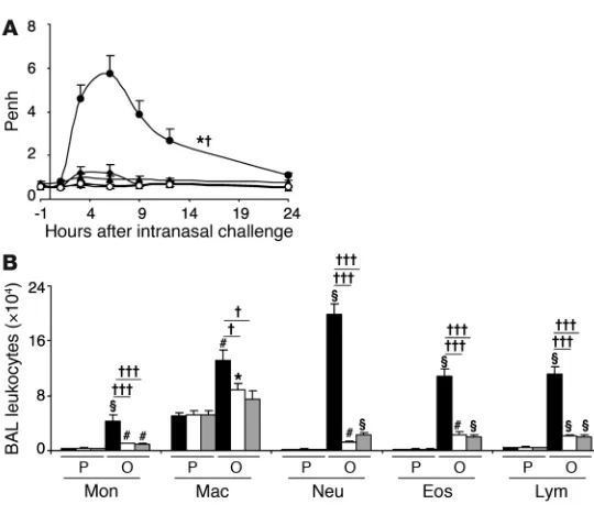
IFN-γ and IFN-γR contribute to this model of chronic allergic inflamma-tion. We first evaluated whether IFN-γ or IFN-γR contributes to airway responsiveness to antigen or airway inflammation in this model (29). As expected (29), WT mice exhibited a significant increase in enhanced pause (Penh, a measure of airway resistance in unrestrained, non-anesthetized mice) after antigen challenge, with the peak of responsiveness occurring between 3 and 9 hours after OVA challenge, whereas Ifng–/– or Ifngr1–/–
mice exhibited sig-nificantly attenuated Penh responses to OVA challenge (Figure 1A). OVA-sensitized Ifng–/– and Ifngr1–/–
mice also exhibited sig-nificantly decreased numbers of total BAL fluid leukocytes after
OVA challenge, as well as significant reductions in individual types of leukocytes, including monocytes, macrophages, neutrophils, eosinophils, and lymphocytes (Figure 1B). We also found that, in WT C57BL/6 mice, i.p. treatment with a single dose of 5 μg IFN-γ, given 30 minutes before or 3 hours after the last of 9 intranasal (i.n.) challenges with OVA, significantly increased both the Penh response to that OVA challenge and the number of neutrophils in the BAL fluid 24 hours after OVA challenge (Supplemental Figure 1; supplemental material available online with this article; doi:10.1172/JCI43598DS1). By contrast, treatment with a neutral-izing antibody to IFN-γ (100 μg per mouse, i.p., at 3 hours after the ninth OVA challenge), compared with treatment with an isotype-matched control antibody, significantly reduced both the Penh response to that OVA challenge and the number of neutrophils, eosinophils, and lymphocytes in the BAL fluid 24 hours after OVA challenge (Supplemental Figure 1). Taken together, these results support the conclusion that IFN-γ and the IFN-γR can significantly enhance these features of the model, but do not clarify which IFN-γR–bearing cell type(s) contributed to these effects.
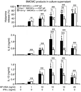
IFN-γ can increase mast cell production of histamine and cytokines in vitro. Because many features of this model require mast cells and mast cell expression of the FcεRIγ chain for full development (29), we next evaluated whether IFN-γ can influence mast cell activation via FcεRI. Bone marrow–derived cultured mast cells (BMCMCs) generated in vitro from WT, IFN-γR1–deficient, or FcεRIγ-deficient mice were sensitized overnight with an anti-dinitrophenol–specific (anti-DNP–specific) monoclonal mouse IgE and then challenged for 1 hour or 24 hours with various concentrations of specific antigen (DNP-HSA) with or without IFN-γ at 1 or 5 ng/ml. Acti-vation of mast cells with IgE+Ag resulted in the dose-dependent secretion of histamine, IL-6, and IL-13 by WT or Ifngr1–/–
BMC-MCs (Figure 2), but no detectable release of IL-4, IFN-γ , or the che-mokines CXCL1, CXCL2, CCL9, CCL11, CCL12, or CCL24. IFN-γ significantly increased the release of histamine, IL-6, and IL-13 by IgE+Ag-stimulated WT BMCMCs, whereas treatment of the cells with IFN-γ alone was without effect (Figure 2 and Supplemental Figure 2). FcεRIγ
-deficient BMCMCs were not detectably respon-Figure 1
[image:3.585.42.312.76.310.2]sive to IgE+Ag challenge, with or without IFN-γ (Figure 2). The ability of IFN-γ to enhance dose-dependently the IgE+Ag-induced mast cell production of IL-13 is of particular interest (Supplemen- tal Figure 2), since IL-13 is thought to contribute to the develop-ment of asthma through such effects as promoting subepithelial fibrosis (in part by upregulating synthesis of arginase-1), increas-ing mucus secretion, and eliciting AHR (35).
Lung expression of IFN-γ. To assess the importance of mast cell signaling via IFN-γR versus FcεRIγ in vivo, we compared features of our model in 5 groups of age-matched mice, all of them on the C57BL/6 background: WT mice, genetically mast cell–defi-cient KitW-sh/W-sh mice, and KitW-sh/W-sh mice engrafted with mast cells
derived from WT, IFN-γR1–deficient, or FcεRIγ–deficient mice. Compared with PBS treatment in control groups, OVA sensitiza-tion and challenge resulted in significant increases in the amounts of IFN-γ in the lungs of each of the 5 types of mice tested (includ-ing the profoundly mast cell–deficient KitW-sh/W-sh mice) (Figure 3A).
By contrast, none of the groups of OVA-sensitized mice exhibited levels of lung IL-4 after OVA challenge that differed significantly
from those in the corresponding PBS-treated groups (Figure 3B). OVA-sensitized and -challenged mice also exhibited elevations in lung levels of IL-17, a cytokine that has been implicated in the development of neutrophilic infiltrates in several settings (36); however, all groups of OVA-challenged mice developed similar elevations in lung levels of IL-17 (Supplemental Figure 3).
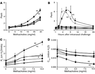
Mast cell IFN-γR can increase airway responses to antigen or methacho-line. OVA-treated WT mice or KitW-sh/W-sh mice engrafted with WT
mast cells (WT BMCMCs→KitW-sh/W-sh mice) exhibited significantly
increased Penh responses to methacholine (a measure of AHR) or to antigen, while little or no enhancement of Penh responses to methacholine or antigen occurred in identically sensitized and challenged mast cell–deficient KitW-sh/W-sh mice or in KitW-sh/W-sh
mice engrafted with Ifngr1–/– or Fcer1g–/– mast cells (Ifngr1–/–
BMC-MCs→KitW-sh/W-sh or Fcer1g–/– BMCMCs→KitW-sh/W-sh mice) (Figure
[image:4.585.55.313.83.376.2]4, A and B, and Supplemental Figure 4, A and B). Similar results were obtained when we measured the effects of methacholine challenge on lung resistance (RL) and dynamic compliance (Cdyn) in anesthetized, tracheostomized mice (Figure 4, C and D, and
Figure 2
[image:4.585.50.382.620.743.2]Influence of IFN-γ on the expression of mast cell function in vitro. Culture supernatants of anti-DNP IgE-sensitized BMC-MCs from WT C57BL/6 (black bars), Ifngr1–/– (white bars), or Fcer1g–/– (gray bars) mice were collected 1 hour (for calcula-tion of % histamine release) or 24 hours (for measurement of secreted cytokines) after addition of DNP-HSA or DNP/IFN-γ. Data were calculated from results pooled from 3 independent experiments, each of which gave similar results. IL-6 levels in supernatants collected from the cultures of WT BMCMCs stimulated with 1 or 5 ng/ml of IFN-γ in the absence of DNP-HSA were 0.009 ± 0.002 or 0.007 ± 0.002 ng/ml; the corre-sponding IL-13 levels were 0.015 ± 0.003 or 0.018 ± 0.004 ng/ml, respectively. *P < 0.05, **P < 0.01, §P < 0.001 versus values from the corresponding BMCMCs stimulated with 1 ng/ml DNP-HSA alone. †P < 0.05, ††P < 0.01, †††P < 0.001 versus group indicated. ‡P < 0.05, ‡‡P < 0.01, #P < 0.001 versus values from the corresponding BMCMCs stimulated with 10 ng/ml DNP-HSA alone. n = 3 per group.
Figure 3
Supplemental Figure 4, C and D). Compared with responses in PBS-treated mice, only OVA-sensitized WT mice and WT BMC-MCs→KitW-sh/W-sh mice exhibited significant reductions in Cdyn in
response to methacholine challenge (Figure 4D and Supplemen-tal Figure 4D). We detected slightly but significantly increased RL following methacholine challenge in OVA-sensitized and -chal-lenged Ifngr1–/– BMCMCs→KitW-sh/W-sh and Fcer1g–/– BMCMCs→
KitW-sh/W-sh mice, but these responses were significantly less intense
than those in the corresponding WT mice and WT BMCMCs→
KitW-sh/W-sh mice (Figure 4C and Supplemental Figure 4C). These
findings indicate that the development of airway responses to antigen and airway hyperreactivity to cholinergic stimulation in this model is largely dependent on mast cell IFN-γR, as well as mast cell FcεRIγ. All 5 groups of OVA-sensitized mice exhibited similar elevations in plasma levels of OVA-specific IgE and IgG1 antibodies (Supplemental Figure 5).
Taken together, our findings indicate that the development of airway reactivity to antigen and airway hyperreactivity to cholin-ergic stimulation in this model is largely dependent on mast cell IFN-γR, as well as mast cell FcεRIγ. Our data also suggest that the
critical mast cell–dependent effects in this model primarily are those that influence end-organ tissue manifestations of anti-gen challenge in sensitized mice, rather than any potential mast cell–dependent effects on sensitization to antigen and/or the generation of antigen-specific IgE or IgG1 antibodies.
Mast cell IFN-γR can increase airway inflam-mation. Histologic analysis (Figure 5, A–E, K, and L) and examination of BAL fluids (Figure 5N) confirmed that mast cells are required for optimal development of anti-gen-induced lung inflammation in this model (29) and showed, unexpectedly, that such leukocyte infiltration was markedly and significantly reduced in mice in which only mast cells lacked IFN-γR, as well as in those in which only mast cells lacked FcεRIγ. Baseline numbers of mast cells/ mm2 of peribronchial tissue were similar in all groups of PBS-treated control mice except KitW-sh/W-sh mice, which totally lacked
mast cells (Figure 5, F–J and M). We did not detect differences in the anatomical distribution of the 3 different types of mast cells (i.e., WT, Ifngr1–/–, and Fcer1g–/–)
that had been adoptively transferred into the KitW-sh/W-sh mice, either in PBS-treated
or OVA sensitized and challenged mice. However, as has been reported for mast cell–engrafted WBB6F1-KitW/W-v (37) and
C57BL/6-KitW-sh/W-sh (38, 39) mice, KitW-sh/W-sh
mice containing adoptively transferred mast cells exhibited more mast cells in the periphery of the lungs than did WT mice. OVA treatment significantly increased the number of peribronchial mast cells in WT BMCMCs→KitW-sh/W-sh and Ifngr1–/–
BMC-MCs→KitW-sh/W-sh mice, but not in Fcer1g–/–
BMCMCs→KitW-sh/W-sh mice (Figure 5, F–J and M). These findings
confirmed that mast cells must express FcεRIγ in order to undergo significant expansion in this model (29) and show that this anti-gen-induced increase in mast cell numbers does not require that the mast cell be able to respond to IFN-γ via IFN-γR.
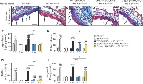
Mast cell IFN-γR can increase airway remodeling. We confirmed that mast cells are required for the full development of features of antigen-induced airway remodeling in this model (29), including subepithelial fibrosis (Figure 6, A–C), increases in lung collagen (Figure 6F), and hyperplasia of mucus-secreting goblet cells (Fig-ure 6G), and that mast cell expression of FcεRIγ contributed more substantially to the development of increased lung collagen than to goblet cell hyperplasia in this model (Figure 6, F and G, and ref. 29). Moreover, mice in which only mast cells lacked IFN-γ R exhib-ited impairment in the antigen-induced development of increased levels of subepithelial fibrosis (Figure 6, A–E), lung collagen (Fig-ure 6F), and airway goblet cells (Figure 6G) that were very similar to those observed in mice in which only mast cells lacked FcεRIγ.
Mast cell expression of each receptor also was required for opti-
mal antigen-induced and mast cell–dependent increases in lung lev-Figure 4
[image:5.585.42.372.79.336.2]els of tissue inhibitor of metalloproteinases–1 (TIMP-1) and inte-grin α7 (Figure 6, H and I). TIMP-1, a major inhibitor of MMP-9, is thought to contribute to a profibrotic environment and airway remodeling by reducing extracellular matrix turnover (40). Integ-rin α7 is thought to regulate smooth muscle cell proliferation and is required for the expression of a contractile phenotype by human airway myocytes (41, 42). In mice in which mast cells lacked either FcεRIγ or IFN-γR, we observed only minimal (albeit statistically significant) antigen-induced increases in TIMP-1 (Figure 6H). By contrast, the development of antigen-dependent increases in lung levels of integrin α7 appeared to be virtually entirely dependent on mast cell expression of INF-γR, whereas this effect was still evident, although at a significantly (~32%) reduced level, in mice whose mast cells lacked FcεRIγ (Figure 6I).
Mast cell IFN-γR is required for optimal expression of lung chemokines.
C-X-C chemokines, including CXCL1 and CXCL2, are predomi-nantly chemotactic for neutrophils, whereas chemokines with C-C motifs are predominantly chemotactic for monocytes, mac-rophages, dendritic cells, lymphocytes, NK cells, eosinophils, and basophils (43). Using a microarray-based analysis of lung gene expression as a screening tool (GEO GSE27066), we found that OVA sensitization and challenge induced markedly increased lung levels of mRNA for CXCL1, CXCL2, CCL11, CCL12, and CCL24 in WT mice and in WT BMCMCs→KitW-sh/W-sh mice, but not in mast
cell–deficient KitW-sh/W-sh mice; we then confirmed these findings
by measuring lung levels of the corresponding proteins (Supple-mental Figure 6). Levels of these chemokines in OVA-treated mice whose mast cells lacked either IFN-γR or FcεRIγ were very similar to those in mast cell–deficient KitW-sh/W-sh mice (there was a minimal,
but statistically significant, OVA-induced increase in levels of lung CXCL1 in Ifngr1–/– BMCMCs→KitW-sh/W-sh
[image:6.585.48.536.86.456.2]mice) (Supplemental Fig-ure 6A). OVA treatment induced an increase in lung CCL9 levels
Figure 5
that was largely, but not entirely, mast cell dependent, and the mast cell–dependent increase in levels of CCL9 in antigen-treated mice required mast cell expression of both IFN-γR and FcεRIγ (Supplemental Figure 6C).
These findings show that mast cells greatly increase lung lev-els of several chemokines in this model, including those that are chemotactic for neutrophils, eosinophils, monocytes, and lympho-cytes, cells whose numbers in the lungs are regulated in a largely mast cell–dependent manner (Figure 5, A–C, K, L, and N). More-over, mast cell transfer studies indicated that the ability of mast cells to enhance lung levels of these chemokines in KitW-sh/W-sh mice
was virtually ablated if the adoptively transferred mast cells lacked either IFN-γR or FcεRIγ (Supplemental Figure 6). By contrast, anti- gen-dependent changes in lung levels of IL-17 occurred indepen-dently of mast cells (Supplemental Figure 3).
Inflammatory cytokines and markers of an alternatively activated mac-rophage response. IFN-γ can increase IgE+Ag-dependent production of IL-6 and IL-13 by mast cells in vitro through effects mediated by IFN-γ R (Figure 2). Consistent with these findings, OVA treat-ment significantly enhanced lung expression of IL-6 and IL-13 in WT or WT BMCMCs→KitW-sh/W-sh mice but not in mast cell–deficient
KitW-sh/W-sh mice or in KitW-sh/W-sh mice containing mast cells that lacked
either IFN-γR or FcεRIγ (Figure 7, A and B). Our microarray-based
screen for increased gene expression identified both IL-33 and mark-ers of an alternatively activated macrophage response as substantially upregulated in this model in OVA-sensitized and challenged WT mice. IL-33 is a member of the IL-1 family that is produced mainly by fibroblasts, epithelial cells, and endothelial cells (44). The recep-tor for IL-33 is composed of two units, the orphan receptor ST2 and IL-1 receptor accessory protein (IL-1RACP) (44). IL-33 can amplify both Th1 and Th2 responses (44) and can directly induce cytokine production by mast cells (including IL-13 production; ref. 45) or basophils, as well as enhance IgE+Ag-dependent activation of these cells (44–46). In the presence of IL-13, IL-33 also can promote polar-ization of alveolar macrophages toward an alternatively activated macrophage phenotype in vitro or in vivo (47). OVA sensitization and challenge markedly increased lung levels of IL-33 in a manner that was largely, but not entirely, mast cell dependent, and the mast cell–dependent component of this response required that mast cells express both IFN-γR and FcεRIγ (Figure 7C).
Th2 responses, including those associated with models of air- way allergic inflammation, are often associated with the develop-ment of an alternatively activated macrophage response (48). Such responses, which contribute to the mitigation or resolution of the pathology associated with allergic inflammation (48), can include
increased lung expression of the macrophage receptor with collage-Figure 6
Features of airway wall remodeling. (A–E) Masson’s trichrome–stained tissue sections demonstrating mucus-secreting goblet cells (blue, indi-cated with arrows) and subepithelial fibrosis (collagen stains blue). (F–I) Collagen levels in the lung (F), numbers of goblet cells along the airway epithelium (G), and levels of lung TIMP-1 (H) and integrin α7 (I). Data are from WT C57BL/6 (gray bars), mast cell–deficient C57BL/6-KitW-sh/W-sh (white bars), C57BL/6 BMCMCs→ C57BL/6-KitW-sh/W-sh (black bars), Ifngr1–/– BMCMCs→ C57BL/6-KitW-sh/W-sh (blue bars), and Fcer1g–/– BMC-MCs→ C57BL/6-KitW-sh/W-sh (yellow bars) mice and the corresponding PBS-treated control mice. **P < 0.01, ***P < 0.001 versus the correspond-ing PBS controls; ‡‡P < 0.01, ‡‡‡P < 0.001 versus OVA-sensitized and -challenged KitW-sh/W-sh mice; †P < 0.05, †††P < 0.001 versus group indicated.
[image:7.585.45.524.86.370.2]nous structure (MARCO) (49) (thought be involved in host defense against modified lipids and tissue repair [ref. 50] and in regulat-ing the tissue distribution and differentiation of macrophages [ref. 51]); the enzyme arginase-1 (48) (which can be increased in aller- gen-induced lung inflammation, but whose role in the pathogen- esis of asthma is unresolved; refs. 52, 53); and the acute-phase pro-tein serum amyloid A3 (SAA3) (54) (which is elevated in a mouse model of allergic inflammation [ref. 55] and may contribute to the accumulation of myeloid cells and to enhancing expression of che- moattractants in this setting [ref. 56]). We found that antigen sen-sitization and challenge resulted in mast cell–dependent increases in lung levels of each of these proteins (Figure 7, D–F). Mast cell responsiveness to IFN-γ, but not mast cell expression of FcεRIγ, was required to effect mast cell–dependent increases in MARCO in anti-gen-challenged mice (Figure 7D). By contrast, mast cell signaling via both IFN-γR and FcεRIγ was required to effect the striking anti-gen- and mast cell–dependent increases in lung levels of arginase-1 protein and Saa3 mRNA (Figure 7, E and F).
Discussion
Our findings reveal an important and previously unsuspected role for IFN-γ–dependent activation of mast cells in the development of the inflammation and tissue pathology associated with this mouse model of chronic allergic inflammation of the airways. As summa-rized in Supplemental Table 1, mast cell expression of IFN-γR is required for optimal development of many features of this model, including AHR to methacholine, inflammation of the airways (with infiltrates containing many neutrophils and eosinophils), airway remodeling (i.e., increases in both numbers of epithelial goblet cells and lung collagen), and markedly increased lung expression of sev- eral cytokines, chemokines, and markers of an alternatively activat-ed macrophage response. It bears emphasis that in KitW-sh/W-sh mice
engrafted with Ifngr1–/– mast cells, mast cells are the only cells that
genetically lack IFN-γR. The poor development of many important features of this model in such mice therefore demonstrates that these features require functions of IFN-γ–responsive mast cells that cannot be provided adequately by any of the other IFN-γ –respon-sive cell types present in this setting.
Mast cell expression of IFN-γR is required for optimal expression of most of the effects of mast cells in this model that also require mast cell expression of FcεRIγ (Supplemental Table 1), and this model is associated with significant, but mast cell–independent, elevations of levels of IFN-γ in the affected lungs (Figure 3A). In vitro, costimu-lation of mouse mast cells with IFN-γ significantly enhanced their ability to release histamine, IL-6, and IL-13 in response to activation with IgE and specific antigen (Figure 2), and IFN-γ dose-depend-ently enhanced the IgE+Ag-dependent release of IL-6 and IL-13 by mast cells in concentrations as low as 0.1 ng/ml (Supplemental Figure 2). In vivo, the significant increases in lung levels of IL-6, IL-13, and IL-33 in this model occurred by wholly (for IL-6 and IL-13) or largely (for IL-33) mast cell–dependent mechanisms (Fig-ure 7, A–C). Moreover, such mast cell–dependent elevations in lung levels of these cytokines required that mast cells express both FcεRIγ and IFN-γ R1 (Figure 7, A–C). Taken together, these findings are con-sistent with the hypothesis that elevations of lung levels of IFN-γ are mechanistically upstream of mast cells in this model and that costimulation of mast cells with IFN-γ (via IFN-γR) and via FcεRIγ results in increased production of IL-6, IL-13, and IL-33, by mast cells (especially for IL-6 and IL-13) and/or other cells that produce these cytokines in a mast cell–dependent manner.
[image:8.585.65.534.83.273.2]Most of the other features of the model that we analyzed — including striking mast cell–dependent increases in airway responses to antigen or airway hyperreactivity to methacholine (Figure 4), tissue neutrophils and eosinophils (Figure 5, K and L,
Figure 7
respectively) and BAL fluid leukocytes (Figure 5), and lung levels of a panel of chemokines (Supplemental Figure 6) — were affected equally (and, in general, markedly diminished) by a lack of either mast cell IFN-γR or mast cell FcεRIγ (Supplemental Table 1). These results indicate that signaling via both IFN-γR and FcεRIγ is required for mast cells to perform the functions important for the development of these mast cell–dependent features of the model. However, some of the in vivo effects of mast cell activation via IFN-γR or FcεRIγ are distinct. The increase in lung mast cells in this model is critically dependent on mast cell FcεRIγ, as revealed both in this (Figure 5M) and in our prior study (29), but this expan-sion of lung mast cells occurs independently of mast cell IFN-γR (Figure 5M). Because mast cells that lack FcεRIγ exhibit ablation of signaling through FcεRIγ and, as a consequence of that, are sig-nificantly impaired in their ability to expand in numbers in the lungs in this model of chronic allergic inflammation, changes in the pathology that are affected by the lack of mast cell expression of FcεRIγ might reflect the lack of FcεRIγ-dependent signaling in those mast cells that are present in such animals, the reduced numbers of lung mast cells in these mice, or both.
Regarding features of lung remodeling, mast cells lacking either IFN-γR or FcεRIγ were equally and markedly impaired in their abil-ity to increase levels of lung collagen (Figure 6F) and TIMP-1 protein (Figure 6H). However, the marked mast cell–dependent increase in lung levels of integrin α 7 protein in this model was completely ablat-ed if mast cells lacked IFN-γR but only modestly, albeit significantly, reduced if mast cells lacked FcεRIγ (Figure 6I). Similarly, the marked and apparently entirely mast cell–dependent increase in lung levels of MARCO protein in this model required that mast cells express IFN-γR but not FcεRIγ (Figure 7D). By contrast, two other mark-ers of an alternatively activated macrophage response, substantially increased lung levels of arginase-1 protein and Saa3 mRNA, were also induced in a largely or entirely mast cell–dependent manner, but in this case the effect was either ablated (in the case of arginase-1 pro-tein; Figure 7E) or markedly reduced (in the case of Saa3 mRNA; Fig-ure 7F) if mast cells lacked either IFN-γR or FcεRIγ. Finally, our data indicate that one important feature of the model, the development of large numbers of intraepithelial goblet cells, is entirely mast cell dependent but that this function can be largely sustained by mast cells lacking either IFN-γR or FcεRIγ (Figure 6G).
Our results support the conclusion that optimal expression of many critical mast cell functions in this model requires costimula-tion of the cells via both IFN-γ/IFN-γR and FcεRIγ, whereas other mast cell functions can be influenced by each receptor independently of the other. We do not know why costimulation of mast cells with IFN-γ is important for the development of so many, but not all, of the features of this model of chronic allergic inflammation. Nor can our mast cell engraftment approach permit one to determine whether particular “mast cell–dependent” features of the model reflect direct actions of mast cells (such as effects of mast cell–derived mediators on leukocytes or structural cells) or indirect effects (such as those mediated by other cells of innate or acquired immunity that are recruited to, and/or influenced in, the lungs in a mast cell–depen-dent manner). However, our study clearly establishes that mast cell responsiveness to IFN-γ, a cytokine classically regarded as a marker of a Th1 response, is as critical for the development of many features of the inflammation, tissue remodeling, and airway functional changes observed in this model of chronic allergic inflammation of the airways as is mast cell expression of FcεRIγ. Moreover, our in vitro findings show that even low concentrations of IFN-γ can enhance
IgE+Ag-dependent release of cytokines by mast cells (Figure 2 and Supplemental Figure 2), suggesting that this mechanism may influ-ence IgE-dependent mast cell functions in many settings, not just those associated with marked elevations of IFN-γ.
Others have shown that various populations of rodent and human mast cells can express IFN-γR or respond to IFN-γ in vitro (31–34), but the importance of mast cell expression of IFN-γR in vivo was not clear. The unexpected identification of an IFN-γ/mast cell axis in this mouse model of airway allergic inflammation chal-lenges the notion that the mast cell’s function in this and perhaps other models of chronic allergic inflammation is entirely or pre-dominantly under the control of the Th2 cell response. Moreover, treatment with a single injection of a neutralizing antibody to IFN-γ, administered to mice 3 hours after the last OVA challenge, reduced the magnitude of both the Penh response and the increases in BAL fluid leukocytes induced by antigen challenge (Supplemen-tal Figure 1), findings indicating that IFN-γ represents a therapeu-tic target in this mouse model.
Are these findings relevant to human asthma? One always must be cautious in attempting to extrapolate findings from any animal model of a disease to its human counterpart, and the relevance of our observations in mice to asthma in humans remains to be deter-mined. For example, while it is well established that the phenotypic features of mast cells can vary based on species, anatomical site, and exposure to cytokines and other factors in the cells’ microenviron-ments (57, 58), little is known about the regulation of the surface expression of IFN-γR by mast cells, whether in the airways or in any other site. Moreover, the relationship of our mouse model of chron- ic allergic inflammation of the airways to particular clinical sub-types of human asthma is not clear. However, as a first step toward addressing the relationship of our mouse model to human asthma, we used gene set enrichment analysis (GSEA) (59) to compare the set of genes that Laprise et al. (60) reported were upregulated in the bronchial biopsy specimens from 4 allergic asthmatic subjects with mild asthma (compared with those from 4 control subjects with-out asthma) and those genes that were upregulated in our mouse model in OVA-sensitized and -challenged (vs. PBS-treated control) mice. We detected significant (i.e., P < 0.05) similarity between genes upregulated in the human asthma specimens compared with those upregulated in our model in WT mice (P ~ 0.021) or WT mast cell–engrafted KitW-sh/W-sh mice (P ~
0.020), but not in identi-cally sensitized and challenged mast cell–deficient KitW-sh/W-sh mice
or in KitW-sh/W-sh mice engrafted with Ifngr1–/– or Fcer1g–/– mast cells
(Supplemental Figures 7–12 and Supplemental Tables 2 and 3). While these findings are intriguing, it will be of interest to repeat such GSEA studies using data sets derived from additional sub-sets of asthma subjects, including those with increased numbers of airway neutrophils and/or increased levels of IFN-γ in the lung, when data of this type become available. Such work, together with other studies in human subjects, will be important in the attempt to decide whether IFN-γ merits investigation as a therapeutic target for reducing the pathology characteristic of IgE-associated allergic inflammation in asthma and perhaps other settings.
Methods
for more than 10 generations. All of the C57BL/6 WT mice and C57BL/6-KitW-sh/W-sh mice used in the experiments reported herein were bred and raised in the same room in the Stanford Laboratory Animal Facility. Fcer1g–/– mice on the C57BL/6 background (B6.129P2-Fcεr1gtm1Rav N12) were purchased from Taconic. Age-matched female mice were used for all experiments. All animal care and experimentation reported herein were conducted in com-pliance with the guidelines of the NIH and with the specific approval of the Institutional Animal Care and Use Committee of Stanford University.
Mast cell engraftment. Engraftment of mast cells in mast cell–deficient C57BL/6-KitW-sh/W-sh mice was performed as described previously (29), with minor modifications. Bone marrow cells derived from 4-week-old female C57BL/6, C57BL/6-Ifngr1–/–, or C57BL/6-Fcer1g–/– mice were cultured in WEHI-3–conditioned medium (ATCC TIB-68), as a source of IL-3, for 4–5 weeks to generate cell populations that contained more than 99% imma-ture mast cells (BMCMCs). BMCMCs (5 × 10⁶) were injected into each mouse via the tail vein, and the recipients (e.g., WT BMCMCs→KitW-sh/W-sh,
Ifngr1–/– BMCMCs→KitW-sh/W-sh, or Fcer1g–/– BMCMCs→KitW-sh/W-sh mice) were used for experiments 18 weeks later.
Production of histamine, cytokines, and chemokines by mast cells in vitro . BMC- MCs were derived as described above and in ref. 29 from femoral bone mar-row cells of C57BL/6 WT, Ifngr1–/–, and Fcer1g–/– mice and were sensitized with 1 μg/ml H1-ε 26 anti-DNP IgE at 37°C overnight. After IgE sensiti- zation, BMCMCs were suspended in DMEM medium (MediaTech) sup-plemented with 10% heat-inactivated FCS (Sigma-Aldrich), 50 μM 2-ME (Sigma-Aldrich), 50 μ g/ml streptomycin (Invitrogen), and 50 U/ml penicil-lin (Invitrogen) at a final concentration of 5 × 10⁵ cells/ml. For activation of the BMCMCs, antigen (DNP-HSA; Sigma-Aldrich) was added to the culture medium at various final concentrations (e.g., 1, 10, or 20 ng/ml). In some groups, we also added rmIFN-γ (PeproTech) at a final concentra-tion of 0.1 to 100 ng/ml. One hour (for calculation of % histamine release) or 24 hours (for measurements of secreted cytokines) after cell activation with antigen with or without rmIFN-γ, BMCMC culture supernatants were collected and stored at –80°C and then were used for measurements of histamine, cytokines, and chemokines.
Immunization and airway challenge with antigen. Mice were immunized by 3 i.p. injections of 50 μg OVA (Sigma-Aldrich) in 0.1 ml PBS on days 1, 4, and 7 (29). Starting on day 12, mice were challenged i.n. with 20 μg OVA in 30 μl PBS weekly for 9 weeks; control mice received i.p. injections and i.n. challenges with PBS on the same schedule (29).
Measurement of airway reactivity to methacholine. Twenty-four hours after the eighth OVA or PBS challenge, responses to aerosolized methacholine were measured using whole-body plethysmography (Buxco Systems). Responses to antigen (OVA) or PBS were assessed by recording Penh over 10-minute time periods at intervals before and after the ninth (final) OVA or PBS challenge (in the absence of methacholine). We also performed inva-sive measurements of airway reactivity in anesthetized, tracheostomized, mechanically ventilated mice. Aerosolized methacholine was administered in increasing concentrations (0, 1.25, 2.5, 5, and 10 mg/ml), with individual doses separated by 2 minutes. RL and Cdyn were continuously computed by fitting flow, volume, and pressure to an equation of motion for each aerosol challenge period, which consisted of a 0.5-minute aerosol exposure and a 1.5-minute period after exposure.
BAL and histology. For BAL, mice were killed by CO2 inhalation 24 hours after the last (ninth) OVA or PBS challenge. The left lung was ligated and removed for measurements of proteins or mRNA (see below), whereas the right lung lobes were lavaged once with ice-cold HBSS (BAL), then fixed (10% formaldehyde) and paraffin-embedded. Paraffin sections of 5 μm were mounted on Superfrost Plus glass slides (Fisher Scientific) and stained with H&E, toluidine blue, or Masson trichrome stains. Numbers of peribron-chial neutrophils, eosinophils, and mast cells were determined using the method described by Pope et al. (61).
Gene expression measurements. Three or 4 mice were randomly selected from each of the 5 different groups of PBS-treated or OVA-sensitized and challenged mice, and specimens of their homogenized left lungs were sub-jected to total RNA isolation using TRIzol Reagent (Invitrogen). Microarray hybridization was performed using the individually isolated RNA samples at the Stanford Protein and Nucleic Acid facility with Affymetrix Mouse Genome 430 2.0 arrays (Affymetrix). The array data were processed using GCRMA (62) and entered into the Gene Expression Omnibus with the accession number GSE27066. A set of 96 upregulated genes in human asthma were identified by quantile normalization and Significance Analy-sis of Microarrays (63) of a publicly available gene expression data set (60), and the enrichment of the 67 genes in this set with homologs measured on the mice arrays was evaluated using GSEA (see Supplemental Methods for additional information about methods used for GSEA) (59).
Extraction of lung tissue protein. Lung tissue protein was extracted using the T-PER Tissue Protein Extraction Reagent purchased from Thermo Scientific. Half of the left lung was placed into 1 ml of protein extraction reagent, followed by homogenization, then the samples were centrifuged at 10,000 g for 5 minutes to pellet cell/tissue debris; the supernatants were collected for further analyses.
ELISA measurements of plasma antibodies. Plasma was collected 24 hours after the ninth OVA or PBS challenge. ELISA kits for total plasma IgE and IgG1 were purchased from Bethyl Laboratories; the ELISA kit for OVA-spe- cific IgE was purchased from MD Biosciences. Concentrations of OVA-spe-cific IgG1 in the plasma were measured by direct ELISA (29).
Measurements of histamine, cytokines, chemokines, and other molecules. Histamine in the BMCMC culture supernatants was measured using an Enzyme Immu-noassay Kit (Beckman Coulter) according to the manufacturer’s instructions, and the results were expressed as percent histamine release (64). ELISA kits for CXCL1, CXCL2, CCL9, CCL11, CCL12, CCL24, IL-33, and TIMP-1 were purchased from R&D Systems; ELISA kits for IL-4, IL-6, IL-17, IL-13, and IFN-γ were purchased from eBioscience. We used direct ELISA methods to measure levels of MARCO, integrin α7, and arginase-1. Primary antibodies against MARCO or integrin α 7 were purchased form R&D Systems, and pri-mary antibodies against arginase-1 were purchased form Novus Biologicals.
Measurement of protein and collagen. The protein levels of tissue samples were measured using a BCA Protein Assay Kit form Thermo Scientific. Tis-sue collagen levels were measured using a Sircol Soluble Collagen Assay Kit from Biocolor Life Science Assays.
Quantitative RT-PCR. Total RNA isolated from the left lungs using TRIzol Reagent (Invitrogen) was reverse transcribed using the High-Capacity cDNA Archive Kit (Applied Biosystems) according to the manufacturer’s instructions. Quantitative real-time PCR was performed using the ABI 7500 Real-Time PCR System (Applied Biosystems) with the following cycling parameters: 10 minutes at 95°C, then 40 biphasic cycles of 15 sec-onds at 95°C, and 1 minute at 60°C. Primers and probes for SAA3 were purchased from Applied Biosystems.
Statistics. Unless otherwise specified, differences in airway response between groups were tested for statistical significance using ANOVA. The unpaired Student’s t test (2 tailed) was used for all other analyses. A P value less than 0.05 was considered statistically significant. Unless otherwise specified, all data are presented as mean ± SEM or mean + SEM.
Acknowledgments
We thank C. Liu and E. Zuo for excellent technical assistance; P. Besmer (Sloan-Kettering Institute) for the gift of C57BL/6-
KitW-sh/W-sh mice; Fu-Tong Liu (University of California, Davis) for
of MARCO; Sally E. Wenzel and Silvana Balzar (University of Pitts-burgh School of Medicine) for helpful discussions regarding IFN-γ in human asthma; and members of the Galli laboratory for discus-sions. This study was supported by grants from the NIH: AI70813, AI23990, CA72074 (to S.J. Galli), and LM009719 (to A.J. Butte).
Received for publication May 4, 2010, and accepted in revised form May 11, 2011.
Address correspondence to: Stephen J. Galli, Department of Pathology, L-235, Stanford University School of Medicine, 300 Pasteur Drive, Stanford, California 94305-5324, USA. Phone: 650.723.7975; Fax: 650.725.6902; E-mail: [email protected]. Or to: Mang Yu, Department of Pathology, Center for Clinical Sciences Research, 269 Campus Dr. #3255, Stanford, Califor-nia 94305-5176, USA. Phone: 650.736.0069; Fax: 650.736.0073; E-mail: [email protected].
1. Busse WW, Lemanske RF Jr. Asthma. N Engl J Med. 2001;344(5):350–362.
2. Umetsu DT, McIntire JJ, Akbari O, Macaubas C, DeKruyff RH. Asthma: an epidemic of dysregu-lated immunity. Nat Immunol. 2002;3(8):715–720. 3. Wills-Karp M. Immunologic basis of
antigen-induced airway hyperresponsiveness. Annu Rev Immunol. 1999;17:255–281.
4. Holgate ST. Novel targets of therapy in asthma.
Curr Opin Pulm Med. 2009;15(1):63–71.
5. Fahy JV. Eosinophilic and neutrophilic inflamma-tion in asthma: insights from clinical studies. Proc Am Thorac Soc. 2009;6(3):256–259.
6. Wenzel SE, Szefler SJ, Leung DY, Sloan SI, Rex MD, Martin RJ. Bronchoscopic evaluation of severe asthma. Persistent inflammation associated with high dose glucocorticoids. Am J Respir Crit Care Med. 1997;156(3 pt 1):737–743.
7. Gibson PG, Simpson JL, Saltos N. Heterogeneity of airway inflammation in persistent asthma: evidence of neutrophilic inflammation and increased spu-tum interleukin-8. Chest. 2001;119(5):1329–1336. 8. Sur S, et al. Sudden-onset fatal asthma. A distinct
entity with few eosinophils and relatively more neutrophils in the airway submucosa? Am Rev Respir Dis. 1993;148(3):713–719.
9. Jatakanon A, Uasuf C, Maziak W, Lim S, Chung KF, Barnes PJ. Neutrophilic inflammation in severe persistent asthma. Am J Respir Crit Care Med. 1999; 160(5 pt 1):1532–1539.
10. Woodruff PG, et al. Relationship between airway inflammation, hyperresponsiveness, and obstruc-tion in asthma. J Allergy Clin Immunol. 2001; 108(5):753–758.
11. Shaw DE, et al. Association between neutrophilic airway inflammation and airflow limitation in adults with asthma. Chest. 2007;132(6):1871–1875. 12. Wenzel SE, et al. Evidence that severe asthma can
be divided pathologically into two inflammatory subtypes with distinct physiologic and clinical characteristics. Am J Respir Crit Care Med. 1999; 160(3):1001–1008.
13. [No authors listed]. The ENFUMOSA cross-sec-tional European multicentre study of the clinical phenotype of chronic severe asthma. European Network for Understanding Mechanisms of Severe Asthma. Eur Respir J. 2003;22(3):470–477. 14. Cembrzynska-Nowak M, Szklarz E, Inglot AD,
Teodorczyk-Injeyan JA. Elevated release of tumor necrosis factor-α and interferon-γ by bronchoal-veolar leukocytes from patients with bronchial asthma. Am Rev Respir Dis. 1993;147(2):291–295. 15. Shannon J, et al. Differences in airway cytokine
profile in severe asthma compared to moderate asthma. Chest. 2008;133(2):420–426.
16. McMillan SJ, Lloyd CM. Prolonged allergen chal- lenge in mice leads to persistent airway remodel-ling. Clin Exp Allergy. 2004;34(3):497–507. 17. Hansen G, Berry G, DeKruyff RH, Umetsu DT.
Allergen-specific Th1 cells fail to counterbalance Th2 cell-induced airway hyperreactivity but cause severe airway inflammation. J Clin Invest. 1999; 103(2):175–183.
18. Cohn L, Homer RJ, Niu N, Bottomly K. T helper 1 cells and interferon γ regulate allergic airway inflam-mation and mucus production. J Exp Med. 1999; 190(9):1309–1318.
19. Huang TJ, et al. Allergen-specific Th1 cells counter- act efferent Th2 cell-dependent bronchial hyperre-sponsiveness and eosinophilic inflammation partly via IFN-γ. J Immunol. 2001;166(1):207–217. 20. Yoshida M, et al. Effect of interferon-γ on allergic
airway responses in interferon-γ-deficient mice. Am J Respir Crit Care Med. 2002;166(4):451–456. 21. Koch M, et al. Role of local pulmonary IFN-γ
expression in murine allergic airway inflammation.
Am J Respir Cell Mol Biol. 2006;35(2):211–219. 22. Coyle AJ, et al. Mice lacking the IFN-γ receptor
have impaired ability to resolve a lung eosinophilic inflammatory response associated with a pro-longed capacity of T cells to exhibit a Th2 cytokine profile. J Immunol. 1996;156(8):2680–2685. 23. Randolph DA, Stephens R, Carruthers CJ, Chaplin
DD. Cooperation between Th1 and Th2 cells in a murine model of eosinophilic airway inflamma-tion. J Clin Invest. 1999;104(8):1021–1029.
24. Fulkerson PC, Zimmermann N, Hassman LM, Fin-kelman FD, Rothenberg ME. Pulmonary chemokine expression is coordinately regulated by STAT1, STAT6, and IFN-γ. J Immunol. 2004;173(12):7565–7574.
25. Boehm U, Klamp T, Groot M, Howard JC. Cellu-lar responses to interferon-γ. Annu Rev Immunol. 1997;15:749–795.
26. Frucht DM, Fukao T, Bogdan C, Schindler H, O’Shea JJ, Koyasu S. IFN-γ production by antigen-presenting cells: mechanisms emerge. Trends Immu-nol. 2001;22(10):556–560.
27. Szabo SJ, Sullivan BM, Peng SL, Glimcher LH. Molecular mechanisms regulating Th1 immune responses. Annu Rev Immunol. 2003;21:713–758. 28. Tau G, Rothman P. Biologic functions of the IFN-γ
receptors. Allergy. 1999;54(12):1233–1251. 29. Yu M, Tsai M, Tam SY, Jones C, Zehnder J, Galli SJ.
Mast cells can promote the development of multi-ple features of chronic asthma in mice. J Clin Invest. 2006;116(6):1633–1641.
30. Carlow DA, Teh SJ, Teh HS. Specific antiviral activ-ity demonstrated by TGTP, a member of a new family of interferon-induced GTPases. J Immunol. 1998;161(5):2348–2355.
31. Nafziger J, Arock M, Guillosson JJ, Wietzerbin J. Specific high-affinity receptors for interferon-γ on mouse bone marrow-derived mast cells: inhibitory effect of interferon-γ on mast cell precursors. Eur J Immunol. 1990;20(1):113–117.
32. Yanagida M, et al. Interferon-γ promotes the survival and FcεRI-mediated histamine release in cultured human mast cells. Immunology. 1996;89(4):547–552. 33. Holliday MR, Banks EM, Dearman RJ, Kimber I,
Coleman JW. Interactions of IFN-γ with IL-3 and IL-4 in the regulation of serotonin and arachi-donate release from mouse peritoneal mast cells.
Immunology. 1994;82(1):70–74.
34. Bissonnette EY, Chin B, Befus AD. Interferons dif-ferentially regulate histamine and TNF-α in rat intestinal mucosal mast cells. Immunology. 1995; 86(1):12–17.
35. Lewis CC, et al. Unique and overlapping gene expres-sion patterns driven by IL-4 and IL-13 in the mouse lung. J Allergy Clin Immunol. 2009;123(4):795–804.
36. Finkelman FD, Hogan SP, Hershey GK, Rothen-berg ME, Wills-Karp M. Importance of cytokines in murine allergic airway disease and human asthma.
J Immunol. 2010;184(4):1663–1674.
37. Williams CM, Galli SJ. Mast cells can amplify air- way reactivity and features of chronic inflamma-tion in an asthma model in mice. J Exp Med. 2000; 192(3):455–462.
38. Wolters PJ, et al. Tissue-selective mast cell recon-stitution and differential lung gene expression in mast cell-deficient KitW-sh/KitW-sh sash mice. Clin Exp
Allergy. 2005;35(1):82–88.
39. Grimbaldeston MA, Chen CC, Piliponsky AM, Tsai M, Tam SY, Galli SJ. Mast cell-deficient W-sash
c-kit mutant KitW-sh/W-sh
mice as a model for investi-gating mast cell biology in vivo. Am J Pathol. 2005; 167(3):835–848.
40. Matsumoto H, et al. Relationship of airway wall thickening to an imbalance between matrix metal-loproteinase-9 and its inhibitor in asthma. Thorax. 2005;60(4):277–281.
41. Liu J, Burkin DJ, Kaufman SJ. Increasing α7β 1- integrin promotes muscle cell proliferation, adhe- sion, and resistance to apoptosis without chang-ing gene expression. Am J Physiol Cell Physiol. 2008; 294(2):C627–C640.
42. Tran T, et al. Laminin-binding integrin α7 is required for contractile phenotype expression by human airway myocytes. Am J Respir Cell Mol Biol. 2007;37(6):668–680.
43. Chin AC, Parkos CA. Pathobiology of neutrophil transepithelial migration: implications in mediating epithelial injury. Annu Rev Pathol. 2007;2:111–143.
44. Liew FY, Pitman NI, McInnes IB. Disease-associ-ated functions of IL-33: the new kid in the IL-1 family. Nat Rev Immunol. 2010;10(2):103–110. 45. Ho LH, et al. IL-33 induces IL-13 production by
mouse mast cells independently of IgE-Fcε RI sig-nals. J Leukoc Biol. 2007;82(6):1481–1490. 46. Silver MR, Margulis A, Wood N, Goldman SJ,
Kasaian M, Chaudhary D. IL-33 synergizes with IgE-dependent and IgE-independent agents to promote mast cell and basophil activation. Inflamm Res. 2010;59(3):207–218.
47. Kurowska-Stolarska M, et al. IL-33 amplifies the polarization of alternatively activated macrophages that contribute to airway inflammation. J Immunol. 2009;183(10):6469–6477.
48. Martinez FO, Helming L, Gordon S. Alternative acti-vation of macrophages: an immunologic functional perspective. Annu Rev Immunol. 2009;27:451–483. 49. Bowdish DM, Gordon S. Conserved domains of the
class A scavenger receptors: evolution and function.
Immunol Rev. 2009;227(1):19–31.
50. Dahl M, et al. Protection against inhaled oxidants through scavenging of oxidized lipids by macro-phage receptors MARCO and SR-AI/II. J Clin Invest. 2007;117(3):757–764.
51. Chen Y, et al. Defective microarchitecture of the spleen marginal zone and impaired response to a thymus-independent type 2 antigen in mice lacking scavenger receptors MARCO and SR-A. J Immunol. 2005;175(12):8173–8180.
52. North ML, Khanna N, Marsden PA, Grasemann H, Scott JA. Functionally important role for arginase 1 in the airway hyperresponsiveness of asthma. Am J Physiol Lung Cell Mol Physiol. 2009;296(6):L911–L920. 53. Niese KA, et al. Bone marrow cell derived arginase I