Introduction
Obstructive sleep apnoea (OSA) is a common and serious condition, affecting approximately 5-15% of adult population in developed countries.1 Beyond poor quality of sleep and its directly related symptoms, OSA is associated with a more adverse cardiovascular risk factor profile and increased risk of coronary artery disease (CAD).2,3 Although OSA patients have increased cardiovascular morbidity and mortality,4 how much of it is due to OSA rather than to other risk factors, such as body mass index (BMI), insulin resistance (IR), age, serum lipid level and cigarette smoking, is yet unknown.
To better understand the relation between OSA and CAD, examining the association between OSA and subclinical coronary artery changes may be valuable.5 The severity of coronary artery stenosis assessed by contrast-enhanced multislice coronary computered tomography angiography (CTA) is a well-established surrogate marker that has been confirmed in numerous studies to be predictive of future coronary events.6,7 However, the risk factors of coronary artery stenosis, such as Prospective Cardiovascular Münster (PROCAM) score8 which provide a truer picture of a person's overall CAD risk, laboratory examinations, such as uric acid and C-reactive protein (CRP), Apnoea Hypopnoea Index (AHI) which aeesees OSA severity, have never been systematically evaluated. The current study was planned to investigate CAD prevalence and characteristics of coronary artery in OSA patient.
Abstract
Objective: To investigate the prevalence of coronary artery disease and characteristics of coronary artery in patients with obstructive sleep apnoea.
Methods: The prospective study was conducted at the Affiliated Huai'an Hospital of Xuzhou Medical University, Huai'an, China, from January, 2012, to June, 2015, and comprised consecutive patients with diagnosed obstructive sleep apnoea. High-resolution 320-slice coronary computed tomography was performed in all the patients. Data was evaluated for the presence of coronary lumen narrowing. Demographic, clinical, laboratory, and echocardiographic characteristics were carefully recorded. Logistic regression was used for multivariate analysis of independent risk factors. SPSS 16 was used for data analysis.
Results: Of the 277 patients, 186(67.14%) were males. The overall mean age was 55.1±14.3 years. Coronary artery disease was found in 41(14.8%) patients. Prospective Cardiovascular Münster score, uric acid, triglyceride, C-reactive protein, apnoea hypopnoea index, Epworth sleepiness scale values were significantly higher in patients with the disease (p<0.05 each). Higher Prospective Cardiovascular Münster score, C-reactive protein, apnoea hyponoea index levels had a significant ability to reflect the occurrence of coronary artery disease (p<0.05 each).
Conclusions: Coronary heart disease occurrence in obstructive sleep apnoea patients was found to be strongly related to Prospective Cardiovascular Münster score, apnoea hyponoea index and C-reactive protein level.
Keywords: Obstructive sleep apnoea, Coronary artery disease, Coronary CT angiography, Risk factors. (JPMA 69: 1610; 2019). doi: 10.5455/JPMA.286541.
Risk factors of coronary artery stenosis in patients with obstructive sleep apnoea:
a prospective study
Yufeng Wan1, Naiquan Yang2, Min XU3, Wenyi Shen4, De HUAI5, Yulong Zheng6
1,4,6Department of Respiratory Diseases, the Affiliated Huai'an Hospital of Xuzhou Medical University; 2Department of Cardiology, the Affiliated Huai'an Hospital of Xuzhou Medical University; 3Department of Radiology, the Affiliated Huai'an Hospital of Xuzhou Medical University; 5ENT Department, the Affiliated Huai'an Hospital of Xuzhou Medical University.
Patients and Methods
The prospective study was conducted at departments of Ear, Nose and Thraot (ENT ), Cardiology and Respirology, Affiliated Huai'an Hospital, Xuzhou Medical University, Huai'an, China, from January, 2012, to June, 2015, and comprised consecutive OSA patients who were recruited after approval was obtained from the institutional ethics committee.
After informed consent was taken from all the subjects, individual information was carefully identified at baseline. Those excluded were subjects with previous adverse reaction to iodinated contrast media, renal dysfunction, t h y r o i d d y s f u n c t i o n , i n c l u d i n g s u b c l i n i c a l hyperthyroidism, history of CAD, valvular heart disease, congestive heart failure (CHF), previous cardiac surgery, myocardial infarction (MI), hypertrophic and dilated cardiomyopathy and history of a disabling cerebral infarction or transient ischaemic attack (TIA), recent infection, autoimmune or inflammatory diseases, respiratory diseases and those consuming alcoholic beverages (Figure 1).
Clinical and demographic profile, including age, gender, smoking status, family history of CAD, medical history, history of drug therapy were recorded. Findings of laboratory examination, including uric acid, serum lipid level, CRP and white blood cell (WBC) count were recorded, and the PROCAM score of the patients was calculated and recorded. Echocardiogram variables and PSG test were carefully recorded.
Laboratory examinations, including complete blood count (CBC) and biochemical investigations, were performed in the fasting state. Levels of total cholesterol
(TC), triglycerides (TG), high-density lipoprotein (HDL) and low-density lipoprotein (LDL) were measured by enzymatic colorimetric methods. The WBC count was determined using a Coulter counter. The levels of serum CRP was assayed with the immunonephelometric method (Dade Behring Marburg, Marburg, Germany). The reference concentrations for CRP were 3mg/L. As a measure of renal function, the baseline estimated glomerular filtration rate (eGFR) was calculated using the abbreviated Modification of Diet in Renal Disease (MDRD) Equation:9 Serum uric acid levels were measured by standard uricase enzymatic test (Normal range: 150-440 mol/L for men; 90-380 mol/L for women).
The subjects slept overnight with the use of Jaeger Sleeplab 1000 (Jaeger, Würzburg, Germany) PSG system. Signals noted included electroencephalogram (EEG), electroocculogram, submental electromyogram, and anterior tibialis electromyogram. Additionally, electrocardiography (ECG) and heart rate (HR) were recorded simultaneously. Snoring was recorded by a microphone placed at the jugular vein, and air flow was recorded by combined oronasal thermistors, while arterial oxyhaemoglobin saturation was recorded by a finger pulse oximeter. Thoracic cage and abdominal motion were recorded by inductive plethysmography. EEG recordings were manually scored according to the standard criteria10. The primary measure was the AHI, quantified as the total number of apnoeas and hypopnoeas per hour of sleep. Apnoea was defined as 90% decrease in airflow from the baseline value for 10 seconds. Apnoeas were further classified as obstructive or central based on the presence or absence of respiratory-related chest wall movement. Hypopnoea was defined as a 30-90% reduction in airflow from the baseline value 10 sec in conjunction with 4% oxygen desaturation. The respiratory event scoring was performed in accordance with the American Academy of Sleep Medicine's 2007 (alternative) guidelines.11 The ESS was used to assess daytime sleepiness.12 OSA diagnosis was made when AHI value exceeded 5, and over 10 points in the ESS.
Two-dimensional echocardiography with M-mode recording was obtained, according to American Society of Echocardiography guidelines13 (iE33 xMATRIX Echocardiography System; Philips, the Netherlands). Data was interpreted by the same operator and recorded on videotape. Left ventricular end-diastolic dimension
358 patients were screened with OSA
Excluded (n=81): a. Acute condition (n=19. b. Prior to CHD or CAD (n=35) c. Refused to participate (n=21) d. Central Sleep Apnea (n=6)
277 patients were eligible for inclusion to the study
Normal appearing (n=202)
Figure-1: Flowchart showing the distribution of the patients eligible for inclusion in the study.
Slightly narrowed (n=34)
Moderately narrowed (n=26)
(LVED), left atrial diameter (LAD) and left ventricular ejection fraction (LVED) were measured. The ejection fraction (EF) was calculated from area measurements using the area-length method14 applied to the average apical area.
All patients were scanned using a 320-slice CT scanner (Aquilion ONE; Toshiba Medical Systems Corp.). CTA images were acquired with a collimation of 320 × 0.5 mm for the 320-slice scanner, a tube rotation time of 350 ms, and a tube current of 100 mA at 120 kV for patients with BMI <30kg/m2. If a patient had a higher BMI, tube current was increased to 100 or 150mA at 135 kV. The acquisition of imaging was prospectively triggered at 75% of the R-R interval. Between 80 and 110 mL of nonionic contrast material (Iomeron 400H; Bracco Atlanta Pharma) was administered with a flow rate of 5 mL/s depending on the total scanning time. The timing of the scan was determined using automated detection of peak enhancement in the aortic root. CTA images were interpreted using transaxial image stacks and curved multiplanar reformatted images. Two reviewers independently evaluated the contrast-enhanced multi-slice CT (MSCT) scans by assessment of the axial multi-slices, of multiplanar reformations and of three thin-slab maximum intensity projections. The reviewers were blinded to all clinical information as well as to the results of endothelial function and intima-media thickness (IMT) testing. Orientated along the heart axis, the thin-slab (5-mm thickness, 1-mm increment) maximum intensity projections were reconstructed perpendicular to each other. The coronary artery tree was segmented according to modified American Heart Association classification15 and the segments were investigated for lumen narrowing. Segments were graded as small
(diameter < 1.5mm), normal appearing (stenosis grade 0-24%), slightly narrowed (stenosis grade 25-49%), moderately narrowed (stenosis grade 50-74%), and severely narrowed (stenosis grade 75%) (Figure 2). CAD was considered in segments of coronary artery lumen graded as moderately and severely narrowed.
Before the study started, a preliminary prevalence for OSA patients who had CAD was conducted. It was estimated that the prevalence of CAD in OSA patients was about 25%. The sample size was confirmed by PASS software.16 Confidence Intervals (CIs) for One Proportion was adopted. A two-sided 95% exact CI was constructed for the population such that the width of the interval was no wider than 0.10. Instead of examining only the interval width of 0.10, widths of 0.08 and 0.12 were also considered. Though in the primary study, the prevalence of CAD in OSA patients was about 25%, a range of values, like 0.15 and 0.35, were included to determine the effect of the proportion estimate on necessary. Parameters above were set in the programme and the minimal sample size 215 was obtained when the proportion was 25% and the width was 0.12 (Figure 3). Continuous variables were presented as mean ± standard deviation. Univariate analysis to assess the predictive value of clinical variables on CAD in OSA patients was computed using the unpaired independent samples t-test for continuous variables and the chi-square test, if necessary, for categorical variables. To test the independence of the risk factors for CAD occurance in OSA patients, the significant variables (p<0.05) in the univariate analyses were entered into a multivariate logistic regression model with backward selection of independent variables. Risk
Figure-2: Grade of Coronary compyted tomography angiography (CTA) features. a.normal appearing, b.slightly narrowed, c.moderately narrowed, d.severely narrowed.
Figure-3: The sample size confirmed by PASS software. N vs C.I. width by P
factors were checked for confounding and co-linearity. All statistical evaluations were made assuming a 2-sided test based on a 5% level of significance using SPSS 16.
Result
Of the 277 patients, 186(67.14%) were males. The overall mean age was 55.1±14.3 years. CAD was found in 41(14.8%) patients (Table 1).
Patients without CAD significantly presented with lower cardiovascular risk profile, resulting in a lower 10-year risk for a cardiovascular event by the PROCAM score. Accordingly, less cardiovascular medications were taken by patients without CAD (p<0.05).
There were no significant differences between the two groups with respect to age, gender, BMI, TC, HDL, LDL, eGFR, WBC count, LAD, LVED and LVEF (p>0.05 each). The prevalences of diabetes and hypertension were significantly higher in OSA patients with CAD than those without CAD (p<0.05).
Patients with CAD were characterised by significantly higher TG levels as well as a trend of having more active smoking (p<0.05). CRP was markedly increased in patients with CAD (p<0.05). Compared to those with non-CAD, OSA patients with CAD were significantly more likely to have higher serum uric acid (p<0.05). Compared with OSA patients with the non-CAD, those with CAD had longer duration of oxygen saturation (SaO2)<90%, higher AHI, ESS scores and values of lowest nocturnal SaO2.
Factors independently associated with CAD in OSA patients included PROCAM score, CRP and AHI (Table 2).
Figure-4: Comparison of the Receover operating characteristics (ROC) plots of different parameters with respect to their ability to reflect significant differences in the area under curve (AUC) for coronary artery disease (CAD). PROCAM: Prospective Cardiovascular Münster; AHI: Apnoea hypopnoea index; CRP: C-reactive protein.
Characteristics With CAD Without CAD Univariate analysis (n=41) (n=236) p-value
Age (years) 54.6±15.5 55.2±14.1 0.81 Male/female 29/12 157/79 0.60 Body mass index 27.5±3.2 27.1±3.8 0.52 Diabetes 13(31.7%) 21(8.9%) <0.001 Arterial hypertension 20(48.8%) 43(18.2%) <0.001 Active smoke 15(36.6%) 52(22.0%) 0.045 Family history of CAD 13(31.7%) 76(32.2%) 0.95 10-yr risk for coronary event 13.3±6.9 2.6±1.6 <0.001 with PROCAM score
Laboratory Examinations
Uric acid (mol/L) 7.04±2.1 5.79±1.77 <0.001 Triglyceride, mg/dL 3.5±1.6 1.8±2.1 <0.001 Total cholesterol, mg/dL 4.5±1.1 46±1.0 0.53 High density lipoprotein, mg/dL 1.3±0.3 1.4±0.3 0.36 Low density lipoprotein, mg/dL 2.5±0.8 2.5±0.7 0.797 eGFR (ml·min-1·m-2) 113.4±79.9 103.8±34.0 0.17
White blood cell count (10*9/L) 6.5±3.0 7.1±2.3 0.13 C reactive protein (mg/L) 7.5±2.6 4.7±2.4 <0.001
Echocardiogram variables(mm)
LAD 35.5±6.0 34.8±5.9 0.43 LVEDD 46.4±5.8 47.2±7.1 0.44 LVEF 63.7±9.3 64.8±9.8 0.55
Drug therapy
Statins 3(7.3%) 11(7.3%) 0.47 Diuretics 4(9.8%) 14(5.9%) 0.36 ACEI/ARBs 10(24.4%) 21(8.9%) 0.004 Beta-blockers 6(14.6%) 14(5.9%) 0.047 CCBs 7(17.1%) 25(10.6%) 0.23
Polysomnography
Apnea hypopnea index, events/h 48.3±12.8 18.9±10.2 <0.001 Lowest SaO2, % 80.6±6.6 70.2±9.8 <0.001 SaO2<90% (%TST) 34.4±11.1 12.6±10.5 <0.001 Epworth sleepiness scale 17.3±3.8 15.7±3.9 0.015 eGFR: estimated glomerular filtration rate; PROCAM: Prospective Cardiovascular Münster; LAVD: Left ventricular end-diastolic dimension; LAD: Left atrial diameter;
LVEF left ventricular ejection fraction (LVEF); ACEI/ARB: Angiotensin converting enzyme inhibitor/ Angiotensin II receptor blockers. CCB: Calcium channel blocker.
Table-1: Clincal characteristics of patients with Obstructive sleep apnoea (OSA).
Variables Wald OR (95% CI) p-value
PROCAM score 18.119 1.593 (1.286-1.974) <0.001 Uric acid mol/L (per 1 unit increase) 1.522 1.004 (.998-1.011) .217 Triglyceride mg/dL (per 1 unit increase) 2.192 1.173 (.950-1.449) .139 Apnoea hypopnoea index (events/h) 7.813 1.091 (1.026-1.159) .005 C reactive protein (mg/L) (per 1 unit increase) 5.369 1.332 (1.045-1.697) .020 Diabetes 2.792 .231 (.042-1.288) .095 Arterial hypertension .849 .466 (.092-2.368) .357 PROCAM: Prospective Cardiovascular Münster; OR: Odds ratio; CI: Cnfidence interval.
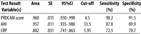
Area under curve (AUC) in ROC curve showed significant differences between CAD andf nn-CAD OSA patients in terms of PROCAM score, AHI and CRP level (Table 3; Figure 4).
Discussion
Intermittent hypoxia, the primary characteristics of OSA have been associated with an increased vulnerability for sudden coronary stenosis, leading to acute ischaemic coronary syndromes. The ability to detect the coronary stenosis at early stages by a noninvasive methodology would probably improve risk stratification of these patients. The current prospective study indicates that such coronary stenosis can be detected in a high proportion of patients investigated by 320-slice CTA for suspected CAD in OSA patients. These patients were characterised by higher PROCAM score, AHI, TG, uric acid and CRP levels and by a trend of having diabetes mellitus. Furthermore, the study demonstrates that PROCAM score, AHI and CRP were the best independent risk factors for CAD development in OSA patients in multivariate regression analysis.
There is increasing evidence, based on large cohorts, that OSA may increase the incidence of CAD.17-19 The identification of OSA patients at increased risk for an potentially life-threatening cardiovascular condition is a very difficult task in sleep medicine. Despite extensive research and testing, traditional cardiovascular risk factors fail to predict development of CAD in a large group of patients.20 With the development of radiological technology and the application of multi-slice CT, coronary CTA has been introduced as noninvasive technique for the reliable detection of coronary stenosis and its micro-structure of coronay artery lesion.21,22 Compared with invasive coronary angiography (ICA), clinical guideline for detecting CAD, noninvasive anatomic assessment by CTA is being increasingly used as an accurate tool for
detecting or excluding CAD.23 The current study assessed the prevalence of coronary artery stenosis in a population of consecutive OSA patients with 320-slice CTA. This population is believed to benefit from noninvasive coronary CTA. The coronary CTA investigations allowed for a separation of the population into two groups: 1) CAD was ruled out in approximately three-fourth of studied patients; 2) CAD with coronary stenosis was present in another one-fourth of investigated patients. There is increasing recognition that noncalcified plaques, defined as clearly discernible lesion with radiodensity greater than the surrounding soft tissue and lower than the luminal contrast, resulted in luminal stenosis of the contrast-enhanced lumen.
Interestingly, OSA patients with CAD had higher TG and CRP levels. The development of systemic inflammation, characterised by elevated levels of certain potent pro-inflammatory mediator, such as CRP, may have an important and direct role in the development of atherosclerotic lesions and in promoting cardiovascular disease. The significantly higher CRP values in OSA patients with coronary stenosis are of particular interest. In OSA patients, the effect of hypoxia and reoxygenation episodes can activate inflammatory cells,24 and ongoing inflammatory responses may also play important roles in coronary atherosclerosis.25 Although CRP is a nonspecific marker of inflammation, meta-analysis have shown that CRP is an independent predictor of risk of future coronary heart disease.26 Furthermore, many studies have confirmed that CRP itself plays a direct role on the expression of inflammatory mediators by vascular endothelium, up-regulation of adhesion molecules and the release of monocyte chemoattractant protein-1 in human endothelial cells.27,28 Therefore, CRP, as a risk factor and an active pathogenic agent, plays a vital role in coronary atherosclerosis. The current study shows that the elevated CRP value was closely related to the development and progression of coronary stenosis.
The evaluation of traditional cardiovascular risk factors for coronary stenosis cannot fully identify the OSA patient population with CAD. The PROCAM study in Europe,29 implemented by statistical procedures such as stepwise logistic regression or the Cox proportional hazards model, may provide a truer picture of a person's overall risk of CAD than clinical classification but may prove comprehensive in clinical practice. This scoring scheme contained nearly all the information present within the
Test Result Area SE 95%CI Cut-off Sensitivity Specificity
Variable(s) (%) (%)
[image:5.595.51.292.134.193.2]PROCAM score .960 .015 .930-.990 4.5 90.2 91.5 AHI .957 .011 .935-.980 33.5 87.8 89.9 CRP .802 .031 .741-.863 5.95 72.5 70.7 PROCAM: Prospective Cardiovascular Münster; AHI: Apnoea hypopnoea index; CRP: C-reactive protein; CAD: Coronary artery disease; OSA: Obstructive sleep apnoea; ROC: Receover operating characteristics; AUC: Area under curve.
Cox function with continuous variables and performed very well in calculating the absolute risk of an acute coronary event. The current study suggests that OSA patients with CAD may have had higher TG values and higher prevalence of hypertension and diabetes mellitus, which are part coefficients of PROCAM scoring scheme. PROCAM score can evaluate person's overall risk of CAD. so all managements of CAD, including quitting smoking, high HDL, low LDL were applied to the OSA patients with CAD.
Data from clinic and community-based studies, although not all, generally suggest that CAD is more frequent in OSA cohorts30 and vice versa that patients with CAD are more likely to have sleep disorders even allowing for the effect of obesity and other confounding factors.31 For example, one such study prospectively followed more than 6,000 participants suffering from the burden of self-reported CAD in the Sleep Heart Health Study among these subjects, and reported the increased prevalence of AHI from 9% in the lowest to 19% in the highest, a relationship that survived adjustment for confounding factors.32 It is unclear if the presence and severity of OSA can actually be used to predict the risk of developing CAD, however. While data from relatively small, but well-organised studies of inpatients suggest a strong dose-response relationship between the severity of OSA and CAD,33 these results from a population-based study cannot be reproduced at the same degree. A community-based study of North American subjects for average follow-up of about 8 years have shown relatively modest relationship between OSA and CAD incidence.29 Within this group of 4,422 (43.6% male) subjects, only severe OSA in men aged 70 or less can increase the risk of developing symptomatic CAD. Furthermore, death analysis has shown that severe OSA served as an independent predictor of mortality of this cohort, and particularly death related to CAD.34 Similar findings were seen about this relationship across the population of the current study.
So far, very little research has addressed the epidemic of CAD by contrast-enhanced multislice coronary ATA in OSA population. The single-centre nature and the relatively small sample size are the main limitations of the current study. Reliability of data would be increased by multicentre study. With a large size, a model of joint AUC of the three variables mentioned would be set up for achieving higher sensitivity and specificity. However,
the study may encourage clinicians to perform early detection and intervention to CAD in OSA patients with high risk. In addition, multivariate analysis was performed to identify inde pendent fact ors of interest.
Conclusions
Coronary heart disease occurrence in OSA patients was found to be strongly related to PROCAM score, AHHI and CRP values. These results might be helpful for monitoring the occurrence of coronary heart disease in OSA patients.
Disclaimer: None.
Conflict of Interest: None. Source of Funding: None.
References
1. Peppard PE, Young T, Barnet JH, Palta M, Hagen EW, Hla KM. Increased prevalence of sleep-disordered breathing in adults. Am J Epidemiol 2013; 177: 1006-14.
2. Somers VK, White DP, Amin R, Abraham WT, Costa F, Culebras A, et al; American Heart Association Council for High Blood Pressure Research Professional Education Committee, Council on Clinical Cardiology; American Heart Association Stroke Council; American Heart Association Council on Cardiovascular Nursing; American College of Cardiology Foundation. Sleep apnea and cardiovascular disease: an American Heart Association/american College Of Cardiology Foundation Scientific Statement from the American Heart Association Council for High Blood Pressure Research Professional Education Committee, Council on Clinical Cardiology, Stroke Council, and Council On Cardiovascular Nursing. In collaboration with the National Heart, Lung, and Blood Institute National Center on Sleep Disorders Research (National Institutes of Health). Circulation 2008 118: 1080-111.
3. Zaigham S, Zaigham Z. Obstructive sleep apnoea: awareness regarding risk factors. J Pak Med Assoc 2010; 60: 74. 4. Lavie P, Lavie L. Cardiovascular morbidity and mortality in obstructive sleep apnea. Curr Pharm Des 2008; 14: 3466-73. 5. Ali SS, Oni ET, Warraich HJ, Blaha MJ, Blumenthal RS, Karim A, et al., Systematic review on noninvasive assessment of subclinical cardiovascular disease in obstructive sleep apnea: new kid on the block! Sleep Med Rev 2014; 18: 379-91.
6. O'Rourke RA, Brundage BH, Froelicher VF, Greenland P, Grundy SM, Hachamovitch R, et al. American College of Cardiology/American Heart Association Expert Consensus document on electron-beam computed tomography for the diagnosis and prognosis of coronary artery disease. Circulation 2000; 102: 126-40.
7. Norgaard BL, Leipsic J, Gaur S, Seneviratne S, Ko BS, Ito H, et al; NXT Trial Study Group. Diagnostic performance of noninvasive fractional flow reserve derived from coronary computed tomography angiography in suspected coronary artery disease: the NXT trial (Analysis of Coronary Blood Flow Using CT Angiography: Next Steps). J Am Coll Cardiol 2014; 63: 1145-55. 8. Assmann G, Cullen P, Schulte H. Simple scoring scheme for calculating the risk of acute coronary events based on the 10-year follow-up of the prospective cardiovascular Munster (PROCAM) study. Circulation 2002; 105: 310-5.
10. Rechtschaffen A, Kales A. A manual of standardized terminology techniques and scoring system for sleep stages of human subjects. Washington, DC: National Institute of Health, 1968. 11. Collop NA, Anderson WM, Boehlecke B, Claman D, Goldberg R, Gottlieb DJ, Hudgel D, et al; Portable Monitoring Task Force of the American Academy of Sleep Medicine. Clinical guidelines for the use of unattended portable monitors in the diagnosis of obstructive sleep apnea in adult patients. Portable Monitoring Task Force of the American Academy of Sleep Medicine. J Clin Sleep Med 2007; 3: 737-47.
12. Johns MW. Daytime sleepiness, snoring, and obstructive sleep apnea. The Epworth Sleepiness Scale. Chest 1993;103: 30-6. 13. Lang RM, Bierig M, Devereux RB, Flachskampf FA, Foster E, Pellikka PA, et al; Chamber Quantification Writing Group; American Society of Echocardiography's Guidelines and Standards Committee; European Association of Echocardiography. Recommendations for chamber quantification: a report from the American Society of Echocardiography's Guidelines and Standards Committee and the Chamber Quantification Writing Group, developed in conjunction with the European Association of Echocardiography, a branch of the European Society of Cardiology. J Am Soc Echocardiogr, 2005; 18: 1440-63.
14. Sievers B, Kirchberg S, Addo M, Bakan A, Brandts B, Trappe HJ. Assessment of left atrial volumes in sinus rhythm and atrial fibrillation using the biplane area-length method and cardiovascular magnetic resonance imaging with TrueFISP. J Cardiovasc Magn Reson 2004; 6: 855-63.
15. Austen WG, Edwards JE, Frye RL, Gensini GG, Gott VL, Griffith LS, et al. A reporting system on patients evaluated for coronary artery disease. Report of the Ad Hoc Committee for Grading of Coronary Artery Disease, Council on Cardiovascular Surgery, American Heart Association. Circulation 1975; 51(4 Suppl): 5-40.
16. Newcombe RG. Two-sided confidence intervals for the single proportion: comparison of seven methods. Stat Med 1998; 17: 857-72.
17. Luyster FS, Kip KE, Aiyer AN, Reis SE, Strollo PJ Jr. Relation of obstructive sleep apnea to coronary artery calcium in non-obese versus obese men and women aged 45-75 years. Am J Cardiol 2014; 114: 1690-4.
18. Glantz H, Thunström E, Johansson MC, Wallentin Guron C, Uzel H, Ejdebäck J, et al. Obstructive sleep apnea is independently associated with worse diastolic function in coronary artery disease. Sleep Med 2015; 16: 160-7.
19. Peker Y, Glantz H, Eulenburg C, Wegscheider K, Herlitz J, Thunström E, et al. Effect of Positive Airway Pressure on Cardiovascular Outcomes in Coronary Artery Disease Patients with Non-Sleepy Obstructive Sleep Apnea: The RICCADSA Randomized Controlled Trial. Am J Respir Crit Care Med 2016; 194: 613-20. 20. Naghavi M, Libby P, Falk E, Casscells SW, Litovsky S, Rumberger J, et al. From vulnerable plaque to vulnerable patient: a call for new definitions and risk assessment strategies: Part II. Circulation 2003; 108:1772-8.
21. Min JK, Koo BK, Erglis A, Doh JH, Daniels DV, Jegere S, et al.
Usefulness of noninvasive fractional flow reserve computed from coronary computed tomographic angiograms for intermediate stenoses confirmed by quantitative coronary angiography. Am J Cardiol 2012; 110: 971-6.
22. Nakazato R, Park HB, Berman DS, Gransar H, Koo BK, Erglis A, et al., Noninvasive fractional flow reserve derived from computed tomography angiography for coronary lesions of intermediate stenosis severity: results from the DeFACTO study. Circ Cardiovasc Imaging 2013; 6: 881-9.
23. Javed S, Shah N. MDCT, an emerging method of investigation for coronary artery disease. J Pak Med Assoc 2011; 61: 206. 24. Ryan S, Taylor CT, McNicholas WT. Selective activation of inflammatory pathways by intermittent hypoxia in obstructive sleep apnea syndrome. Circulation 2005; 112: 2660-7. 25. Thunstrom E, Glantz H, Fu M, Yucel-Lindberg T, Petzold M, Lindberg K, et al. Increased inflammatory activity in nonobese patients with coronary artery disease and obstructive sleep apnea. Sleep 2015; 38: 463-71.
26. Emerging Risk Factors Collaboration, Kaptoge S, Di Angelantonio E, Lowe G, Pepys MB, Thompson SG, Collins R, et al. C-reactive protein concentration and risk of coronary heart disease, stroke, and mortality: an individual participant meta-analysis. Lancet 2010; 375: 132-40.
27. Wei H, Fang L, Song J, Chatterjee S. Lovastatin compromises C-reactive protein induced endothelial dysfunction including altered expression of cell adhesion molecules and increased monocyte recruitment. Atherosclerosis 2005; 178: 399-401.
28. Guo S, Meng S, Chen B, Liu J, Gao L, Wu Y. C-reactive protein can influence the proliferation, apoptosis, and monocyte chemotactic protein-1 production of human umbilical vein endothelial cells. DNA Cell Biol 2011; 30: 157-62.
29. Gottlieb DJ, Yenokyan G, Newman AB, O'Connor GT, Punjabi NM, Quan SF, et al. Prospective study of obstructive sleep apnea and incident coronary heart disease and heart failure: the sleep heart health study. Circulation 2010; 122: 352-60.
30. Kent BD, Garvey JF, Ryan S, Nolan G, Dodd JD, McNicholas WT. Severity of obstructive sleep apnoea predicts coronary artery plaque burden: a coronary computed tomographic angiography study. Eur Respir J 2013; 42: 1263-70.
31. Mooe T, Rabben T, Wiklund U, Franklin KA, Eriksson P. Sleep-disordered breathing in men with coronary artery disease. Chest 1996; 109: 659-63.
32. Shahar E, Whitney CW, Redline S, Lee ET, Newman AB, Nieto FJ, et al. Sleep-disordered breathing and cardiovascular disease: cross-sectional results of the Sleep Heart Health Study. Am J Respir Crit Care Med 2001; 163: 19-25.