0095-1137/10/$12.00
doi:10.1128/JCM.00775-10
Copyright © 2010, American Society for Microbiology. All Rights Reserved.
Characterization of
Staphylococcus aureus
Isolates with a Partial or
Complete Absence of Staphylococcal Cassette
Chromosome Elements
䌤
Henry Wong,
1* Lisa Louie,
1Reggie Y. C. Lo,
2and Andrew E. Simor
1,3Department of Microbiology, Sunnybrook Health Sciences Centre, Toronto, Ontario, Canada
1; Department of Pathobiology,
University of Guelph, Guelph, Ontario, Canada
2; and Department of Laboratory Medicine and
Pathobiology, University of Toronto, Toronto, Ontario, Canada
3Received 16 April 2010/Returned for modification 13 July 2010/Accepted 16 July 2010
Detection of methicillin-resistant
Staphylococcus aureus
(MRSA) by single-locus PCR assays that target the
extremity of the staphylococcal cassette chromosome-
mec
(SCC
mec
) and part of the adjacent
S. aureus
-specific
open reading frame gene (
orfX
) is a significant diagnostic advancement, since it provides real-time detection
directly from screening specimens. However, isolates harboring
mecA
deletions within SCC
mec
may result in
false-positive identification of MRSA in these assays. We characterized 24 methicillin-susceptible
S. aureus
(MSSA) isolates that tested positive in one such assay to investigate this phenomenon. Seven isolates
resem-bled USA100 and carried SCC
mec
II elements with
mecA
deletions that spanned 20 to 46 kbp. The
mecA
excisions in USA100-resembling isolates appeared to be linked with IS
431
transposable elements present in
SCC
mec
II. For 17 isolates that resembled USA400 and/or MSSA476, the identity and possible excision of SCC
elements could not be confirmed. The downstream common sequence (
dcs
) shared by SCC
mec
I, II, and IV
elements was detected in these isolates. Sequence analysis of the chromosomal regions flanking the missing
SCC element revealed an intact SCC integration site, a duplicate
dcs
, and the enterotoxin gene cluster
downstream of
orfX
. An annealing sequence for one of the SCC
mec
-specific primers (mecii574) in the
single-locus PCR assay was identified in the duplicate
dcs
. In the absence of SCC, a 176-bp amplicon can be generated
from this mecii574 annealing sequence to yield a false-positive result. In conclusion, partial SCC
mec
II
excisions via IS
431
elements in strains that resembled USA100 and the presence of a duplicate mecii574
annealing sequence in strains that resembled USA400/MSSA476 were identified as causes for false-positive
results in a single-locus PCR assay that targets the SCC
mec
/
orfX
junction.
Methicillin-resistant
Staphylococcus aureus
(MRSA) is a
multidrug-resistant pathogen associated with significant
mor-bidity, mortality, and hospitalization costs (3, 8, 9, 19, 41).
Since 2003, more than 60% of
S. aureus
infections were caused
by MRSA in intensive care units of hospitals belonging to the
U.S. National Nosocomial Infection Surveillance system (20).
In the last decade, MRSA has also emerged in community
settings, causing skin and soft tissue infections and other
deep-seated infections in individuals without conventional health
care-associated risk factors for MRSA (10). The resistance to

-lactam antibiotics in MRSA is mediated by an altered
peni-cillin-binding protein (PBP2a) encoded by the
mecA
gene (13).
This resistance determinant resides on a mobile genetic
ele-ment termed the staphylococcal cassette chromosome-
mec
(SCC
mec
) that integrates downstream of a
S. aureus
-specific
open reading frame (
orfX
) (11, 14). Eight major SCC
mec
types
ranging from 22 to 64 kbp in size have been described to date
(1, 16-18, 26, 34, 44).
A single-locus PCR assay, utilizing a
S. aureus
-specific
orfX
gene primer (Xsau325) and a combination of SCC
mec
-specific
primers that anneal at the extremity of SCC
mec
to amplify the
SCC
mec
/
orfX
junction, was first proposed by Huletsky et al. for
MRSA detection (15). Since
mecA
resides on SCC
mec
,
detec-tion of the SCC
mec
/
orfX
junction is considered a surrogate for
the detection of MRSA. There are now a number of
commer-cially available assays that identify MRSA based on the
site-specific integration of SCC
mec
at
orfX
(36, 38, 43). Shortly
after the introduction of these tests, there were reports of
assay-positive specimens that only contained
methicillin-sus-ceptible strains of
S. aureus
(2, 4, 12, 32, 35-38, 40, 43).
Al-though investigators mentioned SCC homologues and partial
SCC
mec
deletions as plausible reasons for discrepant results,
few studies have determined why these “false-positive” test
results occurred. In an effort to determine possible causes, we
characterized 24 methicillin-susceptible
S. aureus
(MSSA)
iso-lates, collected from 10 healthcare institutions in the United
States and Canada, which produced “false-positive” results in
one such single-locus PCR assay.
(This study was presented in part at the American Society
for Microbiology 109th General Meeting, Philadelphia, PA, 17
to 21 May 2009.)
MATERIALS AND METHODS
Bacterial isolates.Twenty-fourStaphylococcus aureusthat were identified as MRSA by the BD GeneOhm MRSA assay (version 3; BD Diagnostics, Quebec, Quebec, Canada) originating from hospitals in six states (Illinois, Maryland, Massachusetts, Ohio, Indiana, and North Carolina) and four Canadian hospitals in two provinces (Ontario and Quebec) were examined. TheS. aureusisolates were grown on Columbia agar supplemented with 5% sheep blood (Oxoid,
* Corresponding author. Mailing address: Department of
Microbi-ology, Sunnybrook Health Sciences Centre, B103-2075 Bayview
Ave-nue, Toronto, Ontario M4N 3M5, Canada. Phone: (416) 480-4242.
Fax: (416) 480-6990. E-mail: [email protected].
䌤
Published ahead of print on 28 July 2010.
3525
on May 16, 2020 by guest
http://jcm.asm.org/
Nepean, Ontario, Canada) for 16 to 18 h at 35°C. Suspension cultures were prepared by inoculating a single bacterial colony into 5 ml of BD BBL brain heart infusion broth (Becton Dickinson, Sparks, MD) and grown for 16 to 18 h at 35°C with agitation. TheS. aureuscontrol strains for molecular analyses included MRSA strain N315 (SCCmecII) (17), USA100 MRSA and USA400 MRSA (27), and MSSA476 (an MSSA strain that resembles the USA400 SmaI pulsed-field type) (7).
Determination of methicillin susceptibility.Methicillin susceptibility was de-termined by oxacillin broth microdilution testing in accordance with Clinical and Laboratory Standards Institute guidelines (5, 6). Methicillin susceptibility was further confirmed by PCR detection of themecAdeterminant (25) (see below).
PCR template preparation.DNA templates were prepared by suspendingS. aureuscolonies in 0.5 ml of neutralizing buffer (30 mM Tris [pH 8.4], 2 mM EDTA [pH 9.0]) that contains 50l of 0.1-mm glass beads (Scientific Industries, Bohemia, NY) and heating at 100°C for 2 min. The lysates were vortexed for 2 min and clarified by centrifugation at 20,800⫻gfor 5 s. Templates for long-range PCR were prepared with a High-Pure PCR template purification kit (Roche Diagnostics, Laval, Quebec, Canada).
Primer synthesis and PCR amplification. Custom oligonucleotide primers were purchased from Invitrogen (Burlington, Ontario, Canada) (Table 1). PCR amplifications were performed using the GeneAmp PCR System 9700 (Applied Biosystems, Inc., Foster City, CA). Unless otherwise specified, monoplex and
multiplex PCR assays for amplicons⬍1.5 kbp were performed in 25-l reactions containing 1.25 U of AmpliTaq DNA polymerase, 0.1 mM deoxynucleoside triphosphates, 1.5 mM MgCl2(Roche Diagnostics), and 0.5M concentrations
(each) of forward and reverse primers with the following thermocycling param-eters: 94°C for 2 min, 30 cycles of 94°C for 1 s, 55°C for 15 s, and 72°C for 7 min. Long-range PCRs were performed in 50-l reactions using an Expand Long Template PCR system (Roche Diagnostics) with the following parameters: 92°C for 2 min; 10 cycles of 92°C for 10 s, 50°C for 15 s, and 68°C for 10 min; followed by 20 cycles of 92°C for 10 s, 50°C for 15 s, and 68°C for 10 min (with an additional 20-s extension time incorporated per subsequent cycle), and then 68°C for 7 min. PCR amplification products were resolved on 0.5⫻Tris-borate EDTA agarose gels containing 0.5g of ethidium bromide/ml for digital photography under UV illumination.
MRSA PVL multiplex PCR.Detection of 16S rRNA,mecA,lukS/F, andnuc genes was performed as a multiplex PCR assay with the following concentrations of primer pairs: 0.5Mluk, 0.4MmecA, 0.3M 16S rRNA (internal control), and 0.2Mnuc(23, 25).
SCCmectype determination.SCCmectypes were determined by using a com-bination of SCCmectyping assays as described previously (22, 33). The J1 region of SCCmecII was detected by monoplex PCR using the LIR2 primers.
[image:2.585.46.540.79.504.2]Detection of mobile genetic elements.TheS. aureuspathogenicity islandSa3 present in MW2 (GenBank accession no. BA000033) and the bacteriophage
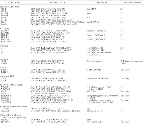
TABLE 1. Primers used in this study
Test and primer Sequence (5⬘–3⬘) Description Source or reference
MRSA/PVL detection
16S-F AGA GTT TGA TCA TGG CTC AG 16S rRNA 25
16S-R GGA CTA CCA GGG TAT CTA AT
mecA-F AAA ATC GAT GGT AAA GGT TGG C mecA 25
mecA-R AGT TCT GCA GTA CCG GAT TTG C
nuc-F GCG ATT GAT GGT GAT ACG GTT nuc 25
nuc-R AGC CAA GCC TTG ACG AAC TAA AGC
luk-F ATC ATT AGG TAA AAT GTC TGG ACA TGA TCC A lukS/F(PVL) 23 luk-R GCA TCA AST GTA TTG GAT AGC AAA AGC
SCC typing
KDP F1 AAT CAT CTG CCA TTG GTG ATG C Locus B (SCCmecII) 33
KDP R1 CGA ATG AAG TGA AAG AAA GTG G
DCS F2 CAT CCT ATG ATA GCT TGG TC Locus D (SCCmecII) 33
DCS R1 CTA AAT CAT AGC CAT GAC CG
IS431 P4 CAG GTC TCT TCA GAT CTA CG Locus G (SCCmecII) 33
pUB110 R1 GAG CCA TAA ACA CCA ATA GCC
ccrtyping
␣1 AAC CTA TAT CAT CAA TCA GTA CGT ccrA1(SCCmecI) 22
␣2 TAA AGG CAT CAA TGC ACA AAC ACT ccrA2 (SCCmecII) 22
c ATT GCC TTG ATA ATA GCC ITC T ccrB1(SCCmecI) andccrB2 (SCCmecII)
22
spatyping
932-F GCC AAA GCG CTA ACC TTT TA Protein A (spa) B. Kreiswirth, unpublished
1740-R TCC AGC TAA TAA CGC TGC AC data
J1 PCR
LIR2-F CGA ATT AAA CAC GCT TGC TC J1 (SCCmecII) This study
LIR2-R CAG AAG TCT ACA GGT CTT GC
Long-range PCR
3.0-R CTC AGA CAG CAA TTT CCC G Downstream of SCCmec This study
4.2-R CCC TAA AAA TAC CTG TAG GGG
Detection of MGE targets
MW756-F TGG TTA GCT ATG AAT GTA GTT GC Hypothetical protein (Sa3), 45
MW756-R GTC CAT CCT CTG TAA TTT TGC unique to MW2
sel2-F ATC ATA CAG TCT TAT CTA ACG GCG sel2(Sa3) This study
sel2-R TCT GGA AGA CCG TAT CCT GTG
SAS0898-F GAA GTA GCA GAT ATA TTG GGC G Intergenic region ofSAS0898/0899 This study
SAS0899-R ATC ACT GGC AAG TCC TAC C (Sa4)
SAS0922-F GAG GGA TAA ACT AAT GAA AGA ACG Hypothetical phage protein This study SAS0922-R TCA TAG AAC CTA AAC GTC CCG (Sa4), unique to MSSA476
SCCmec/orfXjunction detection
Xsau325 GGA TCA AAC GGC CTG CAC A orfX 15
mecii574 GTC AAA AAT CAT GAA CCT CAT TAC TTA TG SCCmecI, II, IV 15
SCCmecII gene elements detection and sequencing
kdpB2 TAA ACT GTG TCA CAC GAT CCA T kdpB 22
II-48600R TCT TAG ATA GTA GCC AAC GGC J3 of SCCmecII This study
on May 16, 2020 by guest
http://jcm.asm.org/
Sa4 associated with MSSA476 (GenBank accession no. BX571857) were de-tected by PCR (24). The gene targetsMW756(nucleotides [nt] 824637 to 825009) andsel2(nt 828744 to 829329) were surrogates forSa3. The intergenic region ofSAS0898/SAS0899(nt 985159 to 985508) andSAS0922(nt 994863 to 995288) were surrogates forSa4.
DNA sequence determination.Long-range PCR products were purified by using a High-Pure PCR product purification kit (Roche Diagnostics) for se-quence determination at the TCAG sequencing facility (Hospital for Sick Chil-dren, Toronto, Ontario, Canada).
spatyping.Strain typing based on the polymorphic X-region of the protein A gene (spa) was performed essentially as described previously (39), except that the primers 932-F and 1740-R were used for amplification and sequencing of thespa amplicon. The Kreiswirthspanomenclature obtained by thespatyping tool (http: //fortinbras.us/cgi-bin/spatyper/spaTyper.pl) was adopted for the present study.
PFGE.The Canadian standardized protocol for pulsed-field gel electro-phoresis (PFGE) typing of MRSA was adopted for MSSA typing in the present study (28). SmaI-restricted DNA profiles were digitized and analyzed with BioNumerics v6.0 (Applied Maths, Austin, TX). XbaI (Roche Diagnos-tics)-digestedSalmonellaserotype Braenderup H9812 DNA was used as the reference standard for band size determination in BioNumerics. Alterna-tively, the Lambda Ladder PFG Marker (New England Biolabs, Pickering, Ontario, Canada) was used as the reference standard in Southern analyses.
Southern hybridization.Southern blot hybridization and detection were per-formed by using an ECL direct nucleic acid labeling and detection system (GE Healthcare, Piscataway, NJ). Briefly, SmaI-digested chromosomal DNA was resolved by PFGE and transferred onto Hybond-N⫹nylon membrane (GE Healthcare). DNA probe corresponding to the 342-bp locus D (33) was used for hybridization, and Kodak BioMax Light film (Rochester, NY) was used for signal detection.
RFLP.Restriction fragment length polymorphism (RFLP) of long-range PCR amplicons was used to assess the similarity between the amplified products. Restriction enzymes BclI, EcoRI, HincII, HindIII, and XbaI (Roche Diagnos-tics) were used according to the manufacturer’s instructions.
RESULTS
Molecular and phenotypic characterization.
The 24 isolates
were determined to be
Staphylococcus aureus
by the presence
of the
S. aureus
-specific
nuc
gene. Susceptibility to methicillin
was determined by broth microdilution and confirmed by the
absence of
mecA
with PCR. These methicillin-susceptible
S.
aureus
(MSSA) did not carry the genes for PVL. The
down-stream common sequence (locus D,
dcs
) shared by SCC
mec
I,
II, and IV was detected in all 24 MSSA isolates (Table 2).
Locus B from the
kdp
gene cluster of SCC
mec
II was detected
in isolates IDI2406, IDI2407, IDI2445, IDI2595, IDI2515, and
IDI2643. In addition, isolates IDI2515 and IDI2683 were also
positive for locus G (linearized pUB110 plasmid) that is
typi-cally present in SCC
mec
II. Apart from IDI2683, all SCC
mec
II isolates were positive for the SCC
mec
II-specific J1 target.
The PCR target for the
ccr
gene complex of SCC
mec
II was
detected in isolates IDI2406, IDI2407, IDI2445, and IDI2595.
Strain typing and genetic deletion determination.
The PFGE
and
spa
typing results are summarized in Table 2. The seven
isolates with remnants of SCC
mec
II resembled USA100
MRSA in their pulsed-field profile and
spa
type; the remaining
17 isolates resembled USA400 MRSA and/or MSSA476 (Fig.
1). Southern blot hybridization with the locus D probe
de-tected genetic deletions of approximately 18 to 45 kbp in the
SCC
mec
II isolates that resembled USA100, and
approxi-mately 20 kbp in the USA400/MSSA476-resembling isolates
(Fig. 1).
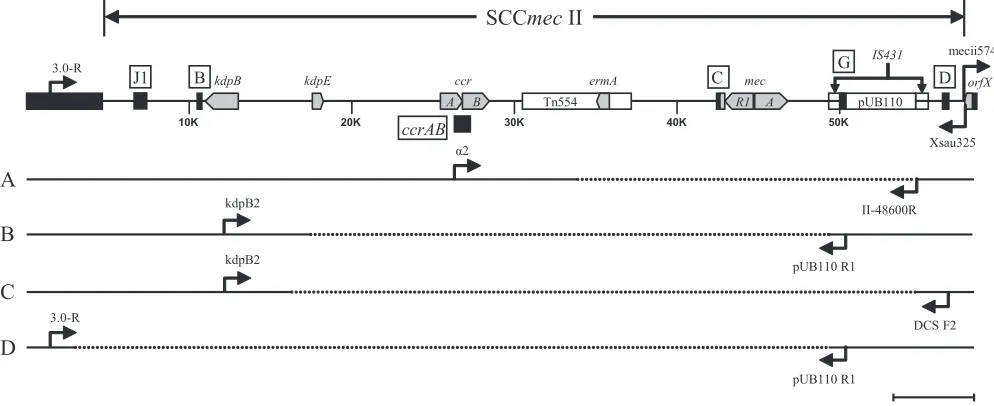
Mapping of deletion junction.
In order to map the deletion
junction, long-range PCR using upstream and downstream
primers based on the positive PCR targets that flanked the
missing
mecA
determinant was performed. Four SCC
mec
II
deletion patterns were observed among the seven isolates that
resembled USA100 (Fig. 2). Long-range PCR with primers
␣
2
[image:3.585.44.540.82.335.2](
ccrAB2
) and II-48600R (pUB110) for isolates IDI2406,
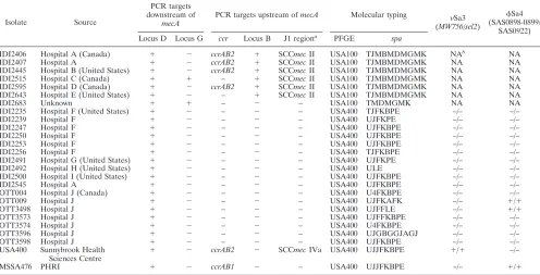
TABLE 2. Characteristics of
S. aureus
isolates
Isolate Source
PCR targets downstream of
mecA
PCR targets upstream ofmecA Molecular typing Sa3 (MW756/sel2)
Sa4 (SAS0898-0899/
SAS0922) Locus D Locus G ccr Locus B J1 regiona PFGE spa
IDI2406 Hospital A (Canada) ⫹ ⫺ ccrAB2 ⫹ SCCmecII USA100 TJMBMDMGMK NAb
NA
IDI2407 Hospital A ⫹ ⫺ ccrAB2 ⫹ SCCmecII USA100 TJMBMDMGMK NA NA
IDI2445 Hospital B (United States) ⫹ ⫺ ccrAB2 ⫹ SCCmecII USA100 TJMBMDMGMK NA NA
IDI2515 Hospital C (Canada) ⫹ ⫹ – ⫹ SCCmecII USA100 TJMBMDMGMK NA NA
IDI2595 Hospital D (Canada) ⫹ ⫺ ccrAB2 ⫹ SCCmecII USA100 TJMBMDMGMK NA NA IDI2643 Hospital E (United States) ⫹ ⫺ – ⫹ SCCmecII USA100 TJMBMDMGMK NA NA
IDI2683 Unknown ⫹ ⫹ – ⫺ – USA100 TMDMGMK NA NA
IDI2235 Hospital F (United States) ⫹ ⫺ – ⫺ – USA400 TJFKBPE –/– –/–
IDI2239 Hospital F ⫹ ⫺ – ⫺ – USA400 UJFKPE –/– –/–
IDI2247 Hospital F ⫹ ⫺ – ⫺ – USA400 UJFKBPE –/– –/–
IDI2250 Hospital F ⫹ ⫺ – ⫺ – USA400 UJFKBPE –/– –/–
IDI2253 Hospital F ⫹ ⫺ – ⫺ – USA400 UJFKBPE –/– –/–
IDI2256 Hospital F ⫹ ⫺ – ⫺ – USA400 TJFKBPE –/– –/–
IDI2491 Hospital G (United States) ⫹ ⫺ – ⫺ – USA400 UJFKPE –/– –/–
IDI2492 Hospital H (United States) ⫹ ⫺ – ⫺ – USA400 ULE –/– –/–
IDI2500 Hospital I (United States) ⫹ ⫺ – ⫺ – USA400 UJFKBPE –/– –/–
IDI2545 Hospital A ⫹ ⫺ – ⫺ – USA400 UJFKBPE –/– –/–
OTT004 Hospital J (Canada) ⫹ ⫺ – ⫺ – USA400 U4FKBPE –/– –/–
OTT009 Hospital J ⫹ ⫺ – ⫺ – USA400 UJFKAFK –/– ⫹/⫹
OTT3498 Hospital J ⫹ ⫺ – ⫺ – USA400 UJFFLE –/– ⫹/⫹
OTT3573 Hospital J ⫹ ⫺ – ⫺ – USA400 UJFFKBPE –/– –/–
OTT3574 Hospital J ⫹ ⫺ – ⫺ – USA400 U4FKBPE –/– –/–
OTT3596 Hospital J ⫹ ⫺ – ⫺ – USA400 UJGBGGJAGJ –/– –/–
OTT3598 Hospital J ⫹ ⫺ – ⫺ – USA400 UJFKBPE –/– –/–
USA400 Sunnybrook Health Sciences Centre
⫹ ⫺ ccrAB2 ⫺ SCCmecIVa USA400 UJJFKBPE ⫹/⫹ –/–
MSSA476 PHRI ⫹ ⫺ ccrAB1 ⫺ – USA400 UJJFKBPE –/– ⫹/⫹
a
J1 region, upstream of theccrgenes. b
NA, not applicable.
on May 16, 2020 by guest
http://jcm.asm.org/
IDI2407, IDI2445, and IDI2595 yielded an
⬃
8-kbp product
(pattern A). RFLP analyses using EcoRI, HindIII, HincII, or
XbaI indicated the PCR product from IDI2595 is different
from the other three isolates (data not shown). Sequence
de-termination revealed nt 33837 to 54641 (20.8 kbp) of SCC
mec
II (GenBank accession no. D86934.2) were missing in IDI2406,
IDI2407, and IDI2445, and nt 34927 to 54641 (19.7 kbp) were
missing in IDI2595. For IDI2515 (pattern B) the primers
kdpB2 (
kdpB
) and pUB110 R1 (locus G) were used to
suc-cessfully amplify a 9-kbp product, and for IDI2643 (pattern C)
the primers kdpB2 (
kdpB
) and DCS F2 (locus D) amplified an
8-kbp product. Deletions from nt 17486 to 49295 (31.8 kbp)
and nt 16339 to 54641 (38.3 kbp) were identified for IDI2515
and IDI2643, respectively. For IDI2683 (pattern D), PCR
re-sults suggested all SCC
mec
II sequences upstream of the
pUB110 plasmid were missing. As such, a chromosomal primer
(3.0-R) upstream of the SCC integration site was designed for
amplification with primer pUB110 R1. A 2.6-kbp product was
obtained; sequence data confirmed that nt 3012 to 49295 (46.3
kbp) was absent in IDI2683.
[image:4.585.83.498.70.239.2]For the 17 isolates that resembled USA400/MSSA476, no
SCC remnant other than locus D was detected. Therefore, a
chromosomal primer (4.2-R) that anneals at nt 65312 to 65292
in the MW2 genome (equivalent to nt 64019 to 63999 in
MSSA476) was designed for long-range PCR in conjunction
with the Xsau325 primer that anneals at nt. 34013 to 34031 of
MW2 and MSSA476. A 7-kbp PCR product with identical
RFLP patterns when digested with BclI, HincII, or XbaI (data
not shown) was obtained. The sequence of the amplicons was
identical to MSSA476 without SCC
far
(nt 34150 to 56985),
MW2 without SCC
mec
IVa (nt 34150 to 58278), the SCC
integration site of MSSA strain 15575 (GenBank accession no.
FIG. 1. PFGE of MSSA isolates that resembled USA100 (A) and USA400/MSSA476 (B) were resolved with the Lambda Ladder PFG Marker
(
) for Southern blotting. SmaI fragments that hybridized with the locus D probe are indicated with an asterisk.
FIG. 2. Internal deletions of SCC
mec
II. The prototypical SCC
mec
II is illustrated with key genetic elements (gray chevron) and locations of
PCR targets (black box) on the top. PCR primers (arrows) used in long-range PCRs to span the deleted region (segmented line) are as indicated
for the four deletion patterns: A (IDI2406, IDI2407, IDI2445, and IDI2595), B (IDI2515), C (IDI2643), and D (IDI2683).
on May 16, 2020 by guest
http://jcm.asm.org/
[image:4.585.43.540.485.688.2]EU272079), MSSA strain NCTC8325, and highly homologous
to several SCC
mec
-excisant strains generated
in vitro
(16, 31)
(Fig. 3).
Detection of mobile genetic elements.
In the absence of their
SCC elements, detection of pathogenicity islands and
bacte-riophages that typically associate with MW2 and MSSA476 was
performed to help identify the 17
USA400/MSSA476-resem-bling strains. The
Sa4-associated genes were detected in
iso-lates OTT009 and OTT3498. None of the gene targets present
on either mobile genetic elements were detected in the
remain-ing 15 isolates (Table 2).
DISCUSSION
Single-locus PCR assays based on the site-specific
integra-tion of SCC
mec
at
orfX
provide real-time identification of
MRSA directly from specimens. There have been reports
de-scribing
S. aureus
isolates containing SCC
mec
“remnants”
(without
mecA
), and misidentified as MRSA in single-locus
PCR assays (15, 35, 36, 40). In most cases, these MSSA isolates
were not fully characterized to elucidate the cause of the
mis-identification. In the report by Shore et al., the molecular
characterization of MSSA isolates with residual SCC
mec
ele-ments provided one explanation of the false-positive reactions
in single-locus PCR assays, although this present study
char-acterized only three MSSA isolates (40).
In the present study, we characterized 24 MSSA isolates,
from geographically diverse regions of North America, which
were detected as MRSA with a single-locus PCR assay, and
identified two possible explanations for the discrepant results.
Molecular typing and PCR analyses identified seven isolates
that resembled USA100 (
spa
, TJMBMDMGMK and related)
with remnants of SCC
mec
II. Internal deletions of SCC
mec
II
terminated at IS
431
sequences (nt 49295 to 50086 and 54642 to
55432 in SCC
mec
II) at the 3
⬘
border. IS
431
elements are
ubiquitous in staphylococci and play a role in gene transfer (21,
42). Partial SCC
mec
II excisions at IS
431
sequences during
in
vitro
exposure to vancomycin have been observed, possibly as a
fitness compensation mechanism (30). Therefore, it is likely
the excision events in SCC
mec
II isolates from the present
study were mediated through IS
431
.
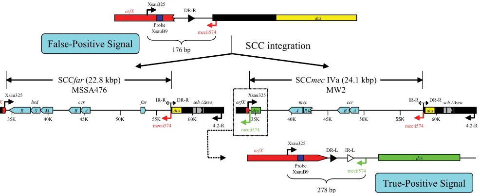
There were 17 isolates that resembled USA400/MSSA476
with undetermined SCC elements (
spa
, UJFKBPE and
re-lated). Characterization of these isolates led to the discovery of
a second cause of false-positive results in the single-locus PCR
assay. The annealing sequence for the SCC
mec
-specific primer
(mecii574) is located in the
dcs
region (GenBank annotation
MW0025
) of SCC
mec
IVa in MW2. A homologue of
MW0025
,
which also contains a mecii574 annealing sequence, exists
ad-jacent to the SCC integration site in both MW2 and MSSA476
(GenBank annotations
MW0048
and
SAS0048
, respectively).
The mecii574 sequence from within SCC
mec
IVa yields the
intended 278-bp amplicon for positive MRSA identification in
the single-locus PCR assay. However, the absence of an intact
SCC
mec
IVa (or SCC
far
) positions the secondary mecii574
annealing sequence immediately downstream of the
orfX
-spe-cific primer (Xsau325). In this configuration, a 176-bp
ampli-con can be generated and detected as a false-positive signal
(Fig. 4).
The identity of the USA400/MSSA476-resembling isolates
remains unclear. Sequence data identified the presence of the
putative transposase and enterotoxin genes (
seh
and
⌬
seo
)
downstream of an intact SCC integration site. The enterotoxin
gene cluster has been proposed to block the excision of SCC
mec
[image:5.585.43.530.74.191.2]IVa in MW2 (29). Apart from two isolates that contained the
MSSA476-associated bacteriophage (
Sa4), mobile genetic
el-ements associated with MW2 (
Sa3) and MSSA476 were not
detected in the USA400/MSSA476 isolates. Therefore, it is
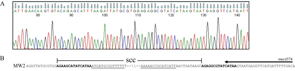
FIG. 3. SCC integration site in isolates that resembled USA400/MSSA476. (A) A representative chromatogram of the Xsau325/4.2-R amplicon
sequenced with the Xsau325 primer. The SCC boundaries are denoted with open and closed circles. (B) Sequence alignment of the SCC borders
delineated from the 17 isolates that resembled USA400/MSSA476 (SCC
⫺) with MW2, MSSA476, and
S. aureus
strain 15575, and NCTC8325
(GenBank accession no. CP000253) illustrating the missing SCC element that encompassed the direct repeat sequence (in boldface) at the left
border to the inverted repeat sequence (underlined) on the right border. The secondary mecii574 annealing sequence is illustrated with an arrow.
The open circle corresponds to nt 34149 (MW2 and MSSA476), 608 (15575), and 34151 (NCTC8325); the closed circle corresponds to nt 58279
(MW2), 56986 (MSSA476), 609 (15575), and 34152 (NTCT8325).
on May 16, 2020 by guest
http://jcm.asm.org/
plausible that the irreversible integration of SCC upstream of
the enterotoxin gene cluster had never occurred, and these
USA400/MSSA476-resembling isolates may be MSSA strains
where the acquisitions of mobile genetic elements such as SCC,
pathogenicity islands, and bacteriophages have not taken place
to derive the contemporary MW2 and MSSA476 strains. A
comprehensive surveillance and characterization of MSSA
iso-lates that test positive in single-locus PCR assays would be
beneficial in determining the frequency of false MRSA
iden-tification due to the presence of
dcs
at the SCC integration site
and in elucidating the identity of these isolates.
In summary, we determined that the single-locus PCR assay
for MRSA detection performed as it was designed, to detect a
SCC
mec
/
orfX
junction. However, detection of the SCC
mec
/
orfX
junction by itself may not be sufficient to confirm the
presence of an intact SCC
mec
element. Our investigation of
SCC
mec
II excisant strains revealed that IS
431
elements may
have been responsible for partial excisions in SCC
mec
type II
that encompassed the
mecA
determinant. In addition, we
de-termined that USA400/MSSA476-resembling isolates where
SCC integration has not taken place would also yield a positive
reaction in the single-locus PCR assay due to the presence of
an additional mecii574 annealing sequence at the SCC
inte-gration site. Ongoing surveillance and molecular
characteriza-tion of such isolates would be beneficial for future
improve-ment in the performance of these MRSA screening tools.
ACKNOWLEDGMENTS
This study was supported by BD Diagnostics.
We thank BD Diagnostics and Marc Desjardins (The Ottawa
Hos-pital, Ontario, Canada) for contributing MSSA strains for this study,
Teruyo Ito (Juntendo University, Tokyo, Japan) for strain N315, and
Barry Kreiswirth (Public Health Research Institute, New Jersey) for
strain MSSA476.
REFERENCES
1.Berglund, C., T. Ito, M. Ikeda, X. X. Ma, B. So¨derquist, and K. Hiramatsu.
2008. Novel type of staphylococcal cassette chromosomemecin a
methicil-lin-resistantStaphylococcus aureusstrain isolated in Sweden. Antimicrob. Agents Chemother.52:3512–3516.
2.Bishop, E. J., E. A. Grabsch, S. A. Ballard, B. Mayall, S. Xie, R. Martin, and M. L. Grayson.2006. Concurrent analysis of nose and groin swab specimens by the IDI-MRSA PCR assay is comparable to analysis by individual-spec-imen PCR and routine culture assays for detection of colonization by methicillin-resistantStaphylococcus aureus. J. Clin. Microbiol.44:2904–2908. 3.Blot, S. I., K. H. Vandewoude, E. A. Hoste, and F. A. Colardyn.2002. Outcome and attributable mortality in critically ill patients with bacteremia involving methicillin-susceptible and methicillin-resistantStaphylococcus au-reus. Arch. Intern. Med.162:2229–2235.
4.Brenwald, N. P., N. Baker, and B. Oppenheim.2010. Feasibility study of a real-time PCR test for methicillin-resistantStaphylococcus aureusin a point of care setting. J. Hosp. Infect.74:245–249.
5.Clinical and Laboratory Standards Institute.2009. Methods for dilution antimicrobial susceptibility tests for bacteria that grow aerobically. CLSI M07–A8. Clinical and Laboratory Standards Institute, Wayne, PA. 6.Clinical and Laboratory Standards Institute.2010. Performance standards
for antimicrobial susceptibility testing. CLSI M100–S20. Clinical and Labo-ratory Standards Institute, Wayne, PA.
7.Corkill, J. E., J. J. Anson, P. Griffiths, and C. A. Hart.2004. Detection of elements of the staphylococcal cassette chromosome (SCC) in a methicillin-susceptible (mecAgene negative) homologue of a fucidin-resistant MRSA. J. Antimicrob. Chemother.54:229–231.
8.Cosgrove, S. E., Y. Qi, K. S. Kaye, S. Harbarth, A. W. Karchmer, and Y. Carmeli.2005. The impact of methicillin resistance inStaphylococcus aureus bacteremia on patient outcomes: mortality, length of stay, and hospital charges. Infect. Control Hosp. Epidemiol.26:166–174.
9.Cosgrove, S. E., G. Sakoulas, E. N. Perencevich, M. J. Schwaber, A. W. Karchmer, and Y. Carmeli.2003. Comparison of mortality associated with methicillin-resistant and methicillin-susceptibleStaphylococcus aureus bac-teremia: a meta-analysis. Clin. Infect. Dis.36:53–59.
10.Deresinski, S.2005. Methicillin-resistantStaphylococcus aureus: an evolu-tionary, epidemiologic, and therapeutic odyssey. Clin. Infect. Dis.40:562– 573.
11.Deurenberg, R. H., C. Vink, S. Kalenic, A. W. Friedrich, C. A. Bruggeman, and E. E. Stobberingh.2007. The molecular evolution of methicillin-resistant Staphylococcus aureus. Clin. Microbiol. Infect.13:222–235.
12.Farley, J. E., P. D. Stamper, T. Ross, M. Cai, S. Speser, and K. C. Carroll.
2008. Comparison of the BD GeneOhm methicillin-resistantStaphylococcus aureus(MRSA) PCR assay to culture by use of BBL CHROMagar MRSA for detection of MRSA in nasal surveillance cultures from an at-risk com-munity population. J. Clin. Microbiol.46:743–746.
13.Hartman, B. J., and A. Tomasz.1984. Low-affinity penicillin-binding protein associated with beta-lactam resistance inStaphylococcus aureus. J. Bacteriol.
158:513–516.
14.Hiramatsu, K., Y. Katayama, H. Yuzawa, and T. Ito.2002. Molecular ge-netics of methicillin-resistantStaphylococcus aureus. Int. J. Med. Microbiol.
[image:6.585.51.544.67.266.2]292:67–74.
FIG. 4. Absence of SCC elements in USA400 or MSSA476 strains. In the absence of an SCC, the secondary mecii574 annealing sequence (in
red) is located 176 bp downstream of the Xsau325 primer to yield a false-positive signal in single-locus PCR assay (top). The acquisition of SCC
in MW2 and MSSA476 prohibits amplification between Xsau325 and the secondary mecii574 sequence (middle). However, the mecii574 sequence
(in green) from within SCC
mec
IVa in MW2 does yield the intended 278-bp amplicon for positive MRSA identification (bottom).
on May 16, 2020 by guest
http://jcm.asm.org/
15.Huletsky, A., R. Giroux, V. Rossbach, M. Gagnon, M. Vaillancourt, M. Bernier, F. Gagnon, K. Truchon, M. Bastien, F. J. Picard, A. van Belkum, M. Ouellette, P. H. Roy, and M. G. Bergeron.2004. New real-time PCR assay for rapid detection of methicillin-resistantStaphylococcus aureusdirectly from specimens containing a mixture of staphylococci. J. Clin. Microbiol.42:1875– 1884.
16.Ito, T., Y. Katayama, K. Asada, N. Mori, K. Tsutsumimoto, C. Tiensasi-torn, and K. Hiramatsu.2001. Structural comparison of three types of staphylococcal cassette chromosomemecintegrated in the chromosome in methicillin-resistantStaphylococcus aureus. Antimicrob. Agents Che-mother.45:1323–1336.
17.Ito, T., Y. Katayama, and K. Hiramatsu. 1999. Cloning and nucleotide sequence determination of the entiremecDNA of pre-methicillin-resistant Staphylococcus aureusN315. Antimicrob. Agents Chemother.43:1449–1458. 18.Ito, T., X. X. Ma, F. Takeuchi, K. Okuma, H. Yuzawa, and K. Hiramatsu.
2004. Novel type V staphylococcal cassette chromosomemecdriven by a novel cassette chromosome recombinase,ccrC. Antimicrob. Agents Che-mother.48:2637–2651.
19.Kim, T., P. I. Oh, and A. E. Simor.2001. The economic impact of methicillin-resistantStaphylococcus aureusin Canadian hospitals. Infect. Control Hosp. Epidemiol.22:99–104.
20.Klevens, R. M., J. R. Edwards, F. C. Tenover, L. C. McDonald, T. Horan, R. Gaynes, and the National Nosocomial Infections Surveillance System.2006. Changes in the epidemiology of methicillin-resistantStaphylococcus aureus in intensive care units in US hospitals, 1992–2003. Clin. Infect. Dis.42:389– 391.
21.Kobayashi, N., M. M. Alam, and S. Urasawa.2001. Genomic rearrangement of themecregulator region mediated by insertion of IS431in methicillin-resistant staphylococci. Antimicrob. Agents Chemother.45:335–338. 22.Kondo, Y., T. Ito, X. X. Ma, S. Watanabe, B. N. Kreiswirth, J. Etienne, and
K. Hiramatsu.2007. Combination of multiplex PCRs for staphylococcal cassette chromosomemectype assignment: rapid identification system for mec,ccr, and major differences in junkyard regions. Antimicrob. Agents Chemother.51:264–274.
23.Lina, G., Y. Pie´mont, F. Godail-Gamot, M. Bes, M. O. Peter, V. Gauduchon, F. Vandenesch, and J. Etienne.1999. Involvement of Panton-Valentine leu-kocidin-producing Staphylococcus aureus in primary skin infections and pneumonia. Clin. Infect. Dis.29:1128–1132.
24.Lindsay, J. A.2008.Staphylococcus aureusevolution: lineages and mobile genetic elements (MGEs), p. 45–69.InJ. A. Lindsay (ed.),Staphylococcus molecular genetics. Caister Academic Press, Norfolk, United Kingdom. 25.Louie, L., J. Goodfellow, P. Mathieu, A. Glatt, M. Louie, and A. E. Simor.
2002. Rapid detection of methicillin-resistant staphylococci from blood cul-ture bottles by using a multiplex PCR assay. J. Clin. Microbiol.40:2786–2790. 26.Ma, X. X., T. Ito, C. Tiensasitorn, M. Jamklang, P. Chongtrakool, S. Boyle-Vavra, R. S. Daum, and K. Hiramatsu.2002. Novel type of staphylococcal cassette chromosomemecidentified in community-acquired methicillin-re-sistantStaphylococcus aureusstrains. Antimicrob. Agents Chemother.46:
1147–1152.
27.McDougal, L. K., C. D. Steward, G. E. Killgore, J. M. Chaitram, S. K. McAllister, and F. C. Tenover.2003. Pulsed-field gel electrophoresis typing of oxacillin-resistantStaphylococcus aureusisolates from the United States: establishing a national database. J. Clin. Microbiol.41:5113–5120. 28.Mulvey, M. R., L. Chui, J. Ismail, L. Louie, C. Murphy, N. Chang, M. Alfa,
and the Canadian Committee for the Standardization of Molecular Meth-ods.2001. Development of a Canadian standardized protocol for subtyping methicillin-resistant Staphylococcus aureususing pulsed-field gel electro-phoresis. J. Clin. Microbiol.39:3481–3485.
29.Noto, M. J., and G. L. Archer.2006. A subset ofStaphylococcus aureusstrains harboring staphylococcal cassette chromosomemec(SCCmec) type IV is deficient in CcrAB-mediated SCCmecexcision. Antimicrob. Agents Che-mother.50:2782–2788.
30.Noto, M. J., P. M. Fox, and G. L. Archer.2008. Spontaneous deletion of the
methicillin resistance determinant,mecA, partially compensates for the fit-ness cost associated with high-level vancomycin resistance inStaphylococcus aureus. Antimicrob. Agents Chemother.52:1221–1229.
31.Noto, M. J., B. N. Kreiswirth, A. B. Monk, and G. L. Archer.2008. Gene acquisition at the insertion site for SCCmec, the genomic island conferring methicillin resistance inStaphylococcus aureus. J. Bacteriol.190:1276–1283. 32.Oberdorfer, K., S. Pohl, M. Frey, K. Heeg, and C. Wendt.2006. Evaluation of a single-locus real-time polymerase chain reaction as a screening test for specific detection of methicillin-resistantStaphylococcus aureusin ICU pa-tients. Eur. J. Clin. Microbiol. Infect. Dis.25:657–663.
33.Oliveira, D. C., and H. de Lencastre.2002. Multiplex PCR strategy for rapid identification of structural types and variants of themecelement in methi-cillin-resistantStaphylococcus aureus. Antimicrob. Agents Chemother.46:
2155–2161.
34.Oliveira, D. C., C. Milheiric¸o, and H. de Lencastre.2006. Redefining a structural variant of staphylococcal cassette chromosomemec, SCCmectype VI. Antimicrob. Agents Chemother.50:3457–3459.
35.Paule, S. M., D. M. Hacek, B. Kufner, K. Truchon, R. B. Thomson, Jr., K. L. Kaul, A. Robicsek, and L. R. Peterson.2007. Performance of the BD GeneOhm methicillin-resistant Staphylococcus aureus test before and during high-volume clinical use. J. Clin. Microbiol.45:2993–2998. 36.Rossney, A. S., C. M. Herra, G. I. Brennan, P. M. Morgan, and B. O’Connell.
2008. Evaluation of the Xpert methicillin-resistantStaphylococcus aureus (MRSA) assay using the GeneXpert real-time PCR platform for rapid de-tection of MRSA from screening specimens. J. Clin. Microbiol.46:3285– 3290.
37.Rossney, A. S., C. M. Herra, M. M. Fitzgibbon, P. M. Morgan, M. J. Lawrence, and B. O’Connell.2007. Evaluation of the IDI-MRSA assay on the SmartCycler real-time PCR platform for rapid detection of MRSA from screening specimens. Eur. J. Clin. Microbiol. Infect. Dis.26:459–466. 38.Sherlock, O., A. Dolan, and H. Humphreys.2010. MRSA screening: can one
swab be used for both culture and rapid testing? An evaluation of chromo-genic culture and subsequent Hain GenoQuick PCR amplification/detection. Clin. Microbiol. Infect.16:955–959.
39.Shopsin, B., M. Gomez, S. O. Montgomery, D. H. Smith, M. Waddington, D. E. Dodge, D. A. Bost, M. Riehman, S. Naidich, and B. N. Kreiswirth.1999. Evaluation of protein A gene polymorphic region DNA sequencing for typing ofStaphylococcus aureusstrains. J. Clin. Microbiol.37:3556–3563. 40.Shore, A. C., A. S. Rossney, B. O’Connell, C. M. Herra, D. J. Sullivan, H.
Humphreys, and D. C. Coleman.2008. Detection of staphylococcal cassette chromosomemec-associated DNA segments in multiresistant methicillin-susceptibleStaphylococcus aureus(MSSA) and identification of Staphylococ-cus epidermidis ccrAB4in both methicillin-resistantS. aureusand MSSA. Antimicrob. Agents Chemother.52:4407–4419.
41.Shorr, A. F., Y. P. Tabak, V. Gupta, R. S. Johannes, L. Z. Liu, and M. H. Kollef.2006. Morbidity and cost burden of methicillin-resistant Staphylococ-cus aureusin early onset ventilator-associated pneumonia. Crit. Care10:R97. 42.Wada, A., Y. Katayama, K. Hiramatsu, and T. Yokota.1991. Southern hybridization analysis of themecAdeletion from methicillin-resistant Staph-ylococcus aureus. Biochem. Biophys. Res. Commun.176:1319–1325. 43.Warren, D. K., R. S. Liao, L. R. Merz, M. Eveland, and W. M. Dunne, Jr.
2004. Detection of methicillin-resistantStaphylococcus aureusdirectly from nasal swab specimens by a real-time PCR assay. J. Clin. Microbiol.42:5578– 5581.
44.Zhang, K., J. A. McClure, S. Elsayed, and J. M. Conly.2009. Novel staph-ylococcal cassette chromosomemectype, tentatively designated type VIII, harboring class Amecand type 4ccrgene complexes in a Canadian epidemic strain of methicillin-resistant Staphylococcus aureus. Antimicrob. Agents Chemother.53:531–540.
45.Zhang, K., J. A. McClure, S. Elsayed, J. Tan, and J. M. Conly.2008. Coexistence of Panton-Valentine leukocidin-positive and -negative commu-nity-associated methicillin-resistantStaphylococcus aureusUSA400 sibling strains in a large Canadian health-care region. J. Infect. Dis.197:195–204.