Development 140, 2130-2138 (2013) doi:10.1242/dev.089409 © 2013. Published by The Company of Biologists Ltd
INTRODUCTION
One of the most intriguing issues in developmental biology is how organ growth and patterning are coordinated during embryogenesis. For example, in vertebrates, the formation of the main body plan is achieved through posterior elongation, and cells ingressing through the primitive streak progressively express more posterior identity genes. Similarly, limb development relies on an exquisite coordination between growth and patterning, but the underlying mechanisms remain elusive. Limb outgrowth requires FGF signaling from the AER (Fallon et al., 1994; Niswander and Martin, 1993), provided by Fgf4,Fgf8,Fgf9and Fgf17(collectively called AER-FGFs). Among them, Fgf8is the only FGF expressed from nascent AER until AER regression and is sufficient to ensure virtually normal limb development in absence of the other AER-FGFs (Boulet et al., 2004; Moon et al., 2000; Sun et al., 2000; Sun et al., 2002). Three mesenchymal factors, Fgf10,Shhand the BMP antagonist Grem1are essential for correct expression of AER-FGFs (Laufer et al., 1994; Ohuchi et al., 1997; Zúñiga et al., 1999). Initially, in the presumptive limb field of the lateral plate mesoderm (LPM), Fgf10triggers the expression of Fgf8in the overlaying ectoderm, which in turn maintains Fgf10expression in the early
limb bud (Ohuchi et al., 1997). Subsequently, further growth along the proximodistal (PD) axis and the anteroposterior (AP) expansion of the distal bud (presumptive hand/foot) is driven by a positive regulatory feedback loop between Shh, Grem1 and AER-FGFs, with Shh signaling being required to maintain Grem1transcription while the latter preserves expression of AER-FGFs by antagonizing BMPs (e.g. Zeller et al., 2009). In mice, expression of Shhand AER-FGFs regresses around E13 and subsequent growth of the limb relies on the elongation of skeletal elements.
The combined inactivation of the HoxA and HoxD gene clusters previously revealed their requirement for the activation and maintenance of Shhexpression (Kmita et al., 2005). These genes are members of the Hox family, which encode transcription factors that control patterning events during embryogenesis. In most vertebrates, Hox genes are grouped into four clusters (HoxA to HoxD) in which the relative order of the genes on the chromosome parallels their serial expression in time and space, a phenomenon referred to as temporal and spatial co-linearity (Duboule and Morata, 1994). For example, in nascent limb buds, genes from the HoxA and HoxD clusters are activated sequentially in time (from group 1 to group 13) and their expression patterns switch from uniform for early activated genes to posteriorly restricted for later activated ones (reviewed by Zakany and Duboule, 2007). Interestingly, only those expressed in the posterior limb mesenchyme (group 10 to 13) are able to activate Shh in vivo (Tarchini et al., 2006) via direct activation of the Shhlimb enhancer (Capellini et al., 2006), thereby establishing a link between the co-linear Hox activation and Shh-dependent AP polarity (Tarchini et al., 2006). Accordingly, ectopic/precocious expression of Hoxd13 and Hoxd12anteriorly results in mirror-image expression of Shh and bilaterally symmetrical limbs (Knezevic et al., 1997; Zákány et al., 2004). Recent studies showed that the activation of Shhactually relies on a Hox-Hand2 protein complex (Galli et al., 2010) and 1Laboratory of Genetics and Development, Institut de Recherches Cliniques de
Montréal (IRCM), 110 avenue des Pins Ouest, H2W1R7, Montréal, Québec, Canada. 2Laboratory of Molecular Genetics, Institut de Recherches Cliniques de Montréal (IRCM), 110 avenue des Pins Ouest, H2W1R7, Montréal, Québec, Canada. 3Instituto de Biomedicina y Biotecnología de Cantabria, CSIC-University of Cantabria-SODERCAN, Herrera Oria s/n, 39011 Santander, Spain. 4Department of Medicine, Université de Montréal, Montréal, Québec, Canada.
*Present address: Institut de Génétique Moléculaire de Montpellier, CNRS UMR5535, 1919 route de Mende, 34293 Montpellier, France
‡Authors for correspondence ([email protected]; [email protected])
Accepted 15 March 2013
SUMMARY
Limb development relies on an exquisite coordination between growth and patterning, but the underlying mechanisms remain elusive. Anterior-posterior and proximal-distal specification initiates in early limb bud concomitantly with the proliferative expansion of limb cells. Previous studies have shown that limb bud growth initially relies on fibroblast growth factors (FGFs) produced in the apical ectodermal ridge (AER-FGFs), the maintenance of which relies on a positive-feedback loop involving sonic hedgehog (Shh) and the BMP antagonist gremlin 1 (Grem1). The positive cross-regulation between Shhand the HoxA and HoxD clustered genes identified an indirect effect of Hox genes on the maintenance of AER-FGFs but the respective function of Shhand Hox genes in this process remains unknown. Here, by uncoupling Hox and Shhfunction, we show that HoxA and HoxD genes are required for proper AER-FGFs expression, independently of their function in controlling Shhexpression. In addition, we provide evidence that the Hox-dependent control of AER-FGF expression is achieved through the regulation of key mesenchymal signals, namely Grem1and Fgf10, ensuring proper epithelial-mesenchymal interactions. Notably, HoxA and HoxD genes contribute to both the initial activation of Grem1and the subsequent anterior expansion of its expression domain. We propose that the intricate interactions between Hox genes and the FGF and Shh signaling pathways act as a molecular network that ensures proper limb bud growth and patterning, probably contributing to the coordination of these two processes.
KEY WORDS: FGF, Gremlin 1, Hox genes, Shh, Limb development, Organ growth, Mouse
Decoupling the function of Hox and Shh in developing limb
reveals multiple inputs of Hox genes on limb growth
Rushikesh Sheth1, Damien Grégoire1,*, Annie Dumouchel1, Martina Scotti1, Jessica My Trang Pham1, Stephen Nemec2, Maria Félix Bastida3, Marian A. Ros3,‡and Marie Kmita1,4,‡
D
E
V
E
LO
P
M
E
N
Hand2itself is activated by paralogous group 9 Hox genes (Xu and Wellik, 2011). Together, these results show both direct and indirect control of Shhexpression by specific members of the Hox family of transcription factors. In turn, Shh signaling is required for the maintenance of HoxA and HoxD gene expression by inhibiting the proteolytic cleavage of the Gli3 protein into a transcriptional repressor (Litingtung et al., 2002; te Welscher et al., 2002).
Inactivation of the HoxA and HoxD clusters eventually results in severe reduction of the limb size, which was initially associated with Shhdownregulation (Kmita et al., 2005). However, the respective role of Shhand HoxA;D genes has remained unclear owing to the Hox-Shh positive cross-regulation. In addition, Shh−/− limb
shortening is significantly less severe compared with the double HoxA;HoxDmutant (Chiang et al., 2001; Kmita et al., 2005; Kraus et al., 2001) suggesting that HoxA and HoxD genes also impact on limb growth independently of their control of Shhexpression. Here, we report on the Hox-dependent limb growth in contexts where Hox and Shhfunctions are uncoupled. First, by investigating HoxA;D function before the maintenance of AER-FGFs becomes associated with Shh signaling. Second, by studying the role of HoxA;D genes in absence of Gli3, which renders Shhfunctionally irrelevant.
MATERIALS AND METHODS Mouse strains
Mutant mouse lines have been described previously: floxed HoxAgene cluster (Kmita et al., 2005), HoxAnull(Scotti and Kmita, 2012), HoxDnull
(Spitz et al., 2001) [also referred to as HoxD–and named TgHd11/lacZDel9
by Spitz et al. (Spitz et al., 2001)], Gli3XtJ(Hui and Joyner, 1993) and
Prx1Cre(Logan et al., 2002). The Shhnullline was derived from crossing the conditional Shh mutant (Lewis et al., 2001) with Mox2Cre mice (Tallquist and Soriano, 2000). Mice and embryos were genotyped by PCR or Southern blot analysis, using genomic DNA extracted from tail biopsies and yolk sacs, respectively.
Optical projection tomography (OPT)
Optical projection tomography (OPT) microscopy (Sharpe et al., 2002) was performed according to manufacturer specifications. Briefly, stained forelimbs skeletons or buds were embedded in 1% low-melt agarose, then dehydrated in 100% methanol and cleared in a mix of Benzyl Alcohol and Benzyl Benzoate (1:2). Scanning was performed using the Bioptonics 3001M OPT Scanner (Bioptonics, UK) with SKYSCAN-3001 (Skyscan, Belgium). Three-dimensional OPT reconstructions were performed with NRecon software (Skyscan) and visualized with Bioptonics viewer (Bioptonics). Measurements were made using Imaris (Bit plane).
Whole-mount in situhybridization
Whole-mount in situhybridizations were performed according to standard procedures using the previously described Shh(Echelard et al., 1993), Fgf8
(Crossley and Martin, 1995), Grem1(Zúñiga et al., 1999), Bmp4(Bénazet et al., 2009), Hoxa11and Hoxa13(Warot et al., 1997) antisense riboprobes.
Luciferase assay
P19 cells were transfected with 250 ng of the luciferase reporter under the control of the minimal promoter with or without enhancer sequences together with 100 ng of either Hoxd9- or Hoxa13-coding sequence or control-GFP DNA. Each plasmid combination was set in triplicates. Cell media were changed 24 hours after transfection and cells were kept in incubation for an additional 24 hours. Cell media was discarded and lysis buffer [100 mM Tris (pH 7.8), 0.5% NP-40, 1 mM DTT] was placed onto cells. Cell lysate was harvested and placed in contact with luciferin (Promega) in order to measure the reporter activity using Promega’s GLOMAX 96 microplate luminometer. Fold activation was measured by comparing mean RLU values for each condition with the mean RLU value obtained for the condition determined as basal level, i.e. control luciferase reporter (no enhancer sequence) in presence of control-GFP DNA.
RESULTS
HoxA and HoxD genes are required for the initial activation of Grem1
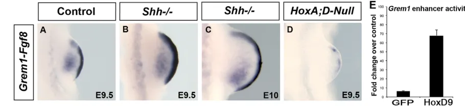
Limb growth initially relies on interactions between the AER and the mesenchyme (referred to as the AER phase hereafter) until the AER regresses. Subsequently, elongation of skeletal elements defines the eventual length of the limb (referred to as the post-AER phase hereafter). We first examined whether the impact of HoxA;D function on the mesenchyme-AER interactions is mediated only by the regulation of Shh expression or whether it includes an independent effect of HoxA;D genes on the other factors implicated in mesenchyme-AER interactions. As HoxA;D function is required for the initial activation of Shhtranscription, there is a narrow time window, at early bud stage, when HoxA and HoxD genes are functional, while Shh signaling is not yet operational. We thus took advantage of this Shh-independent stage to assess whether the lack of HoxA;D genes interferes with the expression of factors known to maintain AER-FGFs.Whole-mount in situhybridization shows readily detectable Grem1expression in wild-type (Fig. 1A) and Shh−/−(Fig. 1B) nascent buds. Grem1expression is actually still
detectable in Shh−/−buds at E10 (Fig. 1C), consistent with previous
[image:2.612.69.524.574.678.2]results showing that Shh is dispensable for the transcriptional activation of Grem1(Panman et al., 2006; Bénazet et al., 2009). By marked contrast, the ubiquitous deletion of the HoxA and HoxD clusters results in the complete lack of Grem1activation in early limb buds (Fig. 1D), indicating that the initial activation of Grem1 requires HoxA;D function. Previous work has revealed that bone morphogenetic protein 4 (Bmp4) is required for the transcriptional
Fig. 1. HoxA;D genes are required for Grem1activation and a proper Fgf8expression pattern, independently of Shh signaling. (A-D) Grem1
and Fgf8expression in control (A), Shh−/−(B,C) and Hox(A;D)null(D) limb buds. Although Grem1expression is absent in Hox(A;D)nullbud at E9.5 (D), it is still readily detectable in E10 Shh−/−bud (C). (E) Transcription assays in P19 cells co-transfected with Grem1enhancer linked to luciferase-coding sequences and either the GFP or Hoxd9-expressing vector. Luciferase activity is indicated as fold change over luciferase activity in absence of the Grem1
enhancer. Error bars represent s.d.
D
E
V
E
LO
P
M
E
N
activation of Grem1in early limb buds (Bénazet et al., 2009), raising the possibility that the absence of Grem1 expression in Hox(A;D)null mutants is secondary to Bmp4 downregulation. However, Bmp4 remains expressed in Hox(A;D)null limbs (supplementary material Fig. S1), thus excluding its downregulation as a cause for the lack of Grem1expression. In turn, these results reveal that Bmp4 signaling is unable to trigger Grem1activation in the absence of HoxA;D genes. To further test the competence of Hox proteins in activating Grem1, we next performed transcription assays in P19 cells, in which the luciferase reporter was under the control of the previously identified Grem1limb enhancer (Zuniga et al., 2012). In these assays, a plasmid encoding Hoxd9 was used as a representative of Hox proteins present in the nascent limb bud. Luciferase quantification reveals robust increase in the activity of the Grem1enhancer in presence of the Hoxd9 protein (Fig. 1E), indicating that Hoxd9 (and most likely the other early Hox genes products) is able to activate Grem1expression. This result, however, does not exclude the requirement of Bmp4together with HoxA;D genes for Grem1 activation, as Bmp4 is expressed in P19 cells (Chang et al., 2010).
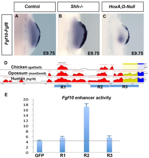
Impaired expression of Fgf8in Hox(A;D)null nascent limb buds is coincident with Fgf10 downregulation
Up to E10, the maintenance of Fgf8expression in the AER does not require Grem1function, as revealed by the analysis of Grem1 -deficient limb buds (Michos et al., 2004). Consequently, the Fgf8 downregulation observed in Hox(A;D)nullnascent buds (Fig. 1D) suggests that another mesenchymal factor maintaining Fgf8 expression is impaired in these mutant buds. We thus analyzed the expression of Fgf10, the function of which triggers Fgf8activation and contributes to Fgf8maintenance (Ohuchi et al., 1997). Our results show that Fgf10is downregulated anteriorly in Hox(A;D)null buds (Fig. 2C) compared with both wild-type (Fig. 2A) and Shh−/−
buds (Fig. 2B), suggesting that HoxA;D genes contribute to Fgf10 regulation in early buds. As Fgf10is initially activated in the lateral plate mesoderm, we also checked Fgf10expression prior to limb budding but failed to assess unambiguously whether its expression is affected or not in the presumptive limb field (not shown). In order to have an independent assessment of the capacity of Hox proteins to positively regulate Fgf10expression, we performed transcription assays in P19 cells. To drive expression of the luciferase reporter, we used a DNA fragment previously identified as containing the Fgf10 limb enhancer (R2 in Fig. 2D,E) (Ohuchi et al., 2005; Sasak et al., 2002). As negative controls, we linked the reporter to the other conserved DNA fragments located upstream Fgf10 but with no enhancer function in limbs (R1 and R3 in Fig. 2D,E), one of them acting as Fgf10enhancer in the inner ear [R1; see also Ohuchi et al. (Ohuchi et al., 2005)]. Upon co-transfection with the Hoxd9-encoding plasmid, there is a significant augmentation of the R2 enhancer activity, as revealed by the reporter expression, whereas the activity of R1 and R3 remained at basal levels (Fig. 2E). These results point to a specific effect of Hoxd9 protein on the activity of the enhancer controlling Fgf10 expression in nascent buds and support the contribution of HoxA;D genes for the proper expression of Fgf10in the limb mesenchyme.
The anterior propagation of Grem1expression involves the function of HoxA;D genes
The requirement of Hox function for Grem1 transcriptional activation raises the possibility that HoxA;D genes are also involved in Grem1regulation at later stages. Investigating this hypothesis
necessitates the inactivation of HoxA;D genes after Grem1initial activation. This can be achieved using the HoxAc/–;D−/−conditional
mutant, in which transcripts from the conditional HoxA allele are transiently generated (up to E10.5) owing to the kinetics of the Prx1-Creactivity (Kmita et al., 2005). Consistent with the residual HoxA transcription, Grem1 is activated in HoxAc/–;D−/− buds
(supplementary material Fig. S2). At E11.5, Grem1 expression remains detectable in HoxAc/–;D−/−mutants, although it fails to
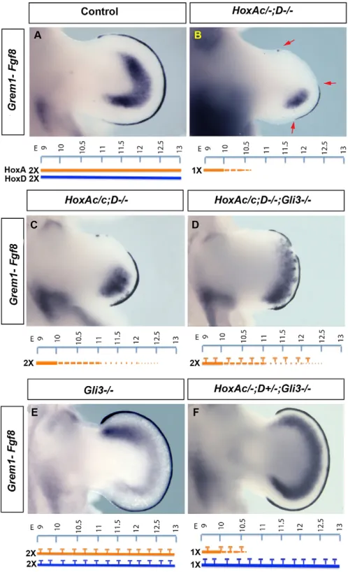
expand anteriorly as it does in wild-type buds (Fig. 3A,B). Interestingly, Fgf8 expression, which is normal at E10.5 (supplementary material Fig. S2C), is subsequently restricted to the part of the AER that parallels the Grem1domain along the AP axis (Fig. 3E, arrows), suggesting that, at this stage, Grem1defines the AP domain of Fgf8expression in the AER.
Although Grem1 expression is posteriorly restricted in the HoxAc/–;D−/−mutant, the respective role of Hox genes and Shhin
Grem1 anterior expansion remains unclear because Shh is downregulated in HoxAc/–;D−/−limb buds (Kmita et al., 2005). To
investigate the Shh-independent function of HoxA;D genes, we needed to remove Gli3 function to render Shh functionally irrelevant (Litingtung et al., 2002; te Welscher et al., 2002). Owing to the lethality associated with Gli3inactivation and HoxAc/– mutation as well as the infertility of HoxD−/−mice, triple mutant
embryos can only be generated from crosses between triple heterozygous mice. However, HoxA+/−;HoxD+/−;Gli3+/−turns out
to be poorly fertile, which drastically reduces the feasibility of studying HoxAc/–;HoxD−/−;Gli3−/− embryos. We could
[image:3.612.320.558.62.314.2]circumvent this limitation using the conditional HoxA allele instead of the null allele and we therefore performed our analysis Fig. 2. Fgf10expression is altered in absence of HoxA;D genes.
(A-C) Fgf10and Fgf8expression in control (A), Shh−/−(B) and HoxA;Dnull (C) limbs. (D) The genomic region upstream Fgf10. Three regions (R1 to R3) are conserved between mouse, chick, opossum and human. Only R2 acts as a limb enhancer. (E) Relative effect of Hoxd9 protein on the transcriptional activity of each conserved region. Luciferase activity is indicated as fold change over control (i.e. luciferase vector without any R regions). Error bars represent s.d.
D
E
V
E
LO
P
M
E
N
with triple mutants in which both HoxAalleles are conditional (HoxAc/c;HoxD−/−;Gli3−/−), which were obtained at the expected
Mendelian ratio (1/128). The difference between HoxAc/cand HoxAc/– is that the completion of Cre-mediated deletion is delayed with two floxed alleles and some HoxA expression remains at least up to E11.5 (supplementary material Fig. S3). Comparison of HoxAc/c;HoxD−/−;Gli3−/−with HoxAc/c;HoxD−/−
buds, at E11.5, reveals an anteriorization of both Grem1and Fgf8 in the triple mutant (Fig. 3C-D). However, expression of both
genes remains clearly impaired compared with Gli3−/−and Shh−/−;
Gli3−/−mutant (Fig. 3E) (see also Litingtung et al., 2002; te
Welscher et al., 2002). Notably, in HoxAc/c;HoxD−/−;Gli3−/−
buds, Grem1expression is mosaic and significantly reduced in the anterior mesenchyme (Fig. 3D compared with 3E). This indicates that Gli3 inactivation is not sufficient to restore the anterior expansion of Grem1in HoxAc/c;HoxD−/−;Gli3−/−as it does in
Shh−/−;Gli3−/−buds and raises the possibility that HoxA;D genes
contribute to this process.
We reasoned that if Hox proteins contribute to Grem1anterior expansion, Grem1expression in HoxAc/c;HoxD−/−;Gli3−/−should
be, at least in part, due to the residual transcription of the conditional HoxA allele, notably that of Hoxa13,which is the most distally expressed HoxA gene. Accordingly, the gain of Grem1upon Gli3 inactivation (Fig. 3D) coincides with the anterior expansion of the residual Hoxa13expression (supplementary material Fig. S4B), which itself is due to the lack of the Gli3R repressor. Grem1 expansion could also be secondary to the lack of Gli3R. However, the correlation between the mosaicism of Grem1expression and that of the residual Hoxa13 expression observed in HoxAc/c;HoxD−/− and HoxAc/c;HoxD−/−;Gli3−/− (Fig. 3C,D;
supplementary material Fig. S5) strongly favors the interpretation that HoxA;D genes are required for the proper anterior expansion of Grem1.Accordingly, a single wild-type allele of the HoxA or HoxD cluster is sufficient to recover the anterior Grem1expression as in Gli3−/−and Shh−/−;Gli3−/−(shown for the HoxD wild-type allele
in Fig. 3F, compare with 3D and 3E).
To test independently the effect of the Hoxa13 protein on Grem1 expression, we performed a transcription assay in P19 cells. Co-transfection of the expression vector encoding for Hoxa13 together with the Grem1 limb enhancer linked to the luciferase reporter resulted in a significantly higher reporter expression, as revealed by luciferase quantification (supplementary material Fig. S6), confirming that Hoxa13 has a positive impact on the activity of the Grem1limb enhancer. Together, these results provide evidence that HoxA;D genes contribute to the anterior expansion of Grem1 expression independently of their function in controlling Shh.
The AP and PD growth in the Gli3−/−background involves the function of HoxA;D genes
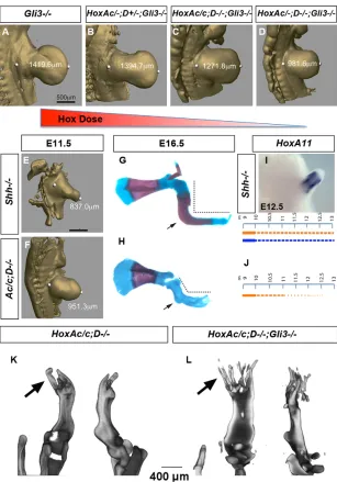
Our data on Grem1expression support the notion that HoxA;D genes promote limb bud growth in parallel to their function in regulating Shh.In turn, it suggests that limb bud growth should vary according to the HoxA;D dose, independently of Shhexpression. We thus performed measurements of limb buds using optical projection tomography (OPT) imaging to characterize how reducing the HoxA;D gene dose in the Gli3−/−background affects limb bud
growth. At E11.5, reduction of the Hox dose to a single HoxA or HoxD wild-type allele results in the shortening of the limb bud along the PD axis (Fig. 4A,B). In addition, this shortening is more pronounced upon further reduction of the HoxA;D dose (Fig. 4C,D). These data reveal a tight link between the HoxA;D dose and the extent of PD bud growth in the Gli3mutant background. Our series of HoxA;D;Gli3mutants also show that one functional allele of either HoxAor HoxDis sufficient to generate the distal ‘fan-shaped’ characteristic of Gli3−/−limb bud (Fig. 4A,B) but this shape is lost with lower Hox doses (Fig. 4C,D). This suggests that the AP expansion of the distal bud observed in Gli3−/−and Shh−/−;Gli3−/−
buds is associated with the function of HoxA;D genes.
[image:4.612.51.300.61.470.2]Together, our series of HoxA;D;Gli3 mutants reveals a link between the HoxA;D dose and the extent of PD and AP bud growth in the Gli3−/−background.
Fig. 3. Grem1expression is altered in the conditional HoxA;D mutants, even in absence of Gli3R. (A-F) Expression pattern of Grem1
and Fgf8at E11.5 in control (A), HoxAc/–;HoxD−/−(B), HoxAc/c;HoxD−/−(C), HoxAc/c;HoxD−/−;Gli3−/−(D), Gli3−/−(E) and HoxAc/–;HoxD+/−;Gli3−/−(F) limb buds. Arrows in B indicate Fgf8expression domains. For each genotype, the HoxA(orange) and HoxD(blue) expression level and timing is schematized. Bold lines represent wild-type expression levels, whereas the Prx1Cre-dependent reduction of HoxAtranscripts are schematized using dotted lines. T bars in D-F represent the anterior gain of Hox expression due to Gli3inactivation. The presence of two HoxA conditional alleles (HoxAc/c) results in more HoxA expression when one of the two alleles is null (supplementary material Fig. S3), leading to more Grem1
expression (compare B with C). The patchy expression pattern of Grem1in
HoxAc/c;HoxD−/−(C), HoxAc/c;HoxD−/−;Gli3−/−(D) coincides with the mosaic expression of Hoxa13(see also supplementary material Fig. S5).
D
E
V
E
LO
P
M
E
N
HoxA;D downregulation and the defective growth of Shh−/−limb
In absence of Shh, the domain of Gli3R, which is normally restricted to the most anterior part of the limb bud, expands towards the posterior part (Wang et al., 2000). This results in the downregulation of several genes in most of the Shh−/−limb bud, including HoxA;D
genes (Chiang et al., 2001; Kraus et al., 2001). The rescue of the Shh−/−limb phenotype upon removal of Gli3indicates that the
downregulation of the direct and/or indirect targets of Gli3R is responsible for the Shh−/−phenotype. Our finding that the HoxA;D
gene dose in the Gli3−/− background correlates with limb bud
growth thus suggests that Shh−/−limb truncation is, at least in part,
the consequence of Gli3R-mediated HoxA;D downregulation. Nonetheless, as Shhremains transiently expressed in the conditional HoxA;Dmutant (Kmita et al., 2005), there is less Gli3R-mediated gene repression, including less repression of the residual HoxA expression, than in the Shhmutant and thus it is possible that HoxAc/c;HoxD−/−buds could develop better than Shh−/−buds until
complete HoxAinactivation. In agreement with this view, at E11.5, Shh−/−buds are smaller than HoxAc/c;HoxD−/−buds (Fig. 4E,F).
The size difference between Shh−/−and the conditional HoxA;D
buds is opposite when skeletons are compared (Fig. 4G,H), indicating that the elongation of the skeletal elements is favored in Shh mutant. Interestingly, around E12, there is a complete deficiency of Hox expression in HoxAc/c;HoxD−/−buds, while Hox expression persists in Shhmutant (Fig. 4I-J). Actually HoxA11 expression in Shhmutant appears even stronger at E12.5 than at E11.5 (Fig. 4I; supplementary material Fig. S7). This sustained Hox expression in Shh−/−(Fig. 4I) could explain the favored elongation
of skeletal elements, consistent with previous reports proposing that the elongation of skeletal elements requires the function of HoxA;D genes (Boulet and Capecchi, 2004; Gross et al., 2012; Zákány et al., 1997). In Shh−/−;Gli3−/− limb, proper elongation of skeletal
[image:5.612.54.361.59.498.2]elements is thus expected to be associated with HoxA;D function. Yet it remains unclear whether other Gli3R targets are implicated. To clarify this issue, we compared the skeleton of the conditional HoxA;Dand HoxA;D;Gli3limbs and found that they are similarly shorten along the PD axis (Fig. 4K-L). This result indicates that among Gli3R targets, HoxA;D genes have the most relevant role for the elongation of skeletal elements.
Fig. 4. Reduction of the HoxA;D dose in the Gli3 mutant background results in the gradual decrease of limb bud size.(A-D) OPT imaging and measurements of limb buds from Gli3−/−(A),
HoxAc/–;HoxD+/−;Gli3−/−(B), HoxAc/c;HoxD−/−;Gli3−/−(C) and HoxAc/–;HoxD−/−;Gli3−/−(D) mutants.
Measurements were made from the distal tip of the limb bud to the ‘groove-type’ shape at the proximal end of the bud (dot). (E,F) OPT imaging and measurements of Shh−/−(E) and HoxAc/c;HoxD−/−(F) limb buds at E11.5. (G,H) Forelimb skeleton of Shh−/− (G) and HoxAc/c;HoxD−/−(H) mutants at E16.5. Arrows indicate the similar bending observed in these mutant skeletons. (I,J) Hoxa11expression at E12.5 in Shh−/− forelimb buds and schemes representing the persistence at late stages of HoxA;D expression in Shh
mutant (I) in contrast to the transient expression of the conditional HoxA allele in the HoxAc/c;HoxD−/−mutant (J). (K,L) OPT imaging of HoxAc/c;HoxD−/−(K) and HoxAc/c;HoxD−/−;Gli3−/−(L) forelimb skeletons at E16.5. Ventral view is on the left part of each panel and lateral view is on the right. Arrows indicate similar banding in
Shh–/–and HoxAc/c;and D–/–skeleton.
D
E
V
E
LO
P
M
E
N
Transient expression of the conditional HoxA allele allows for the realization of a rudimentary but complete PD axis
The HoxAc/c;HoxD−/−;Gli3−/−skeleton at E16.5 is characterized
by the increased number of distal rays when compared with HoxAc/c;HoxD−/−skeletons (Fig. 4K,L). This increased number of
distal rays is reminiscent of the increased number of digits in Gli3−/−
limbs, thus raising the possibility that these distal rays correspond to digit remnants. However, the lack of digit-specific features, such as segmentation into distinct phalanges, makes it difficult to assign an identity unambiguously to these rays. Interestingly, HoxAc/+;HoxD−/−and HoxAc/c;HoxD+/−limbs, although being
significantly smaller than wild-type ones, have the three domains (stylopod, zeugopod and autopod), allowing for unambiguous identification of digits despite their abnormal shape and absence of segmentation into phalanges (Fig. 5A,C). Moreover, the comparison with HoxAc/+;HoxD−/−;Gli3−/−and with HoxAc/c;HoxD+/−;Gli3−/−
limbs (Fig. 5B,D) shows that Gli3inactivation in these Hox mutant backgrounds primarily affects the number of digital condensations. Together, these data support our interpretation that the distal rays in HoxAc/c;HoxD−/−;Gli3−/−limbs are digit remnants.
Interestingly, the bending systematically found in the middle of HoxAc/c;HoxD−/−skeletons (arrow in Fig. 4H) evokes the bending
of wild-type limbs at the transition between stylopod and zeugopod (Fig. 5E). Remarkably, Shhmutant embryos form either a clearly distinct stylopod and zeugopod separated by a rudimentary elbow joint (e.g. Litingtung et al., 2002) or limbs with only the bend
typically associated with the elbow joint (arrow in Fig. 4G) (see also Zhu et al., 2008), supporting that the bending observed in HoxAc/c;HoxD−/−mutant limbs corresponds to a vestige of the
stylopod-zeugopod boundary. Thus, the phenotype of HoxAc/c;HoxD−/− and HoxAc/c;HoxD−/−;Gli3−/− skeletons
suggests that the transient expression of HoxA genes is sufficient to provide cells with a stylopod, zeugopod or digit identity depending of which HoxA genes they express.
DISCUSSION
During limb development, genes of the HoxA and HoxD clusters are expressed in a spatial and temporal co-linear manner. Genetics studies previously established the role of Hox genes in limb skeletal patterning but their role in limb bud growth remains unclear, mainly owing to the crossregulation existing between HoxA;D genes and Shh. The work reported here provides a novel perspective on the role of Hox genes in limb bud growth and the mechanisms involved. First, we show that in nascent bud, prior to the normal activation of Shh, the function of HoxA;D genes is required for correct Fgf8 expression. Moreover, we establish that HoxA;D genes achieve this previously unrecognized role through the transcriptional activation of Grem1 and proper expression of Fgf10in the mesenchyme. Second, taking advantage of Gli3inactivation that renders Shh expression functionally irrelevant, we provide evidence that the anterior expansion of Grem1expression, and thus the propagation of Grem1-mediated maintenance of AER-FGFs, requires the function of HoxA;D genes independently of their activity in regulating Shhexpression. Accordingly, our data show that during the Shh functional phase, the proper AP and PD growth of the limb bud requires the Shh-independent functions of HoxA;D genes. Finally, we also show that HoxA;D genes are subsequently required for the elongation of skeletal elements.
Multiple inputs of Hox genes on the epithelial-mesenchymal interactions triggering limb bud growth
[image:6.612.53.295.369.632.2]Limb bud growth first relies on epithelial-mesenchymal (EM) interactions that trigger and then maintain expression of AER-FGFs, up to AER regression around E13. The initial EM interaction, which induces limb budding, relies on Fgf10 signaling from the lateral plate mesoderm (LPM) that triggers expression of Fgf8 in the overlaying ectoderm (Ohuchi et al., 1997). Activation of Fgf10 expression in the presumptive forelimb field of the LPM relies on Tbx5 (Agarwal et al., 2003; Rallis et al., 2003). Recently, analysis of Tbx5regulation identified Hox proteins from paralogous group 4 and 5 as regulators of Tbx5expression in the LPM (Minguillon et al., 2012). Despite the absence of Hoxd4,Hoxa4and Hoxa5in the LPM of Hox(A;D)null embryos, we did not detect clear modification of Fgf10 expression in the LPM (not shown), suggesting that the absence of HoxA;D genes in the LPM has minor, if any, impact on the Tbx5-mediated regulation of Fgf10. Therefore, the effect of the Hox(A;D)nullmutation on Fgf10expression is primarily associated with the function of HoxA;D genes in the developing limb bud, consistent with the evidence that Hoxd9 protein positively influence the activity of the Fgf10early limb enhancer in cell culture assays. The evidence that Fgf10expression is not abrogated in Hox(A;D)null limb bud but only reduced suggests that Fgf10regulation involves HoxA;D proteins, as well as other transcription factors. Interestingly, switching expression of Hoxd12and Hoxd13from late to early bud stages in absence of Gli3 suppresses Fgf10expression in the limb mesenchyme (Zakany et al., 2007). It was thus proposed that Fgf10downregulation is the Fig. 5. Gli3inactivation does not improve the elongation of skeletal
elements of mutant with reduced dose of HoxA;Dgenes but triggers the formation of additional digits.(A-D) E16.5 forelimb skeletal preparations from HoxAc/+;HoxD−/−(A,B) and HoxAc/c;HoxD+/−(C,D) in the presence (A,C) or absence (B,D) of Gli3. (E) Wild-type forelimb skeleton at E16.5. A single functional copy of the HoxD (C,D) or HoxA (A,B) cluster is sufficient for the formation of the three limb domains, but the reduced Hox dose interferes with the elongation of each domain. In the absence of Gli3(B,D), PD length is not improved but there is a significant increase
in digit number.
D
E
V
E
LO
P
M
E
N
consequence of the functional suppression of early expressed Hox by ‘late’-activated Hox genes (Zakany et al., 2007), which supports the model that Hox genes expressed in early limb buds act as positive regulators of Fgf10. Together, these results point to the implication of Hox genes in the early EM interactions, first through the impact of both group 4 and 5 Hox genes on Fgf10expression in the LPM and second through the function of HoxA;Dgenes in the nascent limb bud.
Although Grem1 is activated in nascent bud, its function in maintaining Fgf8in the AER becomes indispensable around E10, as revealed by the analysis of the Grem1mutant (Michos et al., 2004). Grem1downregulation in Shhmutant and the ability of Shh-soaked beads implanted in the anterior mesenchyme to trigger ectopic Grem1 expression provided evidence that Shh signaling acts positively on Grem1expression (Zúñiga et al., 1999). However, Shh signaling is not needed for the initial activation of Grem1in nascent bud (Panman et al., 2006; Bénazet et al., 2009). Moreover, inactivating Shhtogether with its direct intracellular mediator Gli3 prevents Grem1 downregulation (Litingtung et al., 2002; te Welscher et al., 2002), indicating that Grem1downregulation in Shhmutant is due to Gli3R activity, although Gli3 repression of Grem1has been proposed to be indirect (Vokes et al., 2008). As Gli3R also leads to HoxA;D downregulation, it is possible that Gli3R repression of Grem1is secondary to Hox repression. Our results strongly support this interpretation: first, in contrast to Shh, HoxA;D function is required for the initial activation of Grem1in nascent bud. Second, Gli3 inactivation in the conditional HoxA;Dmutant background is not sufficient to restore Grem1 expression, as reported for the Shh−/−;Gli3−/−mutant. Finally, the mosaicism of Grem1 in the
conditional HoxA;D;Gli3 mutant strongly correlates with the mosaicism of the residual HoxA gene expression in this mutant.
Based on this previously unappreciated function of HoxA;D genes in eliciting Grem1expression, which occurs independently of their role in regulating Shhexpression, we propose a novel model for the control of Grem1expression (Fig. 6). In this model, HoxA;D genes trigger the initial activation of Grem1(either in cooperation or in parallel with Bmp4 signaling) in nascent limb bud. Concomitantly, they activate Shhexpression in the ZPA. In turn, Shh signaling restricts Gli3 proteolytic cleavage into a transcriptional repressor to the most anterior mesenchymal cells, thereby defining a domain permissive for the expression of HoxA;D genes and subsequent propagation of Grem1expression.
HoxA;Dgenes and the control of limb bud growth
By uncoupling Hox and Shhfunctions using Gli3inactivation, which renders Shh functionally irrelevant, we now provide evidence that Hox genes control bud growth in parallel to their activity in controlling Shhexpression. Notably, we found that during the AER phase, reducing the HoxA;Dgene dose in absence of Gli3interferes with limb bud growth in a dose-dependent manner, both along the AP and PD axes. Previous studies suggesting that the elongation of skeletal elements requires HoxA;D function were based on ubiquitous gene inactivation (Boulet and Capecchi, 2004; Gross et al., 2012; Zákány et al., 1997). Our data suggest that at least part of the reported phenotype could have been due to defective growth during the AER phase. Nonetheless, the comparison between the size of Shh−/−and the conditional HoxA;Dmutant at E11.5 and
E16.5 further supports the requirement of Hox products for the elongation of the skeletal elements at the post-AER stages. Moreover, the difference in Fgf8 expression in the conditional HoxA;Dand HoxA;D;Gli3mutants (and thus the difference in the size of the pool of skeletal precursors) has no detectable effect on the
eventual size of the skeleton along the PD axis, further highlighting the importance of HoxA;D function in the elongation of skeletal elements.
Together, these results provide compelling evidence for a Hox -dependent Shh-independent mechanism that promotes limb bud growth during the AER phase, as well as the requirement of HoxA;Dgenes for the subsequent elongation of skeletal elements.
Hox-FGF crossregulation
[image:7.612.322.554.61.252.2]Exhaustive analysis of the function of AER-FGFs suggested that AER-FGF signaling contributes to PD patterning in addition to limb bud growth (Mariani et al., 2008). Interestingly, recent work provided evidence that AER-FGF signaling has a key role for the expression of ‘late/distal’ Hox genes (i.e. group 11 to 13), as revealed by the Fgf8 requirement for the sequential activation of Hoxa11and Hoxa13in limb mesenchymal cells in culture (Cooper et al., 2011; Roselló-Díez et al., 2011) and Hoxd13downregulation associated with a reduced dose of AER-FGFs (Sun et al., 2002). These genes actually instruct cells of their PD identity (zeugopod identity for Hox11and autopod identity for Hox13) and thereby likely represent the effectors through which AER-FGFs influence PD patterning. Our finding that early activated HoxA;D genes promote the maintenance of AER-FGFs suggests that, in addition to providing proximal identity to cells where they are expressed, early activated HoxA;D genes ensure proper AER-FGF-dependent expression of late/distal HoxA;D genes. Accordingly, the Hox-FGF-positive cross-regulation would control limb bud growth and ensure some steadiness between the formation of proximal and distal identity.
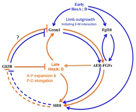
Fig. 6. Model for the sequential inputs of HoxA;D genes on the maintenance of AER-FGFs.In nascent limb buds (blue lines), early activated HoxA;D genes trigger the initial activation of Grem1(either in cooperation or in parallel with Bmp4 signaling) and Shh, and contribute to the proper expression of Fgf10in nascent buds. At this stage, Fgf10
function is sufficient to ensure Fgf8expression in the AER. By the time Shh signaling is activated, it restricts (dashed lines) Gli3 proteolytic cleavage into a transcriptional repressor to the most anterior mesenchymal cells, thereby defining a domain permissive for the expression of HoxA;D genes. Later activated HoxA;D genes (orange) maintain Shhexpression and associated anterior restriction of Gli3R. In parallel, they contribute to the anterior expansion of Grem1, which is required for proper FGF expression in the anterior AER and AP expansion of the distal bud (presumptive autopod). All interactions represent transcriptional inputs except the one indicated by broken lines, which is a post-translational effect.
D
E
V
E
LO
P
M
E
N
Conclusions
In summary, our data uncover a previously unappreciated role of HoxA;D genes in the control of AER-FGFs expression, which provides a novel perspective on the role of HoxA;D genes in limb growth (Fig. 6). First, in nascent limb buds, early-activated HoxA and HoxD genes contribute to the proper expression of Fgf10and as a consequence proper Fgf8 expression in the AER. Concomitantly, Hox proteins are required to activate Grem1and Shhand set up the cross-regulation between Shh, Gli3 and HoxA;D genes as well as the Grem1-dependent maintenance of AER-FGFs. In turn, AER-FGF signaling subsequently triggers the sequential activation of ‘late’ HoxA and HoxD genes, which specify intermediate (zeugopod) and distal (autopod) identity and are essential to sustain growth along both P-D and A-P axis. In parallel, Shh signaling, by restricting the processing of the Gli3 protein into a transcriptional repressor to anterior limb mesenchymal cells, maintains the A-P biased expression of HoxD genes that subsequently translates into A-P patterning. The dual role of HoxA;D genes in controlling growth pathways and patterning, from early limb bud stages onwards, establishes a molecular link between these two processes, which need to be precisely coordinated to ensure robustness of the limb architecture.
Acknowledgements
We are thankful to Aurélie de Ronne, Jessica Barthe and Marisa Junco for technical help, and to Pierre Chambon, Denis Duboule and Rolf Zeller for providing mutant lines. We are particularly grateful to Rolf Zeller and Cliff Tabin for insightful comments on our data.
Funding
This work was supported by the Canadian Institute of Health Research [CIHR-82880 to M.K.], the Canada Research Chairs program (to M.K.) and the Spanish Ministry of Science and Innovation [BFU2011-24972 to M.A.R.]. R.S. was supported by the Angelo Pizzagalli post-doctoral fellowship, D.G. was supported by a post-doctoral fellowship from the Fonds de la Recherche en Santé du Québec and M.S. was supported by a pre-doctoral fellowship from the Molecular Biology program of the University of Montreal.
Competing interests statement
The authors declare no competing financial interests.
Supplementary material
Supplementary material available online at
http://dev.biologists.org/lookup/suppl/doi:10.1242/dev.089409/-/DC1
References
Agarwal, P., Wylie, J. N., Galceran, J., Arkhitko, O., Li, C., Deng, C., Grosschedl, R. and Bruneau, B. G.(2003). Tbx5 is essential for forelimb bud initiation following patterning of the limb field in the mouse embryo. Development130, 623-633.
Bénazet, J. D., Bischofberger, M., Tiecke, E., Gonçalves, A., Martin, J. F., Zuniga, A., Naef, F. and Zeller, R.(2009). A self-regulatory system of interlinked signaling feedback loops controls mouse limb patterning. Science 323, 1050-1053.
Boulet, A. M. and Capecchi, M. R.(2004). Multiple roles of Hoxa11 and Hoxd11
in the formation of the mammalian forelimb zeugopod. Development131,
299-309.
Boulet, A. M., Moon, A. M., Arenkiel, B. R. and Capecchi, M. R.(2004). The roles of Fgf4 and Fgf8 in limb bud initiation and outgrowth. Dev. Biol.273, 361-372.
Capellini, T. D., Di Giacomo, G., Salsi, V., Brendolan, A., Ferretti, E., Srivastava, D., Zappavigna, V. and Selleri, L.(2006). Pbx1/Pbx2 requirement for distal limb patterning is mediated by the hierarchical control of Hox gene spatial distribution and Shh expression. Development133, 2263-2273. Chang, G., Miao, Y. L., Zhang, Y., Liu, S., Kou, Z., Ding, J., Chen, D. Y., Sun, Q.
Y. and Gao, S.(2010). Linking incomplete reprogramming to the improved pluripotency of murine embryonal carcinoma cell-derived pluripotent stem cells. PLoS ONE5, e10320.
Chiang, C., Litingtung, Y., Harris, M. P., Simandl, B. K., Li, Y., Beachy, P. A. and Fallon, J. F.(2001). Manifestation of the limb prepattern: limb development in the absence of sonic hedgehog function. Dev. Biol.236, 421-435.
Cooper, K. L., Hu, J. K., ten Berge, D., Fernandez-Teran, M., Ros, M. A. and Tabin, C. J.(2011). Initiation of proximal-distal patterning in the vertebrate limb by signals and growth. Science332, 1083-1086.
Crossley, P. H. and Martin, G. R.(1995). The mouse Fgf8 gene encodes a family of polypeptides and is expressed in regions that direct outgrowth and
patterning in the developing embryo. Development121, 439-451.
Duboule, D. and Morata, G.(1994). Colinearity and functional hierarchy among
genes of the homeotic complexes. Trends Genet.10, 358-364.
Echelard, Y., Epstein, D. J., St-Jacques, B., Shen, L., Mohler, J., McMahon, J. A. and McMahon, A. P.(1993). Sonic hedgehog, a member of a family of putative signaling molecules, is implicated in the regulation of CNS polarity. Cell75, 1417-1430.
Fallon, J. F., López, A., Ros, M. A., Savage, M. P., Olwin, B. B. and Simandl, B. K.(1994). FGF-2: apical ectodermal ridge growth signal for chick limb
development. Science264, 104-107.
Galli, A., Robay, D., Osterwalder, M., Bao, X., Bénazet, J. D., Tariq, M., Paro, R., Mackem, S. and Zeller, R.(2010). Distinct roles of Hand2 in initiating polarity and posterior Shh expression during the onset of mouse limb bud
development. PLoS Genet.6, e1000901.
Gross, S., Krause, Y., Wuelling, M. and Vortkamp, A.(2012). Hoxa11 and Hoxd11 regulate chondrocyte differentiation upstream of Runx2 and Shox2 in mice. PLoS ONE7, e43553.
Hui, C. C. and Joyner, A. L.(1993). A mouse model of greig
cephalopolysyndactyly syndrome: the extra-toesJ mutation contains an intragenic deletion of the Gli3 gene. Nat. Genet.3, 241-246.
Kmita, M., Tarchini, B., Zàkàny, J., Logan, M., Tabin, C. J. and Duboule, D. (2005). Early developmental arrest of mammalian limbs lacking HoxA/HoxD
gene function. Nature435, 1113-1116.
Knezevic, V., De Santo, R., Schughart, K., Huffstadt, U., Chiang, C., Mahon, K. A. and Mackem, S.(1997). Hoxd-12 differentially affects preaxial and postaxial chondrogenic branches in the limb and regulates Sonic hedgehog
in a positive feedback loop. Development124, 4523-4536.
Kraus, P., Fraidenraich, D. and Loomis, C. A.(2001). Some distal limb structures
develop in mice lacking Sonic hedgehog signaling. Mech. Dev.100, 45-58.
Laufer, E., Nelson, C. E., Johnson, R. L., Morgan, B. A. and Tabin, C.(1994). Sonic hedgehog and Fgf-4 act through a signaling cascade and feedback loop to integrate growth and patterning of the developing limb bud. Cell79, 993-1003.
Lewis, P. M., Dunn, M. P., McMahon, J. A., Logan, M., Martin, J. F., St-Jacques, B. and McMahon, A. P.(2001). Cholesterol modification of sonic hedgehog is required for long-range signaling activity and effective modulation of signaling by Ptc1. Cell105, 599-612.
Litingtung, Y., Dahn, R. D., Li, Y., Fallon, J. F. and Chiang, C.(2002). Shh and Gli3 are dispensable for limb skeleton formation but regulate digit number and identity. Nature418, 979-983.
Logan, M., Martin, J. F., Nagy, A., Lobe, C., Olson, E. N. and Tabin, C. J.(2002). Expression of Cre Recombinase in the developing mouse limb bud driven by a Prxl enhancer. Genesis33, 77-80.
Mariani, F. V., Ahn, C. P. and Martin, G. R.(2008). Genetic evidence that FGFs have an instructive role in limb proximal-distal patterning. Nature453, 401-405.
Michos, O., Panman, L., Vintersten, K., Beier, K., Zeller, R. and Zuniga, A. (2004). Gremlin-mediated BMP antagonism induces the epithelial-mesenchymal feedback signaling controlling metanephric kidney and limb
organogenesis. Development131, 3401-3410.
Minguillon, C., Nishimoto, S., Wood, S., Vendrell, E., Gibson-Brown, J. J. and Logan, M. P.(2012). Hox genes regulate the onset of Tbx5 expression in the
forelimb. Development139, 3180-3188.
Moon, A. M., Boulet, A. M. and Capecchi, M. R.(2000). Normal limb
development in conditional mutants of Fgf4. Development127, 989-996.
Niswander, L. and Martin, G. R.(1993). FGF-4 and BMP-2 have opposite effects on limb growth. Nature361, 68-71.
Ohuchi, H., Nakagawa, T., Yamamoto, A., Araga, A., Ohata, T., Ishimaru, Y., Yoshioka, H., Kuwana, T., Nohno, T., Yamasaki, M. et al.(1997). The mesenchymal factor, FGF10, initiates and maintains the outgrowth of the chick limb bud through interaction with FGF8, an apical ectodermal factor. Development124, 2235-2244.
Ohuchi, H., Yasue, A., Ono, K., Sasaoka, S., Tomonari, S., Takagi, A., Itakura, M., Moriyama, K., Noji, S. and Nohno, T.(2005). Identification of cis-element regulating expression of the mouse Fgf10 gene during inner ear
development. Dev. Dyn.233, 177-187.
Panman, L., Galli, A., Lagarde, N., Michos, O., Soete, G., Zuniga, A. and Zeller, R.(2006). Differential regulation of gene expression in the digit forming area of the mouse limb bud by SHH and gremlin 1/FGF-mediated
epithelial-mesenchymal signalling. Development133, 3419-3428.
Rallis, C., Bruneau, B. G., Del Buono, J., Seidman, C. E., Seidman, J. G., Nissim, S., Tabin, C. J. and Logan, M. P.(2003). Tbx5 is required for forelimb
bud formation and continued outgrowth. Development130, 2741-2751.
D
E
V
E
LO
P
M
E
N
Roselló-Díez, A., Ros, M. A. and Torres, M.(2011). Diffusible signals, not autonomous mechanisms, determine the main proximodistal limb subdivision. Science332, 1086-1088.
Sasak, H., Yamaoka, T., Ohuchi, H., Yasue, A., Nohno, T., Kawano, H., Kato, S., Itakura, M., Nagayama, M. and Noji, S.(2002). Identification of cis-elements regulating expression of Fgf10 during limb development. Int. J. Dev. Biol.46, 963-967.
Scotti, M. and Kmita, M.(2012). Recruitment of 5⬘Hoxa genes in the allantois is essential for proper extra-embryonic function in placental mammals. Development139, 731-739.
Sharpe, J., Ahlgren, U., Perry, P., Hill, B., Ross, A., Hecksher-Sørensen, J., Baldock, R. and Davidson, D.(2002). Optical projection tomography as a
tool for 3D microscopy and gene expression studies. Science296, 541-545.
Spitz, F., Gonzalez, F., Peichel, C., Vogt, T. F., Duboule, D. and Zákány, J. (2001). Large scale transgenic and cluster deletion analysis of the HoxD complex separate an ancestral regulatory module from evolutionary innovations. Genes Dev.15, 2209-2214.
Sun, X., Lewandoski, M., Meyers, E. N., Liu, Y. H., Maxson, R. E., Jr and Martin, G. R.(2000). Conditional inactivation of Fgf4 reveals complexity of signalling during limb bud development. Nat. Genet.25, 83-86.
Sun, X., Mariani, F. V. and Martin, G. R.(2002). Functions of FGF signalling from
the apical ectodermal ridge in limb development. Nature418, 501-508.
Tallquist, M. D. and Soriano, P.(2000). Epiblast-restricted Cre expression in MORE mice: a tool to distinguish embryonic vs. extra-embryonic gene function. Genesis26, 113-115.
Tarchini, B., Duboule, D. and Kmita, M.(2006). Regulatory constraints in the evolution of the tetrapod limb anterior-posterior polarity. Nature443, 985-988. te Welscher, P., Zuniga, A., Kuijper, S., Drenth, T., Goedemans, H. J.,
Meijlink, F. and Zeller, R.(2002). Progression of vertebrate limb development
through SHH-mediated counteraction of GLI3. Science298, 827-830.
Vokes, S. A., Ji, H., Wong, W. H. and McMahon, A. P.(2008). A genome-scale analysis of the cis-regulatory circuitry underlying sonic hedgehog-mediated
patterning of the mammalian limb. Genes Dev.22, 2651-2663.
Wang, B., Fallon, J. F. and Beachy, P. A.(2000). Hedgehog-regulated processing of Gli3 produces an anterior/posterior repressor gradient in the developing vertebrate limb. Cell100, 423-434.
Warot, X., Fromental-Ramain, C., Fraulob, V., Chambon, P. and Dollé, P. (1997). Gene dosage-dependent effects of the Hoxa-13 and Hoxd-13 mutations on morphogenesis of the terminal parts of the digestive and urogenital tracts. Development124, 4781-4791.
Xu, B. and Wellik, D. M.(2011). Axial Hox9 activity establishes the posterior field in the developing forelimb. Proc. Natl. Acad. Sci. USA108, 4888-4891. Zakany, J. and Duboule, D.(2007). The role of Hox genes during vertebrate
limb development. Curr. Opin. Genet. Dev.17, 359-366.
Zákány, J., Fromental-Ramain, C., Warot, X. and Duboule, D.(1997). Regulation of number and size of digits by posterior Hox genes: a
dose-dependent mechanism with potential evolutionary implications. Proc. Natl.
Acad. Sci. USA94, 13695-13700.
Zákány, J., Kmita, M. and Duboule, D.(2004). A dual role for Hox genes in limb anterior-posterior asymmetry. Science304, 1669-1672.
Zakany, J., Zacchetti, G. and Duboule, D.(2007). Interactions between HOXD and Gli3 genes control the limb apical ectodermal ridge via Fgf10. Dev. Biol. 306, 883-893.
Zeller, R., López-Ríos, J. and Zuniga, A.(2009). Vertebrate limb bud
development: moving towards integrative analysis of organogenesis. Nat. Rev.
Genet.10, 845-858.
Zhu, J., Nakamura, E., Nguyen, M. T., Bao, X., Akiyama, H. and Mackem, S. (2008). Uncoupling Sonic hedgehog control of pattern and expansion of the developing limb bud. Dev. Cell14, 624-632.
Zúñiga, A., Haramis, A. P., McMahon, A. P. and Zeller, R.(1999). Signal relay by BMP antagonism controls the SHH/FGF4 feedback loop in vertebrate limb buds. Nature401, 598-602.
Zuniga, A., Laurent, F., Lopez-Rios, J., Klasen, C., Matt, N. and Zeller, R. (2012). Conserved cis-regulatory regions in a large genomic landscape control
SHH and BMP-regulated Gremlin1 expression in mouse limb buds. BMC Dev.
Biol.12, 23.