!
Structure-Function Studies of Nicotinic Acetylcholine
Receptors Using Selective Agonists and Positive
Allosteric Modulators
Thesis by
Christopher Bruno Marotta
In Partial Fulfillment of the Requirements for the Degree of Doctor of Philosophy
CALIFORNIA INSTITUTE OF TECHNOLOGY
Pasadena, California
2015
""!
! 2015
Christopher Bruno Marotta
"""!
Dedicated To My Friends, Family, And
All Of Those Who Have Helped Me Along The Way
Perfection is not attainable, but if we chase perfection we can catch excellence.
"#!
Acknowledgements
During my tenure at Caltech I have met numerous wonderful people who have shaped me into the scientist – and person – I am today. Everyone here has in some way aided me in accomplishing my dissertation; a feat I never thought I would be capable of achieving. I would like to take this time to thank all the positive influences that pushed me to be better in all aspects of my life.
First, I must thank my advisor, Dennis Dougherty. He has provided an enriching environment filled with freedom to experiment and explore new ideas for projects. This has allowed me to venture out to solve unique problems that arise. Although frustrating at times, it has taught me how to look critically at problems and develop new solutions. The constant encouragement to have discussions between lab peers generated numerous useful perspectives on data interpretation. All of these factors have taught me invaluable critical thinking skills and allowed me to embark on numerous endeavors and collaborations. It is satisfying to say my thesis is comprised of projects that I had a significant hand in developing and succeeding in finishing. I am truly grateful for all his guidance and support throughout my time at Caltech.
#!
#"!
#""!
success in the end. I know you will be great at Columbia and will be even more successful at your PhD.
#"""!
few years has been a pleasure. I am glad I got to know you all, and I know you all will be great scientists in this lab. And to Richard Mosesso, even though we only overlapped briefly, I believe you will have an excellent career here, from the work ethic you have already shown.
I have also made many friends in other labs and other departments at Caltech that have been instrumental in my thesis completion. They have provided me with new perspectives and relief when I needed to take a break from my lab work. So many people have been there for me and I am truly grateful for all those that I have met over the years. I lived in a house full of (mostly) GPS students and lived vicariously through their research travel adventures. David Case, my first and continued roommate at Caltech, has been one of my best friends. Thank you for always being there to listen and being supportive though all the ups and downs. I will miss our pick up football and softball as well as our Sunday ritual of watching the NFL in the fall. Brain Kohan, we were the non-GPS people in a house full of them. You’re an extremely talented and loyal friend. I have always got your back and I know you have mine. Also, we will always have Vegas. Elizabeth Trembath-Reichert, you have been an amazing friend and extremely supportive person. I enjoyed all the cooking advice, and I learned a great deal in the kitchen from you. Thank you for always listening and giving great advice. Jeff Prancevic and Sophie Hines, you both are caring and compassionate people and I am glad to have you as friends. Also, thank you for teaching me proper bicycle maintenance and how to change a flat. It has come in handy more than once.
"$!
our joint collaboration and actually getting a hit on a compound made from your synthesis. Thank you Agnes Tong for all your help and support over the years. You are a vital resource to all the chemistry graduate students. Thank you, Amanda Shing, for being a good and inclusive friend. Thank you Sunita Darbe, Kevin Fiedler, and Sam Johnson. It has been a pleasure getting to know you all over the last two years, and I value your friendship immensely. Also, thank you Sam for all the meals you prepared during my proposal exam writing period. You are a wonderful, caring person and a life-long friend I am glad to have. Finally, thank you Gabriela Venturini for being a great friend and an awesome roommate. It has been fun watching all the TV shows and having our vanilla cone moments. Thank you for sharing the “chickens” – Jacob and Nessie – with me over the last two years. I’m sorry the big one loves me so much but he loves you the same. Also, thank you for letting me tag along on your trip back home to Argentina. It was an amazing experience, and I can’t wait to visit again.
$!
Finally, I must thank my family. Their love and support throughout the years have guided me and empowered me to excel in all aspects of my life. I grew up with my extended family and they deserve a lot of credit. It is a wonderful feeling to know I always have support from them and they would do anything for me – just as I would do anything for any one of them. As for my immediate family, my little brother, Derek Marotta, has always pushed me to be a good role model – to study hard and work harder. Despite the natural sibling rivalry, we grew up close and have mutual respect for one another. I know he has the same strength and determination to accomplish his dreams and passions. My stepdad, Bill Fleischer, has always been supportive in all my activities and always provides fun conversations. I am grateful to have him in my life.
My father, Chris Marotta, has always been proud of my achievements but also keeps them from going to my head. He is a hard working individual and always puts family above all else. Of all the things my dad taught me over the years from cooking to car maintenance, the one thing that I am most proud to say I got from him was my dedication to my family. He instilled that passion and love early on and continues to live by it to this day. I know I will always be able to find support, and with that I am not afraid to try anything. No matter when or how many times I fall down, there are always people there to help pick me back up.
$"!
someone else’s face. I saw that from an early age and still see it today. I try to live by the same rules of conduct, treating everyone with respect and compassion. Being able to cheer someone up provides the best feeling in the world. I have made several close, life-long friends from this, which is a true blessing. Thank you all so very much for everything that you have given me. You all mean the world to me. I love you all
$""!
Abstract
This dissertation primarily describes chemical-scale studies of nicotinic acetylcholine receptors (nAChRs) in order to better understand ligand-receptor selectivity and allosteric modulation influences during receptor activation. Electrophysiology coupled with canonical and non-canonical amino acids mutagenesis is used to probe subtle changes in receptor function.
The first half of this dissertation focuses on differential agonist selectivity of
"4#2-containing nAChRs. The "4#2 nAChR can assemble in alternative stoichiometries
as well as assemble with other accessory subunits. Chapter 2 identifies key structural residues that dictate binding and activation of three stoichiometry-dependent "4#2
receptor ligands: sazetidine-A, cytisine, and NS9283. These do not follow previously suggested hydrogen-bonding patterns of selectivity. Instead, three residues on the complementary subunit strongly influence binding ability of a ligand and receptor activation. Chapter 3 involves isolation of a "5"4#2 receptor-enriched population to test
for a potential alternative agonist binding location at the "5-"4 interface. Results
strongly suggest that agonist occupation of this site is not necessary for receptor activation and that the "5 subunit only incorporates at the accessory subunit location.
The second half of this dissertation seeks to identify residue interactions with positive allosteric modulators (PAMs) of the "7 nAChR. Chapter 4 focuses on methods
$"""!
to propagation of PAM effects and/or binding. Chapter 5 investigates "7 receptor
$"#!
Table of Contents
Acknowledgements ... iv
Abstract ... xii
Table of Contents ... xiv
Chapter 1: Introduction ...1
1.1 Neuronal Communication ...1
1.2 Ligand Gated Ion Channels: Nicotinic Acetylcholine Receptors ...2
1.3 Non-Canonical Amino Acid Mutagenesis ...5
1.4 Electrophysiology Assays: The EC50 and Voltage Jump Experiments ...10
1.5 Mutant Cycle Analyses ...11
1.6 Orthosteric vs. Allosteric ...13
1.7 Summary of Dissertation Work ...14
1.8 References ...17
Chapter 2: Selective Ligand Behaviors Provide New Insights into Agonist Activation of Nicotinic Acetylcholine Receptors ...21
2.1 Abstract ...21
2.2 Introduction ...22
2.3 Results and Discussion ...24
2.3.1 Hydrogen Bonding: Non-Canonical Amino Acid Analysis ...24
2.3.2 Sazetidine-A and the #2 Complementary Face ...25
2.3.3 Cytisine and the #2 Complementary Face ...28
2.3.4 NS9283 and the "4 Complementary Face ...31
$#!
Chapter 3: Probing the Non-Canonical Interface for Agonist Interaction with an "5-Containing Nicotinic Acetylcholine Receptor ...43
3.4.1 Expression of an "5-Containing Receptor ...49
3.4.2 The "5V9’S Mutation Confers Distinct Physical Properties Allowing for Definitive Establishment of the Subunit’s Incorporation ...52
3.4.3 Mutational Analysis of the Aromatic Box at the "5-"4 Interface Showed No Functional Impact ...56
$#"!
3.6 Acknowledgments ...62
3.7 References ...63
Chapter 4: Assay Development for Positive Allosteric Modulator Studies: Identification of Necessary Residues for Potentiation of Type I Modulators in "7 Nicotinic Acetylcholine Receptors ...67
4.1 Abstract ...67
4.2 Introduction ...68
4.3 Results and Discussion ...70
4.3.1 Assay Development for PAM Measurements ...70
4.3.2 Screening for Residues Essential in PAM Potentiation ...76
4.4 Conclusions ...83
4.5 Methods...84
4.5.1 Molecular Biology and Homology Models ...84
4.5.2 Injection of Oocytes and Chemical Preparation ...84
4.5.3 Electrophysiology ...85
4.6 References ...87
Chapter 5: An Unaltered Orthosteric Site and a Network of Long-Range Allosteric Interactions for PNU-120596 in "7 Nicotinic Acetylcholine Receptors ...90
5.1 Abstract ...90
5.2 Introduction ...91
5.3 Results ...95
5.3.1 Methodology for Interpretation of Functional Coupling Comparisons ...95
5.3.2 The Orthosteric Site: Binding Interactions are Unaffected by PNU-120596 ...97
$#""!
5.3.4 Measuring the Coupling at the Proposed PNU-120596 Binding Site
...105
Appendix 1: Autosomal Dominant Nocturnal Frontal Lobe Epilepsy Mutation Suppresses Low-Sensitivity ("4)3(#2)2 Nicotinic Acetylcholine Receptor Expression ...121
Appendix 2: Ligand-Gated Ion Channel Screen of Physostigmine Derivatives for Allosteric Modulation and Receptor Agonism ...130
A2.1 Abstract ...130
$#"""!
A2.2.1 Screening Compounds for Functional Properties at Various LGICs
...131
A2.2.1 Physostigmine Analog Inhibition Characterization ...134
A2.3 Conclusions ...136
A2.4 Methods ...137
%!
Chapter 1
Introduction
1.1 Neuronal Communication
&!
properties of the proteins responsible for the continuation of the electrical signal at the synapse.
Figure 1.1 Neuronal communication through synaptic transmission. (A) A diagram of two adjacent nerve cells with close physical contact at the synapse. (B) The synapse is the space between neurons where neurotransmitters are released to diffuse across for adjacent receptor activation. (C) LGICs undergo conformational rearrangement upon neurotransmitter binding to allow ions to traverse the membrane.
!
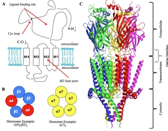
1.2 Ligand Gated Ion Channels: Nicotinic Acetylcholine Receptors
'!
(Figure 1.2). These arrange in a pentameric fashion for ion permeation with the M2
"-helix lining the pore. Neurotransmitters bind at the interface of two subunits and cause
a conformational rearrangement approximately 60 Å away at the receptor gate. The “gating region” comprises several residues along the M2 "-helix that are responsible for
ion permeation. This dissertation focuses mainly on the nAChRs and their structure-function relationships.
nAChRs are aptly named for both their natural agonist (acetylcholine) and for their sensitivity to nicotine, the main addictive component in cigarettes. These receptors are responsible for synaptic transmission in both the peripheral and central nervous systems (1-3). There are 17 nAChR subunits: "1-"10, #1-#4, $, %, and &. However, "8 is
only found in avian species and the “muscle-type” nAChR ("12#1$%/&) is relegated to
(!
only the peripheral system and neuromuscular junctions. "2-"7, "9, "10, and #2-#4
comprise the neuronal nAChR subunits and are prominent throughout the central nervous system in varying assemblies and distributions (Figure 1.2) (2,4). Due to their vast presence, nAChRs are crucial in memory and learning and are prominent targets in neurological disorders such as addiction, Alzheimer’s disease and Parkinson’s disease (2,5-10). The most abundant and widely distributed receptors are the "4#2 and "7
nAChRs (3,4). The "4#2 receptor is a highly sought-after drug target due to its role in
nicotine addiction, as seen with Pfizer’s engineered smoking cessation drug Chantix® (varenicline) (3,4,11,12). The "4#2 receptor has the ability to assemble into two
stoichiometries, the high sensitivity ("4#2)2(#2), and low sensitivity ("4#2)2("4). In
addition, association with other subunits, such as "5, has been seen (9,13-15). The
accessory subunit (the unpaired 5th subunit) can provide distinct tuning of the receptor’s physical properties. The "7 nAChR is a homopentameric channel and also widely
distributed throughout the central nervous system. It has been a constant drug target – both in agonist and allosteric modulator development – due to its association with neurological disorders such as schizophrenia and Alzheimer’s disease (16-23).
)!
(closed) and conducting (open) form of an ion channel (27-31). In the last year, an exciting breakthrough occurred in that the first set of high-resolution crystal structures of vertebrate Cys-loop receptors (5HT3A and GABAA) were achieved (Figure 1.2) (32,33). Although these advances have influenced our structural knowledge, proteins are inherently dynamic, and static representations may not accurately capture motions associated with activation. Thus, structure-function studies still provide vital information regarding interactions necessary for ligand binding and conformational rearrangements required for receptor activation.
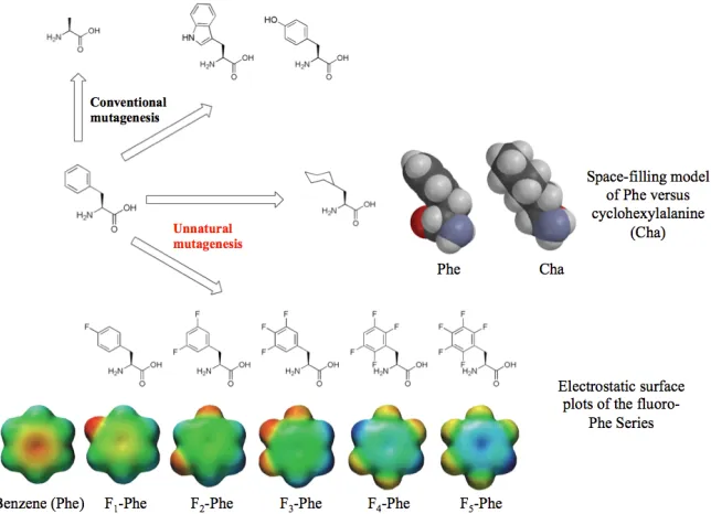
1.3 Non-Canonical Amino Acid Mutagenesis
The goal of the Dougherty lab is to use physical organic chemistry to provide chemical scale structure-function relationships. By identifying distinct non-covalent interactions – cation-', hydrogen bonding, van der Waals, etc. – we can elucidate
contacts that are critical for receptor function, and begin to map important properties to ligand recognition (pharmacophore identification) or functional coupling (conformational motions) on a large and complex protein. We can have a better understanding of neurological diseases and compounds designed for treatment through increased knowledge of the mechanisms associated with receptor activation.
*!
functional distinctions that we aim to tease out. To work around these limitations, the Dougherty lab has established a working protocol to incorporate non-canonical amino acids that has proven to be a powerful technique in studying structure-function relationships in LGICs (Figure 1.3) (34-38). This opens the possibility of probing interactions through the introduction of diverse side chain configurations to study specific interactions of a protein on a chemical scale.
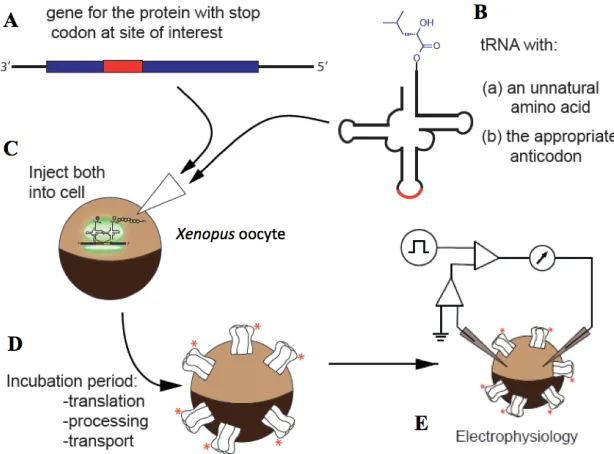
The Dougherty lab has utilized and optimized a method known as in vivo
nonsense suppression to incorporate non-canonical amino acids (Figure 1.4) (35,39-43). This technique allows for site-specific incorporation of a non-canonical amino acid in a relatively quick timeframe. The mRNA codon of the residue to be probed is replaced with a stop or “nonsense” codon (UAG, UGA, or UAA), which is normally used to signify protein synthesis termination. The non-canonical amino acid is chemically
+!
appended to a suppressor tRNA with the appropriate “stop” anticodon. Normally, the nonsense codon generates a truncated protein that is non-functional and is shuttled into degradation pathways. However, in the presence of the chemically ligated suppressor tRNA, protein synthesis continues normally until the full-length protein is generated. Essentially, the ribosome and the remaining machinery of the cell are “hijacked” to synthesize, fold, and post-translationally modify the desired functional protein with a non-canonical amino acid. Xenopus laevis oocytes provide an excellent system for this method due to the extremely large (1 mm in diameter) single cells and the minimal expression of endogenous ion channels. The oocytes are large enough to allow direct injection of the suppressor tRNA/mRNA mixture and contain the cell machinery for proper protein synthesis, folding, assembly, and transport to the membrane (Figure 1.5). The physiology of the expressed ion channels is nearly identical to that of those expressed in other cell systems or found in native neuronal environments (35).
,!
Since evolution of a tRNA synthetase is not needed in this method, incorporation of new non-canonical amino acids can be immediately swapped to probe a large array of side chains. The downside to the in vivo nonsense suppression is the small amount of protein that is actually synthesized. Theoretically, the amount of chemically ligated suppressor tRNA injected translates to the amount of protein synthesized, if efficiency is 100%. Because of reagent limitation and inherent efficiency losses, however, only attomoles of the protein are generated and this is not useful for studying by many of the spectroscopic techniques available. To work around this extremely low yield of protein, we turn to an equally sensitive technique – electrophysiology.
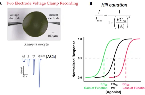
Electrophysiology techniques can be used to indirectly measure receptor-activated currents. Again, the Xenopus oocyte provides an ideal system for electrophysiology
Figure 1.5 Schematic representation of nonsense suppression methodology in Xenopus
-!
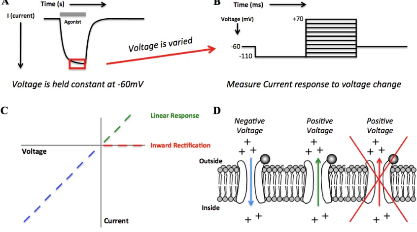
Figure 1.6 The EC50 experiment. (A) Increasing concentration of an agonist shifts more channels to the open state and creates a larger correction current for the TEVC configuration. (B) Responses are normalized to the highest response and plotted against agonist concentration. Fitting the curve to the Hill equation generates a value used for functional comparison of mutated receptors – the EC50. Gain-of-function mutations are depicted as a leftward shift, which decreases the EC50 value. Loss-of-function mutations show the opposite trend: a rightward shift represents an increase in EC50 value.
%.!
1.4 Electrophysiology Assays: The EC50 and Voltage Jump Experiments
%%!
Another electrophysiology test that provides information on receptor properties involves the voltage jump experiment. While the ion channel is open, the negative voltage potential across the membrane drives the flow of cations into the cell. Since we control the membrane potential of the cell, we can vary the voltage by quickly (ms timescale) “jumping” the voltage from large negative potentials to large positive potentials. Switching the membrane potential from negative to positive then drives the flow of ions in the opposite direction – out of the cell. In an idealized pore, plotting voltage vs. current would then provide a linear relationship (Figure 1.7). However, not all receptors act as free flowing pores, and they have a unique property of not allowing ions to flow out of the cell, which is termed inward rectification. The degree of inward rectification can vary between receptor types and in some cases can be used to distinguish stoichiometries of specific subtypes (44,45).
!
1.5 Mutant Cycle Analyses
%&!
mutating one of the residues will change all or most of the interaction in question and perturbation of the second residue should not cause any additional change in function. Thus, the sum of the functional change of the two individual mutations will be different to the collective double mutation. If the mutations are not functionally coupled and are independent of one another, then the collective double mutant and the sums of the individual residues will be identical. EC50 values are used to calculate the coupling coefficient ((), which is converted into a free energy value of ))G (Figure 1.8) (46).
We identify functionally coupled residues or interactions as having values of ))G greater
than 0.5 kcal/mol.
%'!
Figure 1.8 The mutant cycle analysis. (A) Equations used to generate coupling coefficients (() and ))G values. (B) Examples of “uncoupled” and “coupled”
interactions. R is the ideal gas constant and room temperature (25°C) was used for T.
1.6 Orthosteric vs. Allosteric
Discussions of ion channels so far have revolved around the notion that an agonist binds and then the channel gate opens. The location a ligand can occupy and from which it can cause activation of the protein is termed the orthosteric site. Agonists are a class of ligands that activate the receptor by occupying the orthosteric site, whereas antagonists inhibit receptor function by occupying the same orthosteric site without activation. Research involving the orthosteric site has been crucial in developing agonists and antagonists that are selective and potent for a vast array of targets. However, another class of ligands has been garnering more attention and resources in attempts to develop novel compounds for diseases – allosteric modulators.
%(!
positively modulate (increase function) or negatively modulate (decrease function) the protein’s activity. Allosteric modulator properties are desirable as drug discovery targets because they can have high specificity and activity while minimizing adverse side effects from off–target interactions. Thus, more research has been directed towards the development and understanding of both positive and negative allosteric modulators for disease treatments, including neurological disorders.
1.7 Summary of Dissertation Work
In this dissertation, canonical and non-canonical amino acid mutagenesis was used to study structure-function relationships associated with agonists and allosteric modulators of nAChRs. Group methodology of nonsense suppression in Xenopus
oocytes, along with electrophysiological assays, was used to assess functional changes associated with receptor mutations. The "4#2 and the "4#2"5 nAChRs were targeted for
agonist selectivity studies. The "7 receptor was used to probe allosteric modulator
binding and influence on receptor activation.
Chapter 2 details research investigating the interactions involved in the stoichiometry selective compounds of "4#2 receptors: sazetidine-A, cytisine, and
NS9283. Non-canonical amino acid mutagenesis showed that the previously proposed change in hydrogen bonding strengths were not the cause of the selectivity. Instead, the residue composition on the complementary subunit strongly influenced agonist occupation at the binding site. It was concluded that occupation of all primary
%)!
models should incorporate the influence of the complementary side for receptor specificity.
Chapter 3 describes the work done towards producing a pure population of the
"4#2"5 receptor and subsequent tests for agonist binding at the "5-"4 interface.
Generation of the "4#2"5 receptor was aided through the addition of a gain-of-function
pore mutation, which also incorporated a handle for identifying assembly through voltage jump experiments. Mutations of the "5 aromatic box residues resulted in no change to
receptor function. This suggests agonist occupation at the "5-"4 interface is not
necessary for activation as is seen with the "4-"4 interface. In addition, this shows that
the "5 subunit does not replace "4 or #2 subunits and is relegated exclusively to the
auxiliary position.
Chapters 4 and 5 are dedicated to studying positive allosteric modulators of
"7 receptors. Chapter 4 involves assay development for functional screening of allosteric
modulators and adaption of a cooling apparatus for varied temperature control. Chapter 5 revolves around probing the global effects of PNU-120596 on the "7 receptor. It was
shown that the higher potency of acetylcholine in the presence of PNU-120596 is not due to an altered agonist binding site. In addition, several residues were identified in the gating interface that are vital to transmitting the effects of PNU-120596. These results suggest a global propagation through several key residues that influence the receptor gating equilibrium while leaving the agonist binding site unperturbed.
%*!
%+!
1.8 References
1. Corringer, P. J., Novere, N. L., and Changeux, J. P. (2000) Nicotinic Receptors At The Amino Acid Level. Annual Review of Pharmacology and Toxicology 40, 431-458
2. Jensen, A. A., Frolund, B., Lijefors, T., and Krogsgaard-Larsen, P. (2005) Neuronal nicotinic acetylcholine receptors: Structural revelations, target identifications, and therapeutic inspirations. Journal of Medicinal Chemistry 48, 4705-4745
3. Romanelli, M. N., Gratteri, P., Guandalini, L., Martini, E., Bonaccini, C., and Gualtieri, F. (2007) Central nicotinic receptors: structure, function, ligands, and therapeutic potential. ChemMedChem 2, 746-767
4. Gotti, C., Zoli, M., and Clementi, F. (2006) Brain nicotinic acetylcholine receptors: native subtypes and their relevance. Trends in Pharmacological Sciences 27, 482-491
5. Dani, J. A., and Bertrand, D. (2007) Nicotinic acetylcholine receptors and nicotinic cholinergic mechanisms of the central nervous system. Annual Review of
Pharmacology and Toxicology 47, 699-729
6. Miwa, J. M., Freedman, R., and Lester, H. A. (2011) Neural systems governed by nicotinic acetylcholine receptors: emerging hypotheses. Neuron 70, 20-33
7. Taly, A., Corringer, P. J., Guedin, D., Lestage, P., and Changeux, J. P. (2009) Nicotinic receptors: allosteric transitions and therapeutic targets in the nervous system. Nature Reviews. Drug Discovery 8, 733-750
8. Albuquerque, E. X., Pereira, E. F., Alkondon, M., and Rogers, S. W. (2009) Mammalian nicotinic acetylcholine receptors: from structure to function.
Physiological Reviews 89, 73-120
9. Saccone, N. L., Saccone, S. F., Hinrichs, A. L., Stitzel, J. A., Duan, W., Pergadia, M. L., Agrawal, A., Breslau, N., Grucza, R. A., Hatsukami, D., Johnson, E. O., Madden, P. A. F., Swan, G. E., Wang, J., Goate, A. M., Rice, J. P., and Bierut, L. J. (2009) Multiple Distinct Risk Loci for Nicotinic Dependence Identified by Dense Coverage of the Complete Family of Nicotinic Receptor Subunit (CHRN) Genes. American Journal of Medical Genetics Neuropsychiatric Genetics, 453-467
10. Ferini-Strambi, L., Sansoni, V., and Combi, R. (2012) Nocturnal frontal lobe epilepsy and the acetylcholine receptor. The Neurologist 18, 343-349
11. Coe, J. W., Brooks, P. R., Vetelino, M. G., Wirtz, M. C., Arnold, E. P., Huan, J., Sands, S. B., Davis, T. I., Lebel, L. A., Fox, C. B., Shrikhande, A., Heym, J. H., Schaeffer, E., Rollema, H., Lu, Y., Mansbach, R. S., Chambers, L. K., Rovetti, C. C., Schulz, D. W., Tingely III, D., and O'Neil, B. T. (2005) Varenicline: An "4#2
%,!
stoichiometry, and sensitivity to long-term exposure to nicotine. Mol Pharmacol
70, 755-768
14. Nelson, M., Kuryatov, A., Choi, C., Zhou, Y., and Lindstrom, J. (2003) Alternate Stoichiometries of the "4#2 Nicotinic Acetylcholine Receptors. Molecular
Pharmacology 63, 332-342
15. Kuryatov, A., Onksen, J., and Lindstrom, J. (2008) Roles of accessory subunits in
"4#2(*) nicotinic receptors. Molecular Pharmacology 74, 132-143
16. Christopoulos, A. (2002) Allosteric binding sites on cell-surface receptors: novel targets for drug discovery. Nature Reviews. Drug Discovery 1, 198-210
17. Horenstein, N. A., Leonik, F. M., and Papke, R. L. (2008) Multiple pharmacophores for the selective activation of nicotinic "7-type acetylcholine
receptors. Mol Pharmacol 74, 1496-1511
18. Narla, S., Klejbor, I., Birkaya, B., Lee, Y. W., Morys, J., Stachowiak, E. K., Terranova, C., Bencherif, M., and Stachowiak, M. K. (2013) "7 nicotinic receptor
agonist reactivates neurogenesis in adult brain. Biochemical Pharmacology 86, 1099-1104
19. Pandya, A. A., and Yakel, J. L. (2013) Effects of neuronal nicotinic acetylcholine receptor allosteric modulators in animal behavior studies. Biochemical
Pharmacology 86, 1054-1062
20. Parri, H. R., Hernandez, C. M., and Dineley, K. T. (2011) Research update: "7
nicotinic acetylcholine receptor mechanisms in Alzheimer's disease. Biochemical
Pharmacology 82, 931-942
21. Tong, M., Arora, K., White, M. M., and Nichols, R. A. (2011) Role of key aromatic residues in the ligand-binding domain of "7 nicotinic receptors in the
agonist action of #-amyloid. The Journal of Biological Chemistry 286,
34373-34381
22. Williams, D. K., Wang, J., and Papke, R. L. (2011) Positive allosteric modulators as an approach to nicotinic acetylcholine receptor-targeted therapeutics: advantages and limitations. Biochemical Pharmacology 82, 915-930
23. Young, J. W., and Geyer, M. A. (2013) Evaluating the role of the "7 nicotinic
acetylcholine receptor in the pathophysiology and treatment of schizophrenia.
Biochemical Pharmacology 86, 1122-1132
24. Brejc, K., van Dijk, W. J., Klaassen, R. V., Schuurmans, M., van der Oost, J., Smit, A. B., and Sixma, T. K. (2001) Crystal structure of an ACh-binding protein reveals the ligand-binding domain of nicotinic receptors. Nature 411, 269-276 25. Unwin, N. (2005) Refined structure of the nicotinic acetylcholine receptor at 4Å
resolution. J Mol Biol 346, 967-989
26. Miyazawa, A., Fujiyoshi, Y., Stowell, M., and Unwin, N. (1999) Nicotinic Acetylcholine Receptor at 4.6 Å Resolution: Transverse Tunnels in the Channel Wall. J Mol. Biol. 288, 765-786
27. Bocquet, N., Nury, H., Baaden, M., Le Poupon, C., Changeux, J. P., Delarue, M., and Corringer, P. J. (2009) X-ray structure of a pentameric ligand-gated ion channel in an apparently open conformation. Nature 457, 111-114
%-!
29. Hilf, R. J., and Dutzler, R. (2008) X-ray structure of a prokaryotic pentameric ligand-gated ion channel. Nature 452, 375-379
30. Hilf, R. J., and Dutzler, R. (2009) Structure of a potentially open state of a proton-activated pentameric ligand-gated ion channel. Nature 457, 115-118
31. Nury, H., Van Renterghem, C., Weng, Y., Tran, A., Baaden, M., Dufresne, V., Changeux, J. P., Sonner, J. M., Delarue, M., and Corringer, P. J. (2011) X-ray structures of general anaesthetics bound to a pentameric ligand-gated ion channel.
Nature 469, 428-431
32. Hassaine, G., Deluz, C., Grasso, L., Wyss, R., Tol, M. B., Hovius, R., Graff, A., Stahlberg, H., Tomizaki, T., Desmyter, A., Moreau, C., Li, X. D., Poitevin, F., Vogel, H., and Nury, H. (2014) X-ray structure of the mouse serotonin 5-HT3 receptor. Nature 512, 276-281
33. Miller, P. S., and Aricescu, A. R. (2014) Crystal structure of a human GABAA receptor. Nature 512, 270-275 (2012) Ligand-gated ion channels: new insights into neurological disorders and ligand recognition. Chem Rev 112, 6285-6318
37. Pless, S. A., and Ahern, C. A. (2013) Unnatural amino acids as probes of ligand-receptor interactions and their conformational consequences. Annual Review of
Pharmacology and Toxicology 53, 211-229
38. Dougherty, D. A., and Van Arnam, E. B. (2014) In vivo incorporation of non-canonical amino acids by using the chemical aminoacylation strategy: a broadly applicable mechanistic tool. Chembiochem : a European Journal of Chemical Biology 15, 1710-1720
39. Nowak, M., Gallivan, J. P., Silverman, S., Labarca, C. G., Dougherty, D. A., and Lester, H. A. (1998) In Vivo Incorporation of Unnatural Amino Acids into Ion Channels in Xenopus Oocyte Expression System. Methods Enzymol 293, 504-530 40. Rodriguez, E. A., Lester, H. A., and Dougherty, D. A. (2007) Improved amber
and opal suppressor tRNAs for incorporation of unnatural amino acids in vivo. Part 2: evaluating suppression efficiency. RNA 13, 1715-1722
41. Rodriguez, E. A., Lester, H. A., and Dougherty, D. A. (2007) Improved amber and opal suppressor tRNAs for incorporation of unnatural amino acids in vivo. Part 1: minimizing misacylation. RNA 13, 1703-1714
42. Rodriguez, E. A., Lester, H. A., and Dougherty, D. A. (2006) In vivo incorporation of multiple unnatural amino acids through nonsense and frameshift suppression. Proceedings of the National Academy of Sciences of the United States of America 103, 8650-8655
&.!
44. Marotta, C. B., Dilworth, C. N., Lester, H. A., and Dougherty, D. A. (2013) Probing the non-canonical interface for agonist interaction with an "5 containing
nicotinic acetylcholine receptor. Neuropharmacology 77C, 342-349
45. Xiu, X., Puskar, N. L., Shanata, J. A., Lester, H. A., and Dougherty, D. A. (2009) Nicotine binding to brain receptors requires a strong cation-' interaction. Nature
458, 534-537
46. Horovitz, A. (1996) Double-mutant cycles: a powerful tool for analyzing protein structure and function. Folding and Design 1, R121-R126
47. Blum, A. P., Gleitsman, K. R., Lester, H. A., and Dougherty, D. A. (2011) Evidence for an extended hydrogen bond network in the binding site of the nicotinic receptor: role of the vicinal disulfide of the "1 subunit. The Journal of
Biological Chemistry 286, 32251-32258
48. Blum, A. P., Lester, H. A., and Dougherty, D. A. (2010) Nicotinic pharmacophore: the pyridine N of nicotine and carbonyl of acetylcholine hydrogen bond across a subunit interface to a backbone NH. Proceedings of the
National Academy of Sciences of the United States of America 107, 13206-13211
49. Gleitsman, K. R., Kedrowski, S. M., Lester, H. A., and Dougherty, D. A. (2008) An intersubunit hydrogen bond in the nicotinic acetylcholine receptor that contributes to channel gating. The Journal of Biological Chemistry 283, 35638-35643
50. Gleitsman, K. R., Shanata, J. A., Frazier, S. J., Lester, H. A., and Dougherty, D. A. (2009) Long-range coupling in an allosteric receptor revealed by mutant cycle analysis. Biophysical Journal 96, 3168-3178
51. Kash, T. L., Jenkins, A., Kelly, J. C., Trudell, J. R., and Harrison, N. L. (2003) Coupling of agonist binding to channel gating in the GABAA receptor. Nature
421, 272-275
52. Price, K. L., Millen, K. S., and Lummis, S. C. (2007) Transducing agonist binding to channel gating involves different interactions in 5-HT3 and GABAC receptors.
The Journal of Biological Chemistry 282, 25623-25630
53. Venkatachalan, S. P., and Czajkowski, C. (2008) A conserved salt bridge critical for GABA(A) receptor function and loop C dynamics. Proceedings of the
National Academy of Sciences of the United States of America 105, 13604-13609
54. Daeffler, K. N., Lester, H. A., and Dougherty, D. A. (2012) Functionally important aromatic-aromatic and sulfur-' interactions in the D2 dopamine
receptor. Journal of the American Chemical Society 134, 14890-14896
&%!
Chapter 2
Selective Ligand Behaviors Provide New Insights into
Agonist Activation of Nicotinic Acetylcholine Receptors*
*Reproduced with permission from: (DOI: 10.1021/cb400937d) Christopher B. Marotta,
Iva Rreza, Henry A. Lester, and Dennis A. Dougherty. Selective ligand behaviors provide new insights into agonist activation of nicotinic acetylcholine receptors. ACS Chem. Biol., 2014, 9 (5), pp 1153-1159. Copyright 2014 American Chemical Society. The work described in this chapter was done in collaboration with Iva Rreza.
!"#$%&'%()&"*+,:10.1021/cb400937d
2.1 Abstract
Nicotinic acetylcholine receptors are a diverse set of ion channels that are essential to everyday brain function. Contemporary research studies selective activation of individual subtypes of receptors, with the hope of increasing our understanding of behavioral responses and neurodegenerative diseases. Here, we aim to expand current binding models to help explain the specificity seen among three activators of "4#2
receptors: sazetidine-A, cytisine, and NS9283. Through mutational analysis, we can interchange the activation profiles of the stoichiometry-selective compounds sazetidine-A and cytisine. In addition, mutations render NS9283 – currently identified as a positive allosteric modulator – into an agonist. These results lead to two conclusions: (1) occupation at each primary face of an " subunit is needed to activate the channel and
&&!
2.2 Introduction
Nicotinic acetylcholine receptors (nAChRs) are a diverse family of the larger Cys-loop superfamily of ligand gated ion channels (LIGCs). These channels are found throughout the brain and CNS and play vital roles in the chemical and electrical communication between neurons, contributing to memory, learning, and other neural functions (1-3). Because of their diverse properties and widespread distribution throughout the brain, these LGICs are prominent targets in neurological disorders, such as drug addiction, Alzheimer’s disease, and Parkinson’s disease (4,5).
Neuronal nAChRs have been extensively studied, and much is known about their assembly and structure (2,6-8). There are two classes of subunits, identified as "2 – "9
and #2 – #4, which assemble into homomeric (" only) or heteromeric (" and #) channels
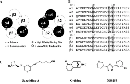
consisting of five subunits in total (9). For each subunit, there is a main agonist binding pocket, denoted as the primary (+) face, which includes four of the five residues that make up the canonical “aromatic box” (10). The fifth aromatic box residue and other key binding residues, including backbone contacts, are found on the complementary (–) face of the adjacent subunit (10). The five subunits then arrange in an alternating + and – fashion to form a functional receptor (Figure 2.1). For decades it has been assumed that the (+) face of the agonist binding site is provided by " subunits and the (–) face by
# subunits. However, recent evidence establishes the ability of " subunits to also
contribute to the (–) face.
&'!
the primary subunit, along with hydrogen bonding interactions on both the primary and complementary subunits (12,13). Based on this model, a number of studies have aimed to explain the efficacies and selectivities of agonists to different nAChRs of varying subunits and stoichiometries (12-14).
Here, we studied the activation profiles of "4#2 receptors and their responses to
mutations for the following compounds: sazetidine-A, cytisine, and NS9283 (Figure 2.1). Our conclusions lead us to propose an expansion of the published structural
circles at the interfaces of "4-#2 subunits and "4-"4 subunits. (B) Sequence alignment of
&(!
hydrophobic in some subunits and hydrophilic in others and (2) an agonist must be bound at all " subunits in a given receptor to favor the activated channel. This expansion aids in
our understanding of subunit- and stoichiometry-selective agents and can provide valuable insight for further development and application towards therapeutic strategies.
2.3 Results and Discussion
2.3.1 Hydrogen Bonding: Non-Canonical Amino Acid Analysis
Sazetidine-A has a unique activation profile, in that it selectively activates the
("4)2(#2)3 stoichiometry over the ("4)3(#2)2; these stoichiometries will be abbreviated
A2B3 and A3B2, respectively (17). Unnatural amino acids are useful tools used to parse out specific chemical interactions between ligand and receptor. Previous structure-function studies of cytisine, an agonist that has the opposite activation profile for "4#2
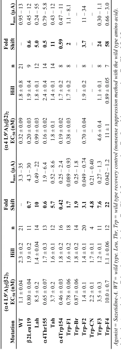
receptors, showed that the active drug-receptor combination (A3B2) favored the hydrogen bond to the TrpB backbone CO (“donor”), while the inactive form favored the hydrogen bond to the backbone NH on the complementary face (“acceptor”) (Table 2.1, Figure 2.2) (12). We proposed that this difference could explain the stoichiometric selectivity of the drug. Through unnatural amino acid incorporation, we were able to characterize the cation-' binding, hydrogen-bond donating, and hydrogen-bond accepting
&)!
50 corrections. b Ratio of Imax of compound divided by Imax of acetylcholine. c Ratio of EC
50 values for 4,5,6,7-tetraflouro-Trp and Trp incorporation at !4 W154 (Fig. 2A). d Ratio of EC50 values for Thr-!-hydroxy and Thr incorporation at !4 T155 (Fig. 2A). e Ratio of EC50 values for Leu !-hydroxy and Leu incorporation at "2 L119 (Fig. 2A). f Previously reported values from
Tavares et al. (12). Measured EC50 values reported in Table 2.2.
2.3.2 Sazetidine-Aand the "2Complementary Face
It has been shown that the unique hydrophobic appendage off of the pyridine ring of sazetidine-A gives the compound its subunit and receptor selectivity, and that the alcohol group at the end of the appendage does not play a significant role (15,18,19). Because this aliphatic adjunct interacts mostly with the complementary side, we began by focusing on the known differences between "4 and #2 subunits in this region (16).
Previous investigations identified an "4-"4 binding site and suggested the differences
between the “high” affinity ("4-#2) and “low” affinity ("4-"4) binding pockets are due
to three key residues that reside on the complementary face (20-22). The #2(–) face
residues (V109, F117, and L119) generate a hydrophobic pocket for the high affinity case, while the aligning "4(–) face residues (H114, Q122, and T124) create a
&*!
A
B
Figure 2.2 Binding models of sazetidine-A and analogs. (A) Binding model for sazetidine-A based on established interactions seen with nicotine (12). The cation-'
interaction is in purple, the hydrogen bond donor is in red, and the hydrogen bond acceptor is in green. (B) Crystal structure showing a sazetidine-A analog bound to Ct-AChBP (PDB: 4B5D) (15). The three key residues identified for the hydrophobic pocket associated with the #2 subunit (V109, F117, & L119) are shown, as is the TrpB
residue from the "4 subunit. These residues were mutated into the crystal structure to
show general spatial locations (no residue minimizations calculated).
&,!
to make them resemble the #2(–)face. We were able to generate receptor responses and
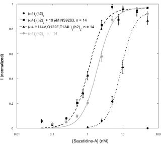
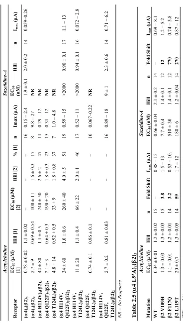
measure an EC50 curve for sazetidine-A in the A3B2 receptor, which was not possible with the wild type "4 subunit (Table 2.3, Figure 2.3). The EC50 value for the triple
mutant was about five-fold larger than the wild type A2B3 response, a small difference compared to having zero response in the wild type A3B2 receptor. Single mutations at
the "4 subunit did not give rise to sazetidine-A response at low µM doses (Table 2.4).
Combinations of double mutations saw some response towards low µM doses of
sazetidine-A (Table 2.4). Mutations to make the # subunit more like the " subunit
resulted in a large loss of function for sazetidine-A (Table 2.5).
Table 2.3 Sazetidine-A EC50 (nM) Values
Receptor EC50 (nM) Hill n Imax (µA)
2.3.3 Cytisine and the "2Complementary Face
Since these three residues had a large affect on receptor agonist selectivity and activation for sazetidine-A, we considered cytisine in an attempt to explain its selectivity for A3B2 over A2B3 receptors. Early chimera analysis showed that cytisine selectivity for human #4 over #2 subunits is strongly influenced by the extracellular region (23), and
&-!
#4 L117 for the human subunits. For the rodent subunits considered here, #4 is Q117,
which is identical to the "4 residue at this same position (Figure 2.1). In the rodent wild
type "4#2 receptors, cytisine is a partial agonist with a biphasic response for the A3B2
receptor. No response is observed for the A2B3 receptor (Table 2.6), although signal can be obtained in receptors with hypersensitive mutations (see 2.5 Methods). However, with the single mutation of F117Q in the #2 subunit, cytisine generated a sizable
response for the A2B3 receptor. The mutation also raised the efficacy of the A3B2 receptor compared to the wild type response (Table 2.6).
Figure 2.3 Sazetidine-A EC50 curves. ("4)3(#2)2 cannot be activated by sazetidine-A.
Responses can be obtained by mutating the complementary side of the "4 subunit to
resemble the #2 subunit as seen with ("4 H114V, Q122F, T124L)3(#2)2 receptor. Also
shown is wild type ("4)3(#2)2 receptor exposed to a combination of sazetidine-A and
'%!
2.3.4 NS9283 and the !4Complementary Face
We next considered NS9283, which has a binding preference for the "4-"4
interface (25). This compound has been previously characterized as a benzodiazepine-like positive allosteric modulator (PAM) for only the A3B2 stoichiometry of receptors containing either "2 or "4 subunits (25,26). In addition, its
effects are lost when the "4(–) face is mutated to resemble the #2(–) face in the region of
the classical agonist binding site (27). Since we have molecules that selectively associate with "4-"4 (NS9283) and "4-#2 (sazetidine-A) interfaces, co-application should
generate an A3B2 receptor response. As shown in Figures 2.3, 2.4 and Table 2.3, we find that individual applications of NS9283 and sazetidine-A show essentially no activation of wild type A3B2 receptors. However, co-application generates full activation of the receptor, compared to acetylcholine. The A3B2 "4 triple mutant (H114V, Q122F,
T124L) was then exposed to similar conditions. The sazetidine-A response for the mutant was preserved, but the effect of NS9283 was completely lost (Table 2.3, Figure 2.4). The mutations eliminate the ability of NS9283 to bind at the "4-"4 interface and allow
sazetidine-A to replace it in binding. These data suggest that occupation of an agonist at each " subunit is necessary for a receptor response. We generated the A3B2 #2 triple
'&!
expression, the corresponding A2B3 #2 triple mutant (V109H, F117Q, L119T) was
inconclusive with regard to activation via NS9283. A
B
C
Figure 2.4 Sample traces of responses to acetylcholine (ACh), sazetidine-A (Saz-A), and NS9283 (NS) to A3B2 receptors. Solid gray bars indicate drug application and dashed bars indicate a pause where drug remains present but the buffer wash has not started. Gaps between traces indicate buffer washes (see methods for duration of drug application and buffer washes). (A) Activation of wild type receptor by ACh at its EC50 value, and Saz-A and NS at the concentrations shown. (B) Activation of ("4 H144V, Q122F,
T124L)3(#2)2. (C) Application of Imax concentrations of acetylcholine and two
concentrations of NS to ("4)3(#2 V109H, F117Q, L119T)2. The * indicates a 1% by
''!
2.4 Conclusions
The present work confirms and expands upon recent studies on the (–), complementary face of the agonist binding site of nAChRs. In particular, the long-held belief that agonist binding sites are formed only at "(+)/#(–) interfaces has been
challenged by increasing evidence for a viable agonist binding site at "(+)/"(–)
interfaces. Here we use several drugs that show some subtype specificity to probe this issue. The novel agonist sazetidine-A can only activate "4#2 receptors with an A2B3
stoichiometry. In the alternative A3B2 stoichiometry, an "(+)/"(–) interface exists. The
(–) face of such an interface is relatively polar, and evidently it is incompatible with the hydrophobic side chain of sazetidine-A that is expected to project into this region (recall that the OH group of sazetidine-A is not necessary for function). By mutating three residues in this region to be more hydrophobic and thus more like a #2(–) face, we can
prepare A3B2 receptors that are quite responsive to sazetidine-A (Table 2.3).
In a complementary series of experiments, we considered the drug NS9283, which binds only to "(+)/"(–) interfaces. It is unable to activate the receptor on its own,
and it is thus an allosteric modulator. We reasoned that a combination of NS9283 and sazetidine-A would activate A3B2 receptors, with the former binding to the "(+)/"(–)
interface and the latter to the "(+)/#(–) interfaces. Indeed, a mixture of NS9283 and
sazetidine-A is quite potent at A3B2 receptors, while neither compound alone can activate the receptor. Taking this one step further, by mutating all interfaces so they resemble "(+)/"(–) interfaces, NS9283 becomes an agonist, rather than the allosteric
'(!
We also applied this interface concept to cytisine, which has the reverse activation profile of sazetidine-A, in that it cannot activate the A2B3 receptor. By mutating one residue of the #2 subunit to that of the "4 subunit, we find that cytisine can activate the
A2B3 receptor. In addition, this same mutation increased efficacy of cytisine for the A3B2 receptor from 7% to 16% (Table 2.6).
In sum, this work shows the relevance of the "(+)/"(–) interface of nAChRs to
achieving full receptor activation. This knowledge could be of great value to efforts to
pGEMhe (natural mutagenesis) vectors. Site-directed mutagenesis was performed using
the QuikChange protocol (Stratagene). Circular DNA of "4 and #2 in pAMV was
linearized with the NotI restriction enzyme and the plasmids in pGEMhe were linearized
with the SbfI restriction enzyme. After purification (Qiagen), the T7 mMessage Machine
')!
QIAGEN’s RNeasy RNA purification kit was used to isolate the transcribed mRNA product.
For unnatural amino acid incorporation, the amber (UAG) stop codon was used for all "4 subunit incorporation and the opal (UGA) stop codon was used for the #2
subunit incorporation. 74-nucleotide THG73 tRNA (for UAG) and 74-nucleotide TQOpS’ tRNA (for UGA) were in vitro transcribed using the MEGAshortscript T7 (Ambion) kit and isolated using Chroma Spin DEPC-H2O columns (Clontech). Synthesized unnatural amino acids coupled to the dinucleotide dCA were enzymatically ligated to the appropriate 74-nucleotide tRNA as previously described (13,28).
2.5.2 Oocyte Preparation and Injection
Xenopus laevis stage V and VI oocytes were harvested via standard protocols (28). For unnatural amino acid incorporation to the " subunit, the "4 and #2 mRNAs
were mixed in a 3:1 ratio by mass to obtain the A2B3 receptor, and in a 100:1 ratio to obtain the A3B2 receptor. Unnatural amino acid incorporation to the # subunit used "4
and #2 mRNA ratios of 1:20 and 10:1 to obtain the A2B3 and A3B2 receptors,
respectively. mRNA mixtures and deprotected (photolysis) tRNA were mixed in a 1:1 volume ratio, and 50 nL were injected into each oocyte. After injection, the oocytes were incubated at 18° C in ND96+ medium for 24 h. For the unnatural amino acids with reduced cation-' binding ability, a second round of injections following the same
'*!
For the natural mutagenesis experiments, the "4 and #2 mRNAs were mixed in
1:2 or 10:1 ratios by mass to obtain the A2B3 and A3B2 receptors, respectively (29). A total of 50 nL were injected to each oocyte, delivering an mRNA mass total of 25 ng. Oocytes were incubated at 18° C in ND96+ medium for 24-72 h.
2.5.3 Chemical Preparation
Acetylcholine chloride was purchased from Sigma-Aldrich and dissolved to 1 M stock solutions in ND96 Ca2+ free buffer (96 mM NaCl, 2 mM KCl, 1 mM MgCl2, 5 mM HEPES at pH 7.5). Sazetidine-A dihydrochloride and (-)-cytisine were purchased from Tocris Bioscience and dissolved to 10 mM stock solutions in ND96 Ca2+ free buffer.
NS9283 was synthesized following a patented protocol (30). 3-pyridylamidoxime and 3-cyanobenzoyl chloride were purchase from Sigma-Aldrich. 0.5 g of 3-pyridylamidoxime was dissolved in 5.4 mL of pyridine. Then 0.6 g of 3-cyanobenzoyl chloride was added while stirring. The mixture was heated at reflux for 90 min and then cooled to room temperature. 200 mL of water was added and the white powder was filtered with two subsequent washes with water. The resulting powder was lyophilized overnight to remove the excess water. The reaction resulted in 60% yield, and the product was pure by LC-MS and NMR. 1H NMR (300 MHz, DMSO-d6) ! 9.26 (dd, J = 2.2, 0.9
Hz, 1H), 8.82 (dd, J = 4.8, 1.6 Hz, 1H), 8.64 (td, J = 1.7, 0.7 Hz, 1H), 8.50 (ddd, J = 8.0,
1.9, 1.1 Hz, 1H), 8.47 (dt, J = 6.0, 2.1 Hz, 1H), 8.22 (dt, J = 7.9, 1.4 Hz, 1H), 7.88 (td, J =
7.9, 0.7 Hz, 1H), 7.65 (ddd, J = 7.9, 4.8, 0.9 Hz, 1H); MS (+ES-API) m/z 249 (M+H)+.
'+!
exception of the 100 mM dose, which had 1% DMSO (v/v). Appropriate controls of 1% DMSO (v/v) in ND96 Ca2+ free buffer only were applied to expressing cells to show no receptor response to the higher DMSO concentration.
2.5.4 Electrophysiology
The OpusXpress 6000A (Axon Instruments) in two-electrode voltage clamp mode was used for all electrophysiological recordings. The holding potential was set to -60 mV and the running buffer used was ND96 Ca2+ free solution for all experiments. All acetylcholine drug applications used 1 mL of drug solution applied over 15 s followed by a 2.5 min buffer wash at a rate of 3 mL min-1. All sazetidine-A, cytisine, NS9283, and co-applications used 1 mL of drug solution applied over 8 s with a 30 s pause before a 5 min buffer wash at a rate of 3 mL min-1. Dose-response measurements utilized a series of approximately three-fold concentration steps, spanning several orders of magnitude, for a total of eight to eighteen doses. Data were sampled at 50 Hz and then low-pass filtered at 5 Hz. Experiments testing activity of compounds involved two to three acetylcholine doses of either EC50 or Imax values, followed by the test doses of compounds being probed, followed by one to two doses of the previous acetylcholine concentrations.
',!
In the case of unnatural amino acid incorporation and mutagenesis scanning, EC50 values were obtained using a hypersensitive mutation in the "4 subunit (L9'A). This
'-!
2.6 Acknowledgments
(.!
2.7 References
1. Miwa, J. M., Freedman, R., and Lester, H. A. (2011) Neural systems governed by nicotinic acetylcholine receptors: emerging hypotheses. Neuron 70, 20-33
2. Albuquerque, E. X., Pereira, E. F., Alkondon, M., and Rogers, S. W. (2009) Mammalian nicotinic acetylcholine receptors: from structure to function.
Physiological Reviews 89, 73-120
3. Dani, J. A., and Bertrand, D. (2007) Nicotinic acetylcholine receptors and nicotinic cholinergic mechanisms of the central nervous system. Annual Review of
Pharmacology and Toxicology 47, 699-729
4. Jensen, A. A., Frolund, B., Lijefors, T., and Krogsgaard-Larsen, P. (2005) Neuronal nicotinic acetylcholine receptors: Structural revelations, target identifications, and therapeutic inspirations. Journal of Medicinal Chemistry 48, 4705-4745
5. Taly, A., Corringer, P. J., Guedin, D., Lestage, P., and Changeux, J. P. (2009) Nicotinic receptors: allosteric transitions and therapeutic targets in the nervous system. Nature Reviews. Drug Discovery 8, 733-750
6. Dougherty, D. A. (2008) Cys-Loop Neuroreceptors: Structure to the Rescue?
Chemical Reviews 108, 1642-1654
7. Dougherty, D. A. (2008) Physical Organic Chemistry on the Brain. Journal of
Organic Chemistry 73, 3667-3674
8. Unwin, N. (2005) Refined structure of the nicotinic acetylcholine receptor at 4Å resolution. J Mol Biol 346, 967-989
9. Gotti, C., Zoli, M., and Clementi, F. (2006) Brain nicotinic acetylcholine receptors: native subtypes and their relevance. Trends in Pharmacological Sciences 27, 482-491
10. Brejc, K., van Dijk, W. J., Klaassen, R. V., Schuurmans, M., van der Oost, J., Smit, A. B., and Sixma, T. K. (2001) Crystal structure of an ACh-binding protein reveals the ligand-binding domain of nicotinic receptors. Nature 411, 269-276 11. Beers, W. H., and Reich, E. (1970) Structure And Activity Of Acetylcholine.
Nature 228, 917-922
12. Tavares Xda, S., Blum, A. P., Nakamura, D. T., Puskar, N. L., Shanata, J. A., Lester, H. A., and Dougherty, D. A. (2012) Variations in binding among several agonists at two stoichiometries of the neuronal, "4#2 nicotinic receptor. Journal
of the American Chemical Society 134, 11474-11480
13. Xiu, X., Puskar, N. L., Shanata, J. A., Lester, H. A., and Dougherty, D. A. (2009) Nicotine binding to brain receptors requires a strong cation-pi interaction. Nature
458, 534-537
14. Puskar, N. L., Xiu, X., Lester, H. A., and Dougherty, D. A. (2011) Two neuronal nicotinic acetylcholine receptors, "4#4 and "7, show differential agonist binding
modes. The Journal of Biological Chemistry 286, 14618-14627
cyclopropane-(%!
containing ligands to "4#2-nicotinic acetylcholine receptors: an integrated
approach to behaviorally active nicotinic ligands. J Med Chem 55, 8028-8037 16. Billen, B., Spurny, R., Brams, M., van Elk, R., Valera-Kummer, S., Yakel, J. L.,
Voets, T., Bertrand, D., Smit, A. B., and Ulens, C. (2012) Molecular actions of smoking cessation drugs at "4#2 nicotinic receptors defined in crystal structures
of a homologous binding protein. Proc. Natl. Acad. Sci. U. S. A. 109, 9173-9178 17. Zwart, R., Carbone, A. L., Moroni, M., Bermudez, I., Mogg, A. J., Folly, E. A.,
Broad, L. M., Williams, A. C., Zhang, D., Ding, C., Heinz, B. A., and Sher, E. (2008) Sazetidine-A is a potent and selective agonist at native and recombinant
"4#2 nicotinic acetylcholine receptors. Molecular Pharmacology 73, 1838-1843
18. Liu, Y., Richardson, J., Tran, T., Al-Muhtasib, N., Xie, T., Yenugonda, V. M., Sexton, H. G., Rezvani, A. H., Levin, E. D., Sahibzada, N., Kellar, K. J., Brown, M. L., Xiao, Y., and Paige, M. (2013) Chemistry and pharmacological studies of 3-alkoxy-2,5-disubstituted-pyridinyl compounds as novel selective "4#2 nicotinic
acetylcholine receptor ligands that reduce alcohol intake in rats. J Med Chem 56, 3000-3011
19. Liu, J., Yu, L. F., Eaton, J. B., Caldarone, B., Cavino, K., Ruiz, C., Terry, M., Fedolak, A., Wang, D., Ghavami, A., Lowe, D. A., Brunner, D., Lukas, R. J., and Kozikowski, A. P. (2011) Discovery of isoxazole analogues of sazetidine-A as selective "4#2-nicotinic acetylcholine receptor partial agonists for the treatment
of depression. J Med Chem 54, 7280-7288
20. Eaton, J. B., Lucero, L. M., Stratton, H., Chang, Y., Cooper, J. F., Lindstrom, J. M., Lukas, R. J., and Whiteaker, P. (2014) The unique "4(+)/(-)"4 agonist
binding site in ("4)3(#)2 subtype nicotinic acetylcholine receptors permits
differential agonist desensitization pharmacology vs. the ("4)2(#2)3 subtype. The
Journal of Pharmacology and Experimental Therapeutics 348, 46-58
21. Mazzaferro, S., Benallegue, N., Carbone, A., Gasparri, F., Vijayan, R., Biggin, P. C., Moroni, M., and Bermudez, I. (2011) Additional acetylcholine (ACh) binding site at "4/"4 interface of ("4#2)2"4 nicotinic receptor influences agonist
sensitivity. The Journal of Biological Chemistry 286, 31043-31054
22. Harpsoe, K., Ahring, P. K., Christensen, J. K., Jensen, M. L., Peters, D., and Balle, T. (2011) Unraveling the high- and low-sensitivity agonist responses of nicotinic acetylcholine receptors. The Journal of Neuroscience: the Official Journal of the Society for Neuroscience 31, 10759-10766
23. Figl, A., Cohen, B. N., Quick, M. W., Davidson, N., and Lester, H. A. (1992) Regions of #4-#2 subunit chimeras that contribute to the agonist selectivity of
neuronal nicotinic receptors. FEBS letters 308, 245-248
24. Harpsoe, K., Hald, H., Timmermann, D. B., Jensen, M. L., Dyhring, T., Nielsen, E. O., Peters, D., Balle, T., Gajhede, M., Kastrup, J. S., and Ahring, P. K. (2013) Molecular determinants of subtype-selective efficacies of cytisine and the novel compound NS3861 at heteromeric nicotinic acetylcholine receptors. The Journal of Biological Chemistry 288, 2559-2570
4-(&!
containing nicotinic acetylcholine receptors. British Journal of Pharmacology
167, 164-182 Activity Associated Linkage at Cys-Loop Receptors. The Journal of Biological
Chemistry 288, 35997-36006
28. Nowak, M., Gallivan, J. P., Silverman, S., Labarca, C. G., Dougherty, D. A., and Lester, H. A. (1998) In Vivo Incorporation of Unnatural Amino Acids into Ion Channels in Xenopus Oocyte Expression System. Methods Enzymol 293, 26 29. Nelson, M., Kuryatov, A., Choi, C., Zhou, Y., and Lindstrom, J. (2003) Alternate
Stoichiometries of the "4#2 Nicotinic Acetylcholine Receptors. Molecular
Pharmacology 63, 332-342
30. Ji, J., Lee, C.-H., Sippy, K. B., Li, T., and Gopalakrishnan, M. (2009) Novel [1,2,4]oxadiazole compounds as "4#2 positive allosteric modulators and their
preparation and use in the treatment of diseases. (Laboratories, A. ed., USA) 31. Marotta, C. B., Dilworth, C. N., Lester, H. A., and Dougherty, D. A. (2013)
Probing the non-canonical interface for agonist interaction with an "5 containing
nicotinic acetylcholine receptor. Neuropharmacology 77C, 342-349
32. Moroni, M., Zwart, R., Sher, E., Cassels, B. K., and Bermudez, I. (2006) "4#2
nicotinic receptors with high and low acetylcholine sensitivity: pharmacology, stoichiometry, and sensitivity to long-term exposure to nicotine. Molecular
('!
Chapter 3
Probing the Non-Canonical Interface for Agonist Interaction
with an
!
5 Containing Nicotinic Acetylcholine Receptor*
*This chapter is adapted from: Christopher B. Marotta, Crystal N. Dilworth, Henry A. Lester, and Dennis A. Dougherty. Probing the Non-Canonical Interface for Agonist Interaction with an "5 Containing Nicotinic Acetylcholine Receptor. Neuropharmacology, 2014, 77, pages 342-349. Copyright 2013 Elsevier Ltd. The work described in this chapter was done in collaboration with Dr. Crystal N. Dilworth.
%
!"#$%&'%()&"*+,: doi:10.1016/j.neuropharm.2013.09.028
3.1 Abstract
Nicotinic acetylcholine receptors (nAChRs) containing the "5 subunit are of
interest because genome-wide association studies and candidate gene studies have identified polymorphisms in the "5 gene that are linked to an increased risk for nicotine
dependence, lung cancer, and/or alcohol addiction. To probe the functional impact of an
"5 subunit on nAChRs, a method to prepare a homogeneous population of "5-containing
receptors must be developed. Here we use a gain of function (9’) mutation to isolate populations of "5-containing nAChRs for characterization by electrophysiology. We find
that the "5 subunit modulates nAChR rectification when co-assembled with "4 and #2
subunits. We also probe the "5–"4 interface for possible ligand binding interactions. We
find that mutations expected to ablate an agonist binding site involving the "5 subunit
have no impact on receptor function. The most straightforward interpretation of this observation is that agonists do not bind at the "5–"4 interface, in contrast to what has