University of South Carolina
Scholar Commons
Theses and Dissertations
2014
Cellular and Biochemical Effects of Sparstolonin B
on Endothelial Cells to Inhibit Angiogenesis
Marwa Belhaj
University of South Carolina - Columbia
Follow this and additional works at:https://scholarcommons.sc.edu/etd
Part of theMedicine and Health Sciences Commons
This Open Access Thesis is brought to you by Scholar Commons. It has been accepted for inclusion in Theses and Dissertations by an authorized administrator of Scholar Commons. For more information, please [email protected].
Recommended Citation
Cellular and Biochemical Effects of Sparstolonin B on Endothelial Cells to
Inhibit Angiogenesis
by
Marwa Belhaj
Bachelor in Pharmaceutical Sciences Al-Fateh University, 2005
Submitted in Partial Fulfillment of the Requirements
For the Degree of Master of Science in
Biomedical Science
School of Medicine
University of South Carolina
2014
Accepted by:
Susan Lessner, Director of Thesis
Holly LaVoie, Reader
Daping Fan, Reader
ii
© Copyright by Marwa Belhaj 2014
iii
DEDICATION
This thesis is dedicated to my lovely husband, Suliman, who has been a constant
source of support and encouragement during the challenges of graduate school and life. I
am truly thankful for having you in my life, and for helping me figuring out my path for
bright future. This work is also dedicated to my parents who have always loved me,
supported me and who were role models to work hard for the things that I aspire to
achieve. They were always paying for my safety and success.
I would also to thank my daughters and sons who I really faced with them the
hardest time that we may face far away from our home country. They were patient
enough to postpone all of their fun plans in order for me to complete my mission; we
have always being passionate to support each other with love and caring. It is my
pleasure also to thank my sister for her support and love, and I thank my brothers, nieces
and nephews for their care and love.
I dedicate this work to the memory of my beloved nephew, Feras Belhaj, who was
iv
ACKNOWLEDGEMENT
Foremost, I would like to express my sincere gratitude to my advisor Dr. Susan
Lessner for the continuous support of my Master’s study and research, for her patience,
motivation, and immense knowledge. Her guidance helped me in all the time of research
and writing of this thesis.
Also, I would like to thank the rest of my thesis committee: Dr. Daping Fan for
providing me with Sparstolonin B and Dr. Holly LaVoie for helping me with Western
blotting and PCR. Also, I would like to thank them for their encouragement, insightful
comments, and continuous help.
I would like to acknowledge all of my friends in Dr. Lessner’s lab for their
support, help, and answering my continuous questions. My friends in the lab including
Henry Bateman, John Johnson, Shana Watson,Mohamed Gabr, and Lindsey Davis.
My sincere thanks also go to Dr. Jay Potts and the IRF facility staff for providing
me with needed materials and instruments to conduct my research. Also, I thank my
v
ABSTRACT
Angiogenesis is the process of new blood vessel development by endothelial cells
from pre-existing vasculature; however, abnormal angiogenesis contributes to the
pathogenesis of many disorders such as cardiovascular diseases, cancer, and chronic
inflammation. Angiogenesis in atherosclerotic plaques contributes to their instability and
therefore increases the risk of plaque rupture and thrombus formation. Sparstolonin B
(SsnB) is a novel bioactive compound isolated from Sparganium stoloniferum, an herb
that has been used historically in the Chinese herbal medicine “SanLeng” as an herbal
remedy for the treatment of several inflammatory diseases. In this study, we have
explored the anti-angiogenic properties of SsnB in vitro. In cell culture, SsnB induced
rapid changes in the morphology of human umbilical vein endothelial cells (HUVECs).
After 6 hours, SsnB induced endothelial cell actin stress fibers, increased cell
perimeter/area ratio, and enhanced formation of focal adhesions. These effects occurred
in a dose-dependent manner in which the maximum effect was at 100M SsnB. In
addition, we have also analyzed early response gene expression in response to 100 M
SsnB. Our data show that SsnB blocked the up-regulation c-Myc and c-Fos, which
occurred in response to addition of vehicle control (DMSO). These results imply that
alteration of endothelial cell morphology and change of early response gene regulation
vi
TABLE OF CONTENTS
DEDICATION ... iii
ACKNOWLEDGEMENT ... iv
ABSTRACT ... v
LIST OF FIGURES ... viii
LIST OF ABBREVIATIONS ... ix
CHAPTER 1: INTRODUCTION ... 1
1.1 Atherosclerosis ... 1
1.2 Plaque Formation ... 3
1.3 Endothelial Cell Function... 5
1.4 Physiological and Pathological Angiogenesis ... 6
1.5 Anti-angiogenic Therapy and Sparstolonin B Effect ... 7
1.6 Published Research Studies ... 10
1.7 Specific Aims ... 12
CHAPTER 2: THE EFFECTS OF SPARSTOLONIN B ON CYTOSKELETON ARRANGEMENT AND MORPHOLOGY OF ENDOTHELIAL CELLS ... 13
2.1 Introduction ... 13
2.2 Materials and Methods ... 14
2.3 Results ... 16
vii
3.1 Introduction ... 22
3.2 Materials and Methods ... 23
3.3 Results ... 26
CHAPTER 4: DISCUSSION ... 29
REFERENCES ... 33
viii
LIST OF FIGURES
Figure 1.1 Illustration of vasa vasorum in large blood vessel ... 2
Figure 1.2 Illustration of atherosclerotic plaque formation ... 4
Figure 1.3 Illustration of angiogenesis process... 7
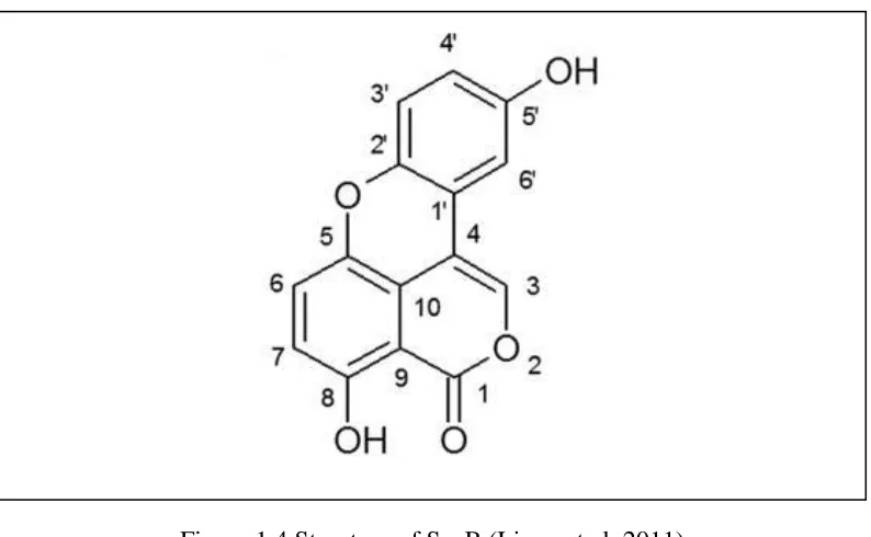
Figure 1.4 Structure of SsnB ... 9
Figure 2.1 Microtubules in HUVECs. ... 17
Figure 2.2 Representative example of SsnB effect on cytoskeletal organization of endothelial cells. ... 18
Figure 2.3 SsnB effects on HUVEC morphology ... 19
Figure 2.4 SsnB effect on HUVEC shape. ... 19
Figure 2.5 Focal adhesions in HUVECs ... 20
Figure 2.6 SsnB promotes focal adhesion formation ... 21
Figure 3.1 SsnB effect on c-Myc protein and mRNA expression ... 27
ix
LIST OF ABBREVIATIONS
AP-1 ... Activator Protein-1
CAM ... Chorioallantoic Membrane
CCNB1 ... Cyclin B1
CCNE2 ... Cyclin E2
CDC2 ... Cell Dependent Kinase 1
CDC6 ... Cell Division Cycle 6
CRP...C-Reactive Protein
CT...Computed Tomography
DMSO ... Dimethyl Sulfoxide
FBS ...Fetal Bovine Serum
Figf ... c-Fos-induced growth factor
GAPDH ... Glyceraldehyde-3-Phosphate Dehydrogenase
HASMCs ... Human Aortic Smooth Muscle Cells
HUVEC ... Human Umbilical Vein Endothelial Cells
LDL ...Low Density Lipoprotein
IL... Interleukin
LPS...Lipopolysacharide
x
NF-B...Nuclear Factor B
NMR...Nuclear Magnetic Resonance
ROS ...Reactive Oxygen Species
SsnB ... Sparstolonin B
THP-1 ... Human Monocytic Leukemia Cell line
TIR...Toll/Interleukin-1 Receptor
TLR ... Toll-like Receptor
TSP-1... Thrombospondin-1
VC ... Vehicle Control
VEGF ...Vascular Endothelial Growth Factor
VEGF-D ... Vascular Endothelial Growth Factor D
VEGFR...Vascular Endothelial Growth Factor Receptor
1 CHAPTER 1
INTRODUCTION
1.1 Atherosclerosis
Atherosclerotic cardiovascular disease is the first leading cause of morbidity and
mortality in the United States, where cardiovascular risk assessment accounts for <50%
of the variability in risk. Approximately 85% of middle-aged susceptible subjects have
significant coronary atherosclerosis, which remains silent for decades until it finally
presents with plaque rupture and thrombosis. In fact, myocardial infarction (MI) occurs in
62% of men and 42% of women with coronary atherosclerosis regardless of race or
ethnicity (Weintraub et al. 2008). Many risk factors have been determined to be
associated with progressive atherosclerotic disease, including hypertension, obesity,
diabetes, smoking, and hyperlipidemia. Hyperlipidemia is defined as an elevation of
low-density lipoprotein (LDL) cholesterol level, which is directly related to plaque formation
(Victor et al. 2009).
In the normal physiological state, many of the large human arteries possess a
microvasculature in their adventitial layer called the vasa vasorum. Large blood vessels
are composed of three layers: tunica media, tunica intima, and tunica adventia. The tunica
intima is the inner layer of the blood vessel wall and consists of endothelium. The middle
layer of the vessel wall, tunica media, contains circularly arranged smooth muscle cells.
2
fibers, lymphatic vessels, and microvasculature. Normal vasa vasorum originate from
coronary artery branch points at regular intervals and run longitudinally along the vessel
wall as is illustrated in (Figure 1.1).
Detection of early development of atherosclerosis can be accomplished using a
variety of screening and diagnostic tests such as electrocardiogram, stress test, CT scan or
arteriography. Moreover, biomarkers levels within the plasma can be helpful to diagnose
atherosclerosis; for example elevated plasma concentrations of both fibrinogen and
C-reactive protein (CRP), which are acute phase proteins released in response to
inflammation (Kaperonis et al. 2006).
3
1.2 Plaque Formation
Atherosclerosis mainly affects wall thickness of medium and large arterial blood
vessels. Endothelial dysfunction has been thought to have a significant role in
pathogenesis of atherosclerosis. Also, overproduction of reactive oxygen species (ROS)
has been determined to be related to the development of atherosclerosis (Davignon and
Ganz 2004).
Endothelial damage or dysfunction will increase the permeability of the
endothelium, known as endothelial activation. The infiltration of LDL into the inner wall
of blood vessels, the arterial intima, will increase due to impaired endothelial function.
Over time, substances such as LDL, cholesterol and fatty materials will accumulate at
damaged areas as shown schematically in (Figure 1.2). LDL will be oxidized by reactive
oxygen species to form oxidized-LDL (ox-LDL). Then, ECs from the damaged sites will
initiate immune responses and release chemoattractants to attract monocytes from the
blood and induce an inflammatory reaction. Monocytes will differentiate into
macrophages in response to oxidized-LDL stimulation. The first immune response will
trigger a further cycle of immune responses. Macrophages will continue to take up and
digest the modified cholesterol molecules. Macrophages are not able to clear the
oxidized-LDL from the intima, although these white blood cells are intended to play a
protective role. This process ends with formation of foam cells and eventual generation of
fatty streaks, which are found in human aorta during the first decade of life (Lusis 2000).
Vascular smooth muscle cells (VSMC) begin to proliferate and migrate from the
media to the intima. The initial lesion progresses into an advanced plaque with a fibrous
4
lesion and the lumen of the blood vessel. The media and neo-intima will be subjected to a
restriction of oxygen supply and nutrients, and the hypoxic environment may lead to
expression of Vascular Endothelial Growth Factor (VEGF) and other angiogenic
regulators that stimulate angiogenesis, thereby promoting plaque growth. Various
research studies have identified intra-plaque hemorrhage as a critical factor in
atherosclerotic plaque growth and destabilization (Sluimer etal.2009; Khurana et al.
2005; Di Stefano et al. 2009; Virmani et al. 2005; Seeley et al. 2007).
The composition of a plaque largely determines the risk of rupture for fibrous
caps, which may cause an acute ischemic event, for example a myocardial infarction or
stroke (Owens et al. 2004). Thrombus formation is a significant clinical complication of
5
formation of microvessels (angiogenesis) contributes to the development of plaques and
increases the risk of plaque rupture (Sluimer et al. 2009).
1.3 Endothelial Cell Function
Endothelial cells play a wide variety of critical roles in controlling vascular
function. It has been proposed that hemangioblasts, as common progenitors of
endothelial, hematopoietic cells, and angioblasts, differentiate from the mesoderm in the
embryo and VEGF receptor is expressed early in these progenitors (Hirashima 2009).
VEGF is the most critical driver of vascular formation, as it is required to initiate the
formation of immature vessels by vasculogenesis or angiogenic sprouting. Endothelial
cells interact with circulating cells and cells present in the vascular wall including smooth
muscle cells, during homeostasis. Being at the interface between blood and tissue,
endothelial cells are most susceptible to changes in blood composition and in blood flow.
Endothelial dysfunction could dispose specific areas of the vasculature to coagulation,
inflammation and vasoconstriction. Endothelial dysfunction is certainly a key initiating
event in several pathological conditions, including atherosclerosis (Michiels 2003).
VEGF plays a major role in all aspects of vascular development, including
endothelial cell proliferation, migration, and survival. Endothelial cells are the main
regulators of vascular development. The biological effects of VEGF are mediated by two
receptor tyrosine kinases (RTKs), VEGFR-1 and VEGFR-2. It has been determined that
VEGF is a survival factor for endothelial cells, both in vitro and in vivo. In vitro,VEGF
prevents apoptosis induced by serum starvation where this activity is mediated by the
phosphatidylinositol (PI)-3 kinase–Akt pathway. VEGF also induces expression of the
6
of VEGF are developmentally regulated. It has also been shown that inhibition of VEGF
results in extensive apoptotic changes in the vasculature of neonatal but not adult mice
(Ferrara,Gerber and LeCouter 2003).
1.4 Physiological and Pathological Angiogenesis
Angiogenesis is the process of new blood vessel development from pre-existing
vasculature. This process involves endothelial cell proliferation, degradation of the
basement membrane and surrounding extracellular matrix, cell migration, and
tubulogenesis. Angiogenesis consists of four stages: initiation, progression,
differentiation, and maturation. It is controlled by a complex regulatory system composed
of pro-angiogenic and anti-angiogenic factors, among which the VEGF family plays a
key role through its pro-angiogenic activity, in addition to other factors and enzymes
(Hoeben et al. 2004; Bussolino et al. 1997; Carmeliet et al. 2003). De novo blood vessel
formation, or vasculogenesis, is an important process for the survival of living organisms
in the embryonic state. In the adult state, physiological angiogenesis plays an essential
role in wound healing, growth of the uterine lining, and the female reproductive cycle.
However, excessive, insufficient or abnormal angiogenesis contributes to the
pathogenesis of many disorders such as cardiovascular diseases, cancer, arthritis,
psoriasis, retinopathy, and inflammation (Khurana et al. 2005; Carmeliet et al. 2003;
7
1.5 Anti-angiogenic Therapy and Sparstolonin B Effect
Anti-angiogenic therapy has been recognized as a promising approach recently
particularly in cancer treatment. This approach has advantages over conventional
anticancer therapy because the therapy does not directly target the tumor but inhibits the
development of blood vessels by targeting the endothelial cells. Thus, anti-angiogenic
therapy is indirectly cytotoxic to the tumor cells (Hoeben et al. 2004; Somani et al. 2013).
This new approach is still questionable as a treatment for atherosclerotic disease because
angiogenesis in atherosclerosis is more complex and depends on disease severity
(Khurana et al. 2005); however, some previous work has demonstrated effective
treatment procedures to inhibit neovascularization in atherosclerotic plaques that will
8
Most chemotherapeutic drugs are considered have some anti-angiogenic activity.
This activity was demonstrated at a lower dose of chemotherapeutic drugs, including
cyclophosphamide, paclitaxel, doxorubicin, and vincristine, than would be required to
target tumor cells. In addition, many anti-angiogenic agents showed a successful
suppression of angiogenesis especially within tumors such as anti-VEGF monoclonal
antibody and tyrosine kinase inhibitors. Moreover, EGF receptor inhibitors have been
shown to suppress VEGF expression through blocking the VEGF-R2 receptor signaling
(Scappaticci 2002). Angiogenesis gene therapy is another promising approach that is
directed to tumor endothelial cells and their microenvironment. The use of gene therapy
that delivers anti-angiogenesis genes has shown promise in preclinical models in mice.
The expression of EC specific cell surface molecules like vascular endothelial growth
factor receptor (VEGFR), E selectin or angiogenic growth factors (VEGF, FGF, PDGF)
produced by tumor cells can be inhibited by specific antibodies, antisense RNA, or gene
specific siRNA. The targeted gene therapy can be achieved by using either viral vectors
or nanoparticles to target anti-angiogenic genes to tumor vasculature (Tandle, Blazer, and
Libutti 2002).
The Sparganium stoloniferum plant has been used traditionally in Chinese
medicine for the treatment of cancer. Extracts and other isolated chemical compounds
from this herb, including a sucrose ester, a phenylpropanoid glycerol, carboxylic acid
esters, and a phenylpropanoid glycoside, possess potent anti-cancer effects. It is already
known that anti-tumor agents often show the ability to inhibit angiogenesis, and
9
activity. Also, SsnB has been identified as a compound that possesses anti-angiogenic
activity in addition to its anti-inflammatory properties (Bateman et al. 2013).
SsnB is a newly described polyphenolic compound isolated from the tubers of
Sparganium stoloniferum. It has been identified as a compound containing the core
structures of both xanthone and isocoumarin using NMR spectroscopy and X-ray
crystallography (Liang et al. 2011) as shown in (Figure 1.5.1). The naturally occurring
xanthones and isocoumarins possess anti-inflammatory, antioxidant, anti-atherosclerotic,
and antitumor activities (El-Seedi et al. 2010; Fylaktakidou et al. 2004). Sparganium
stoloniferum has been used historically in the Chinese herbal medicine “SanLeng” as an
herbal remedy for the treatment of several inflammatory diseases.
Cytotoxicity measurements of SsnB show that the compound is safe to use in
10
macrophages, human monocytic THP-1 cells, HUVECs, and human aortic smooth
muscle cells (HASMCs).
Recently, SsnB has been successfully synthesized based upon its well- known
xanthone core structure through development of a practical method consisting of
sequential chemical reactions to yield the final SsnB product. It has been shown that the
synthetic SsnB shares identical structure with the plant-derived SsnB using HPLC and
NMR analysis. The final product has purity greater than 99%, which has been achieved
using HPLC and mass spectrometry techniques to maintain reproducibility and to control
the purity (Hu et al. 2012).
1.6 Published Research Studies
In previous research, it has been demonstrated that SsnB is a selective Toll-like
receptor antagonist (TLR2 and TLR4); it blocks TLR2- and TLR4-triggered
inflammatory signaling in macrophages by inhibiting the recruitment of MyD88 to TIR
domains of TLR2 and TLR4 but does not block signaling in response to TLR3 and TLR9
ligands (Liang et al. 2011). Toll-like receptors are expressed by macrophages, dendritic
cells, and many other cell types. TLRs are known for their activity as the first line of
defense against invading pathogens such as bacteria and viruses (Kumar, Kawai and
Akira 2011). The therapeutic consequences of blockage of excessive TLR signaling are
being demonstrated for many inflammatory diseases including inflammatory
cardiovascular diseases. In addition, it has been shown that SsnB suppressed multiple
signaling pathways downstream of TLR2 and TLR4 activation including MAPK and NF
11
According to a previously published paper, it has been demonstrated that SsnB
possesses anti-inflammatory effects on vascular endothelial cells. SsnB showed a
suppressive effect on lipopolysaccharide (LPS)-induced expression of interleukin (IL)-1b
and monocyte chemoattractant protein 1 at the transcriptional and translational levels in
HUVECs (Liang et al. 2013). LPS is considered the major portion of the outer membrane
of Gram-negative bacteria, which is an important pathogenic stimulus, in addition to
being a ligand for Toll-like receptor (TLR4) (Wiedermann et al. 1999). Moreover, LPS in
the blood directly promotes vascular inflammation via the activation of resident cells
such as monocytes and endothelial cells (Roth et al.).
Published data from a previous study suggests that SsnB targets endothelial cells
and inhibits angiogenesis by interfering with crucial steps in the process (Bateman et al.
2013). The data shows that SsnB is an inhibitor of endothelial cell morphogenesis and
cell migration. In addition, the results suggest that SsnB is able to inhibit cell
proliferation through arresting the cell cycle in the G0/G1 phase. Microarray data showed
differential expression of important genes involved in angiogenesis in response to SsnB
treatment, including genes in pathways associated with the cytoskeleton, cell
proliferation, and cell cycle. Using the chick chorioallantoic membrane (CAM) assay, an
ex vivo angiogenesis assay, it was demonstrated that SsnB causes an inhibition of blood
vessel formation in the chick embryos, where the vessels in SsnB-treated embryos are
shorter in length and show reduced branch formation compared to control groups
12
1.7 Specific Aims
We hypothesize that SsnB will exert its anti-angiogenic effects by changing the
cell cytoskeleton, enhancing focal adhesion formation, and interfering with early
response gene up-regulation.
Specific Aim 1
To test the hypothesis that SsnB influences endothelial cell morphology and
cytoskeletal organization. For this purpose, we will use immunofluorescence analysis of
actin filaments and microtubules.
Specific Aim 2
To test the hypothesis that SsnB promotes focal adhesion formation in HUVECs.
Immunofluorescence analysis for focal adhesions will be used to investigate the
anti-angiogenic effect of SsnB.
Specific Aim 3
To test the hypothesis that SsnB interferes with early response gene up-regulation.
Immunoblot analysis and real time qRT-PCR will be used to examine the changes in
protein and mRNA expression of c-Myc, c-Fos, and c-Jun due to the effect of SsnB
13
CHAPTER 2
THE EFFECTS OF SPARSTOLONIN B ON CYTOCKELETON
ARRANGEMENT AND MORPHOLOGY OF ENDOTHELIAL CELLS
2.1 Introduction
Since inhibition of angiogenesis is associated with changes in cell motility and cell
proliferation, we investigated in this work the effect of SsnB on the cytoskeleton
including actin filaments, microtubules, and focal adhesions. The actin cytoskeleton has a
vital role in various cellular processes such as migration and cytokinesis. In addition,
abnormalities in actin dynamics are associated with many pathological disorders such as
cancer. Actin filaments form stress fibers that contribute to cell migration; however, other
studies observed that stress fibers are more prominent in stationary cells suggesting that
stress fibers may inhibit cell migration under specific conditions (Tojkander et al 2012;
Burridge 1981). Microtubules are structural components of the mitotic spindle, and
microtubule dynamics play an important role in several cellular processes including
intracellular trafficking, cell migration and cell division. Inhibition of microtubules
results in blocking of cell cycle progression in mitosis, whereas prolonged mitotic arrest
triggers various apoptotic pathways (Rai et al. 2012). In addition, focal adhesions,
dynamic protein complexes that connect the cytoskeleton of a cell to the extracellular
matrix, play an essential role in vital biological processes including cell motility, cell
14
cell migration and to increase attachment of cells to their substrate (Petit and Thiery
2000).
2.2 Materials and Methods
Compounds—Sparstolonin B (SsnB) was purified from the plant Sparganium
stoloniferum and its structure identified with isocoumarin core using NMR spectroscopy
and X-ray crystallography (Liang et al. 2011). Recently, the compound has been
synthesized and tested for efficacy (Hu et al. 2012). For the in vitro experiments, SsnB
was dissolved in DMSO (74.5 mM and 100 mM stock solutions) and freshly diluted in
HUVEC medium before use.
Cells—Human umbilical vein endothelial cells (HUVECs) were obtained from
Lonza (Hopkinton, MA) and cultured on petri plates (100 X 20 mm) coated with 0.1%
gelatin. HUVECs were cultured in endothelial cell medium supplemented with 10% fetal
bovine serum (FBS) and endothelial cell mitogen / growth supplement (Biomedical
Technologies, Stoughton, MA). The endothelial cell medium was replaced every 2-3
days, and the cells were passaged after complete confluence was reached. Cells were
used between third and fifth passage.
Immunofluorescence analysis of the cytoskeleton—HUVECs were trypsinized
from confluent plates and then grown overnight on 0.1% gelatin-coated multi-well
chamber slides with initial seeding density of 5,000 cells per well. Next, various
concentrations of SsnB (1, 10, and 100 µM) or vehicle control (0.1% DMSO) were added
to the cells in assigned wells. After 6 hours of treatment, cells were fixed with 2%
formaldehyde in, cytoskeleton buffer with sucrose (CBS) for 20 minutes and
15
incubated at room temperature for 30 minutes with rabbit anti- /-Tubulin antibody
(Cell Signaling Technologies), 1:50. After washes, cells were incubated with 1:100
Rhodamine Red X (RRX)-conjugated donkey anti-rabbit antibody (Jackson
ImmunoResearch Laboratories, West Grove, PA) together with the F-actin- binding
Alexa Fluor® 488 phalloidin (Invitrogen/ Life Technologies). Coverslips were then
mounted with Dako fluorescent mounting medium. Samples were analyzed on a laser
scanning confocal microscope (Zeiss LSM 510 META) in the Instrumentation Resource
Facility (IRF) at the USC School of Medicine. Cells were observed with a 40X objective
lens, using a FITC filter to visualize F-actin and a CY3 filter for tubulin. Results were
observed in four separate experiments.
Immunofluorescence analysis of focal adhesions—HUVECs were trypsinized
from confluent plates and then grown overnight on 0.1% gelatin-coated multi-well
chamber slides with initial seeding of 5,000 cells per well. SsnB treatment of various
concentrations (1, 10, and 100 µM) or vehicle control (0.1% DMSO) was added to the
cells in assigned wells. After 6 hours of SsnB treatment, the cells were fixed with 4%
formaldehyde/CBS for 20 minutes and permeabilized with PBS- 0.5% Triton X-100 for
10 minutes. After several washings, cells were incubated with mouse anti-vinculin
antibody (hVIN-1; Novus Biologicals), 1:400, at room temperature for 30 minutes to
stain focal adhesions. Then, cells were incubated with 1:100 RRX-conjugated donkey
anti-mouse IgG antibody (Jackson ImmunoResearch Laboratories, West Grove, PA) in
addition to actin staining as described previously. The slide was then cover slipped using
Dako fluorescent mounting medium, and the cells were observed on a laser scanning
16
filter for F-actin and the CY3 filter for vinculin. Image-Pro Plus software was used to
analyze the images. First, the brightness and contrast of images were uniformly adjusted.
Images were thresholded by histogram-based segmentation to identify focal adhesion
pixels, the bright objects. The size of objects counted as focal adhesions was between 20
and 250 pixels in area, and the total area of focal adhesions was quantified in
approximately 100 cells for each treatment. Also, the same software was used to quantify
cell area and cell perimeter.The threshold for images was identified to separate cell body
and background region. After thresholding the image, the regions less than 10 pixels in
area and perimeter are discarded. The brightness and contrast were maintained constant
for all images for each measurement (focal adhesion area or cell perimeter/cell area).
Results are representative of those obtained in three separate experiments.
Statistical Analysis—Data were represented as mean ± SD for each group.
Comparisons among mean values of the treatment groups and control group were done
using one-way and two-way ANOVA. Holm-Sidak test was used for comparisons
between groups for the data that showed significance by ANOVA (p<0.05).
2.3 Results
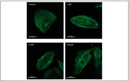
SsnB influences endothelial cell morphology and cytoskeleton organization—
SsnB has been shown to interfere with endothelial cell migration and proliferation
(Bateman et al 2013). Since cytoskeleton is directly related to migration and proliferation,
we investigated the effect of SsnB on endothelial cell cytoskeleton. In addition, since
changes in cell shape are often associated with changes in motility, the effects of SsnB on
endothelial cell morphology were investigated. We also attempted to examine
17
[Figure 2.1]. Both control cells and SsnB treated cells showed disconnected microtubules
(B) whereas cells used for / tubulin antibody titration showed well organized
microtubules (A). SsnB-treated cells had more actin stress fibers across the cell body than
control cells, and this effect was more intense at 100M concentration as can be observed
qualitatively in [Figure 2.2]. Also, SsnB treatment resulted in a significant dose
dependent increase in cell perimeter/cell area at concentrations 1, 10, 100 M (p < 0.05
ANOVA, Sidak’s test) [Figure 2.3].
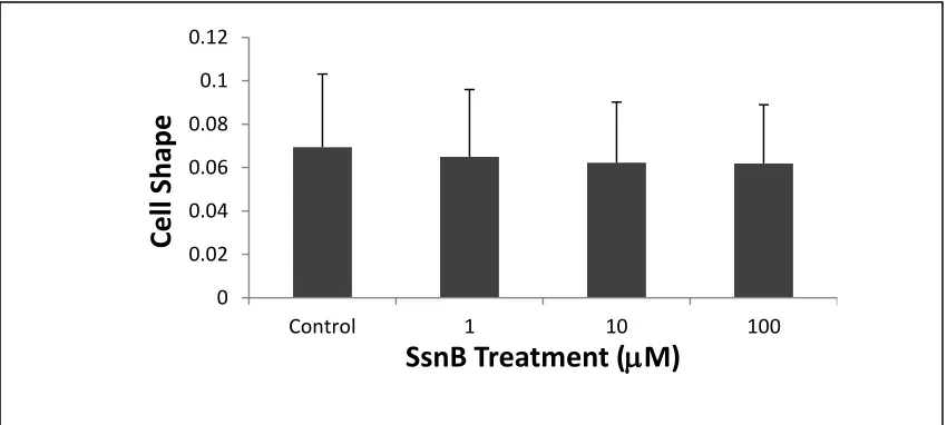
In addition, we investigated cell shape and found that SsnB-treated cells tended to
be more irregular in shape at high concentrations than control cells [Figure 2.4]. Cell
shape was defined using the equation (4 x cell area/ (cell perimeter) 2), where 1 indicates
A B
18
a perfect circle and smaller values indicate a more irregular shape. Our data suggest that
SsnB may affect endothelial cell migration by altering cytoskeletal organization.
Figure 2.2 Representative example of SsnB effect on cytoskeletal organization of endothelial cells. HUVECs adherent to gelatin were exposed to 1 M, 10 M, and 100
19
** ** ***
Figure 2.3 SsnB effect on HUVEC morphology. Treated cells exhibit a significant increase in cell perimeter to area ratio compared to control. **p<0.05 vs. vehicle control, Sidak’s test. ***p<0.001 vs. vehicle control, Sidak’s test. n = 100 cells per group, error bars correspond to +1 SD.
0 0.02 0.04 0.06 0.08 0.1 0.12
Control 1 10 100
Cel
l Shape
SsnB Treatment (
M)
20
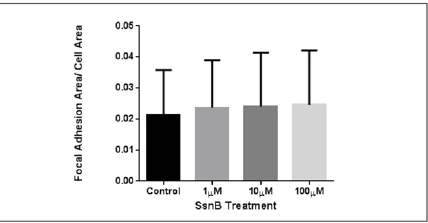
SsnB promotes focal adhesion formation—We investigated the effect of SsnB on
focal adhesions due to their important role in cell migration. As presented in [Figure 2.5],
endothelial cells show staining for focal adhesions in addition to actin filaments. Figure
2.6 shows representative results from focal adhesion staining of HUVECs. Cells treated
with SsnB showed an enhanced formation of focal adhesions in a dose-dependent manner
at concentrations of 1, 10, and 100M. The results are statistically significant (p<0.05
ANOVA) even though they show high variability. These data suggest that SsnB
treatment may increase attachment of endothelial cells to their substrate by increased
formation of focal adhesions, consistent with previously published observations of
impaired cell migration in SsnB treated HUVECs after 6 hours of treatment (Bateman et
al. 2013).
21
22
CHAPTER 3
EFFECTS OF SPARSTOLONIN B ON THE BIOCHEMICAL
COMPONENTS OF ENDOTHELIAL CELLS
3.1 Introduction
In an effort to determine how SsnB inhibits angiogenesis at the molecular level,
we have investigated the expression of immediate early response genes such as Fos,
c-Jun, and c-Myc. Early response genes are a set of genes that are induced in response to
both cell-extrinsic and cell-intrinsic signals and do not require de novo protein synthesis
for their expression (Fowler et al. 2011). The AP-1 protein family, including Fos and
c-Jun, is associated with cell proliferation through their ability to regulate the expression
and function of cell cycle regulators. The expression of c-Myc is significant for
endothelial cell proliferation, migration, and therefore angiogenesis. Previously published
cancer studies demonstrate an important rule of c-Myc and c-Jun in tumor angiogenesis;
these oncogenes possess the ability to suppress the expression of anti-angiogenic factor
thrombospondin-1 (TSP-1) to provoke angiogenesis in progressed tumors. These genes
contribute effectively to angiogenesis in various tumor types, and the same concept can
be extended to angiogenesis in plaques (Vleugel et al. 2006; Baudino et al. 2002;
23
3.2 Materials and Methods
Compounds—Sparstolonin B (SsnB) was purified from the plant Sparganium
stoloniferum as described in a previously published paper (Liang et al. 2011). Recently,
the compound has been synthesized and tested for efficacy (Hu et al. 2012). For the in
vitro experiments, SsnB was dissolved in DMSO (74.5 mM and 100 mM stock solutions)
and freshly diluted in HUVEC medium before use.
Cells—Human umbilical vein endothelial cells (HUVECs) were obtained from
Lonza (Hopkinton, MA) and cultured on petri plates (100 X 20 mm) coated with 0.1%
gelatin. HUVECs were cultured in endothelial cell medium supplemented with 10% fetal
bovine serum (FBS) and endothelial cell mitogen / growth supplement (Biomedical
Technologies, Stoughton, MA). The endothelial cell medium was replaced every 2-3
days, and the cells were passaged after complete confluence was reached. Cells were
used between third and fifth passage.
Immunoblot analysis— HUVECs were cultured in 100 X 20 mm polystyrene
culture plates coated with 0.1% gelatin to approximately 80% confluence. Cells were
treated with 100M SsnB for different incubation times (1 hr, 2 hrs, and 3 hrs), and
control cells were incubated with0.1% DMSO vehicle for the same time intervals. After
lysing cells of each group with 200 l Berk’s lysis buffer (BLB), total protein amount
was measured using a DC (detergent compatible) protein assay (Bio-Rad Laboratories) in
which bovine serum albumin (BSA) was used as the protein standard. Thirty micrograms
of total protein from each sample were mixed with loading dye buffer, boiled, and
electrophoretically separated on 10% SDS polyacrylamide gels. Proteins were
24
nonfat milk in Tris-buffered saline containing 0.05% Tween (TTBS) and incubated
overnight with primary rabbit antibodies against c-Myc, c-Fos (Cell Signaling
Technologies) or c-Jun (Santa Cruz Biotech), at 1:1000 dilutions in 1% milk-TTBS. After
washings, membranes were incubated in 1:3000 dilution of horse-radish peroxidase
(HRP)-conjugated goat anti-rabbit antibody (Bio-Rad) in 1% milk-TTBS.
Immunoreactive band specificity was determined by enhanced chemiluminescence
detection (Pierce). After initial probing, membranes were stripped with a buffer
containing 0.6M Tris-Cl, 2% SDS, 0.7% -mercaptoethanol at room temperature for 30
min, and then reprobed with (1:1000) -actin antibody (MediMabs, Canada) as a loading
control. Results are representative of three separate experiments.
Densitometric measurements of Western blots—Image Pro Plus 7 software was
used to measure the density of the imaged bands. The brightness and contrast of the blot
image were adjusted to separate the bands, appearing as bright objects, from the
background. The average intensity of each band was measured. The experimental bands
were normalized to -actin bands, and then the ratio between the normalized value of
each band and the vehicle control (at 0 hour) was obtained. The ratio represents a
quantitative measurement of c-Myc protein expression.
Real Time- PCR— HUVECs were chosen for real time PCR from 90% confluent
plates (100 X 20 mm polystyrene, tissue culture-treated petri plates coated with 0.1%
gelatin). Cells were trypsinized and seeded (400,000 cells per well) overnight in 6 well
polystyrene culture plates coated with 0.1% gelatin. Half of the wells received HUVEC
medium containing 100 M SsnB, and the remaining plates received vehicle control
25
intervals (1 hr, 2 hrs, and 3 hrs) to allow SsnB to have an effect on the expression of early
response genes. Following incubation, the cells were lysed using Trizol reagent (Life
Technologies) according to the manufacturer’s instructions. Total RNA was extracted
using chloroform, and then samples were centrifuged (13,000 rpm) for 15 min at 4C.
Approximately 350 l of aqueous layer was collected, and isopropanol was added to
precipitate the RNA followed by centrifugation for 10 min under the same conditions.
The RNA pellet was washed by addition of 1 ml of 75% ethanol and centrifuged at
13,000 rpm for 5 min. The pellet was re-suspended in 30 l nuclease free water. The
concentration of RNA was determined spectrophotometrically at 260 nm and the purity
of RNA was assessed by measuring the 260 nm/ 280 nm ratio. First strand cDNA was
prepared from the RNA with reverse transcriptase (iScript cDNA synthesis kit, BioRad).
The RNA was amplified using c-Myc, c-Fos, or c-Jun primers (Qiagen) and iQ SYBR
Green super mix (BioRad). One-step RT-PCR reactions were completed on the BioRad
CFX Connect thermal cycler system in the Instrumentation Resource Facility at the USC
School of Medicine. The expression levels were normalized to the housekeeping gene
GAPDH, and RNA levels were quantified and compared between groups with the
relative Pfaffl method for c-Myc (Pfaffl 2001). In addition, we used the CT method for
c-Fos and c-Jun to compare RNA levels between groups. Representative results from
three separate experiments are shown.
Statistical Analysis—Data were represented as mean +SD for each group.
Comparisons among mean values of the treatment groups and control group were done
using one-way and two-way ANOVA. Holm-Sidak test was used for pairwise
26
3.3 Results
SsnB effects on early response gene expression— Western blot analysis was used
to investigate the effect of SsnB on c-Fos, c-Jun, and c-Myc protein levels. We did not
detect any signal for c-Fos protein, and it was hard to detect c-Jun signal because of the
appearance of several nonspecific bands near the expected position of c-Jun. However,
Western blot analysis showed that treatment with vehicle control (0.1% DMSO) resulted
in increased expression of c-Myc protein which peaked at 2 hours and then diminished.
This effect appeared to be abrogated by treatment with 100 M SsnB; however, the data
did not reach statistical significance. For verification purposes we investigated the effect
of SsnB on c-Myc mRNA levels. qRT- PCR analysis demonstrates a decrease in c-Myc
gene expression after SsnB treatment compared to the control group, which supports the
results obtained from immunoblot analysis (p<0.05 ANOVA, Sidak’s test) [Figure 3.1].
In addition, we investigated the effect of SsnB on c-Fos and c-Jun mRNA level. Our
preliminary data shows that SsnB treated cells reveal a decrease in expression of both
genes compared to the control group [Figure 3.2]. This reduction effect was significant
for c-Fos (p<0.05 ANOVA, Sidak’s test), but it was insignificant for c-Jun. The data
suggests that SsnB treatment may interfere with or inhibit up-regulation of c-Myc, c-Fos
and c-Jun. This effect may relate to the inhibition of cell cycle progression by SsnB
27
0 h r 1h r 2h rs 3h rs 0 .0 0 .5 1 .0 1 .5 2 .0 2 .5
T im e
R e la t iv e D e n s it y o f c -M y c / A c t in
C o n tro l T re a te d
1h r 2h rs 3h rs 0 .0 0 .5 1 .0 1 .5 2 .0
T im e
c -M y c / G A P D H m R N
A C o n tr o l
T re a tm e n t
*
A
B
Figure 3.1 SsnB effect on c-Myc expression. (A) Western blot analysis and quantitative densitometry of c-Myc protein expression. When data are normalized for protein loading (actin) and compared to control, c-Myc shows an apparent decrease in expression in SsnB-treated cells relative to control, which did not reach statistical significance. (B) Real Time-PCR shows a decrease of c-Myc mRNA expression (normalized to housekeeping gene GAPDH) in response to SsnB treatment (*p<0.05 ANOVA, Sidak’s test). All data represent three observations for each group and are expressed as mean values +SD; all groups were compared to 1hr vehicle control.
A
28
*
29
CHAPTER 4
DISCUSSION
In the present study, we sought to assess the anti-angiogenic effect of SsnB at the
cellular and molecular level of endothelial cells. Our results demonstrate that HUVECs
treated with high concentrations of SsnB (10 and 100 M) show an increase in formation
of actin stress fibers compared to controls. Furthermore, our data shows that focal
adhesion area, which is related to stress fiber formation, increased in cells treated with
high SsnB concentrations. Qualitative analysis of focal adhesion formation was
unreliable because of cell population variability; some cells were bigger in size than
others, so it was hard to conclude a difference in focal adhesion formation between
control and treated groups. Quantitative analysis was used where focal adhesion area per
cell was determined and statistically represented as shown previously in [Figure 2.3.6].
These changes affect the morphology of endothelial cells. SsnB-treated cells demonstrate
larger perimeter/cell area ratio than control cells, and become irregular in shape. Taken
together, the results suggest that cells treated with SsnB increase their attachment to the
substrate, which may play a role in inhibiting their migration, an essential process in
angiogenesis. Actin stress fibers play an important role in cell migration; however, stress
fibers contribute to cell adhesion as well through formation of stable actin bundles and
focal adhesions (Tojkander et al. 2012). Previous work has shown that the tension and
30
maturation and dynamics (Johnson et al. 2007). These results support the data shown by
Bateman et al. in a previous study that demonstrated inhibition of endothelial cell
migration in a Transwell migration assay in response to SsnB treatment (Bateman et al.
2013).
Microtubules are structural components of the mitotic spindle, and microtubule
dynamics play an important role in several cellular processes including intracellular
trafficking, cell migration and cell division (Müsch 2004). The inhibition of microtubule
assembly dynamics has been shown to be the mode of action for several clinically
successful anticancer drugs including vinblastine and vincristine (Dumontet and Jordan
2010; Singh et al. 2008). SsnB showed the ability to interfere with cell cycle progression
in mitosis through down regulating some cyclins and cyclin-dependent kinases,
regulatory proteins that control cell cycle progression, including CCNE2, CCNB1,
CDC6, and CDC2. In our study, we examined whether SsnB could perturb microtubule
assembly in vitro, but we did not obtain any meaningful results. The microtubules in
stained cells, both control and SsnB treated, were fragmented and disconnected. We tried
several approaches to determine why the anti-tubulin staining was unsuccessful. We tried
to modify the staining procedure by using cold methanol as a fixative instead of
formaldehyde. We also tried to use different concentrations of permeabilization solution,
PBS- 0.5%- 0.1 %Triton X-100. Moreover, we were careful to ensure that the cells were
healthy by using a new passage for each experiment, but all ended with the same
conclusion. However, we will try to determine whether the antibody itself is the source of
31
division, which SsnB interferes with, we are planning to continue to investigate the effect
of SsnB on microtubule assembly in the future.
We observed either reduced upregulation or downregulation of the early response
genes, specifically c-Myc, c-Fos, and c-Jun, in HUVECs after SsnB treatment. The effect
of SsnB on c-Myc was more powerful in this study because it affected both c-Myc
protein and mRNA. Immunoblotting of c-Myc protein revealed a decrease of expressed
protein in response to SsnB treatment relative to vehicle control, and data were confirmed
by qRT-PCR. The c-Myc mRNA level was reduced. This could be due either to a
decrease in transcription or to an increase in its mRNA degradation, which remains to be
determined. c-Myc functions are necessary and sufficient for the entry of most cells into
the DNA synthetic (S) phase of the cell cycle (De Alboran et al. 2001). Studies of c-Myc
in cancer showed that c-Myc is essential for vasculogenesis and angiogenesis during
development and tumor progression. Deletion of c-Myc has a lethal effect on
c-Myc-deficient embryos. c-Myc is also required for the proper expression of angiogenic factors
such as VEGF; however, the precise mechanism by which c-Myc controls their
expression is not resolved. Also, it is assumed that VEGF regulation by c-Myc to be
indirect (Baudino et al. 2002).
qRT-PCR analysis demonstrates a reduction of c-Fos and c-Jun mRNA levels
between 1 and 2 hours after SsnB treatment, even though no corresponding protein
expression data were obtained by immunoblotting. c-Fos and c-Jun, members of the AP-1
protein family, have biological functions in controlling cell proliferation, survival and
death. One of the major mechanisms that modulates AP-1 activity is the differential
32
2001). A study revealed new ways to block tumor angiogenesis by targeting Jun/AP-1,
and established a functional, in vitro link between activated c-Jun and angiogenesis in
breast cancer (Vleugel et al. 2006). Another study showed that the nuclear oncogene
c-Fos regulates c-c-Fos-induced growth factor/vascular endothelial growth factor D
(Figf/VEGF-D). This regulation is involved in transformation and in regulation of cell
growth and differentiation of various tissues. Also, it has been demonstrated that
developed tumors in c-Fos deficient mice appear lacking in vascularization (Marconcini
et al. 1999).
In conclusion, our data demonstrates that early effects of SsnB on endothelial
cells include promoting formation of stress fibers and focal adhesions, and reduction of
early response geneexpression. These events may contribute to the anti-angiogenic effect
of SsnB. Future additional studies are required to fully understand the effect of SsnB on
microtubule assembly, and to examine whether SsnB disrupts mitotic spindle and
microtubule dynamics. Also, we would like to examine whether the effect on
cytoskeleton is reversible after removing SsnB treatment in vitro. Moreover, we will
investigate the effect of SsnB on c-Fos and c-Jun gene expression by using the Pfaffl
33
REFERENCES
Bateman H, Liang Q, Fan D, Rodriguez V, Lessner S. Sparstolonin B Inhibits Pro-Angiogenic Functions and Blocks Cell Cycle Progression in Endothelial Cells. Plos ONE. August 2013;8(8):1-9.
Baudino T, McKay C, Pendeville-Samain H, Nilsson J, Maclean K, White E, Cleveland J. c-Myc is essential for vasculogenesis and angiogenesis during development and tumor progression. Genes & Development. October 1, 2002;16(19):2530-2543.
Burridge, K.. Are stress fibres contractile? Nature. 1981:294, 691-692.
Bussolino F, Mantovani A, Persico G. Molecular mechanism of blood vessel formation. Trends in Biochemical Sciences. July 1997;22(7):251.
Carine M. Endothelial Cell Functions. Journal of Cellular Physiology. 196: 430–443, 2003Carmeliet P. Angiogenesis in health and disease. Nature Medicine. June 2003;9(6):653.
Choudhury R, Fuster V, Fayad Z. Molecular, cellular and functional imaging of atherothrombosis. Nature Reviews Drug Discovery. November 2004;3(11):913-925.
Davignon, J. and Ganz P. Role of endothelial dysfunction in atherosclerosis. Circulation, 2004. 109(23): p. 27-32.
De Alboran I, O'Hagan R, Gärtner F, Malynn B, Davidson L, Rickert R, Rajewsky K, DePinho R, Alt F. Analysis of C-MYC function in normal cells via conditional gene-targeted mutation. Immunity. January 2001;14(1):45-55.
Deed R, Rooney P, Kumar P, Norton J, Smith J, Freemont A, Kumar S. Early-response gene signalling is induced by angiogenic oligosaccharides of hyaluronan in endothelial cells. Inhibition by non-angiogenic, high-molecular-weight hyaluronan. International Journal off Cancer. Journal International Du Cancer. April 10, 1997;71(2):251-256.
34
El-Seedi H, El-Barbary M, El-Ghorab D, Bohlin L, Borg-Karlson A, Göransson U, Verpoorte R. Recent insights into the biosynthesis and biological activities of natural xanthones. Current Medicinal Chemistry. 2010;17(9):854-901.
Ferrara N, Gerber H, LeCouter J. The biology of VEGF and its receptors. Nature Medicine. June 2003;9(6):669.
Fowler T, Sen R, Roy A. Regulation of Primary Response Genes. Molecular Cell. November 4, 2011;44(3):348-360.
Fylaktakidou K, Hadjipavlou-Litina D, Litinas K, Nicolaides D. Natural and synthetic coumarin derivatives with anti-inflammatory/ antioxidant activities. Current Pharmaceutical Design. 2004;10(30):3813-3833.
Griffioen A, Molema G. Angiogenesis: potentials for pharmacologic intervention in the treatment of cancer, cardiovascular diseases, and chronic inflammation. Pharmacological Reviews. June 2000;52(2):237-268.
Hirashima M. Regulation of endothelial cell differentiation and arterial specification by VEGF and Notch signaling. Anatomical Science International. September 2009;84(3):95-101.
Hoeben A, Landuyt B, Highley MS, Wildiers H, Va Oosterom AT, and De Bruijn EA. Vascular Endothelial Growth Factor and Angiogenesis. Pharmacol Rev. December 2004:56:549-580.
Hu J, Adogla E, Ju Y, Fan D, Wang Q. Copper-catalyzed ortho-acylation of phenols with aryl aldehydes and its application in one-step preparation of xanthones. Chemical Communications (Cambridge, England). November 25, 2012;48(91):11256-11258.
Johnson C, Tang H, Carag C, Speicher D, Discher D. Forced unfolding of proteins within cells. Science (New York, N.Y.). August 3, 2007;317(5838):663-666.
Kaiser M, Younge B, Björnsson J, Goronzy J, Weyand C. Formation of new vasa vasorum in vasculitis. Production of angiogenic cytokines by multinucleated giant cells. The American Journal of Pathology. September 1999;155(3):765-774.
Kaperonis, E.A.,Liapis CD, Kakisis JD, Dimitroulis D, Papavassiliou VG., Inflammation and atherosclerosis. European Journal of Vascular and Endovascular Surgery, 2006. 31(4): p. 386-393.
35
Kumar H, Kawai T, Akira S. Pathogen Recognition by the Innate Immune System. International Reviews of Immunology. February 2011;30(1):16-34.
Liang Q, Wu Q, Jiang J, Duan J, Wang C, Smith M, Fan D.Characterization of sparstolonin B, a Chinese herb-derived compound, as a selective Toll-like receptor antagonist with potent anti-inflammatory properties. The Journal of Biological Chemistry. July 29, 2011;286(30):26470-26479.
Liang Q, Yu F, Cui X, Duan J, Wu Q, Nagarkatti P, Fan D. Sparstolonin B suppresses lipopolysaccharide-induced inflammation in human umbilical vein endothelial cells. Archives of Pharmacal Research. July 2013;36(7):890-896.
Lusis, A.J., Atherosclerosis. Nature, 2000. 407(6801): p. 233-241.
Marconcini L, Marchio S, Morbidelli L, Cartocci E, Albini A, Ziche M, Oliviero S. c-fos-induced growth factor/vascular endothelial growth factor D induces angiogenesis
in vivo and in vitro. Proceedings Of The National Academy Of Sciences Of The
United States Of America. August 17, 1999;96(17):9671-9676.
Müsch A. Microtubule Organization and Function in Epithelial Cells. Traffic. January 2004;5(1):1-9.
Owens, G.K., M.S. Kumar, and B.R. Wamhoff, Molecular regulation of vascular smooth muscle cell differentiation in development and disease. Physiological Reviews, 2004. 84(3): p.767-801.
Petit V, Thiery J. Focal adhesions: structure and dynamics. Biology of the Cell / Under the Auspices of the European Cell Biology Organization. October 2000;92(7):477-494.
Pfaffl M. A new mathematical model for relative quantification in real-time RT-PCR. Nucleic Acids Research. May 1, 2001;29(9):e45.
Rai A, Surolia A, Panda D, Kellermayer M. An Antitubulin Agent BCFMT Inhibits Proliferation of Cancer Cells and Induces Cell Death by Inhibiting Microtubule Dynamics. Plos ONE. August 2012;7(8):1-12.
Roth, G.A., B. Moser, F. Roth-Walter, M.B. Giacona, E. Harja, P.N. Papapanou, A.M. Schmidt, and E. Lalla. 2007. Infection with a periodontal pathogen increases mononuclear cell adhesion to human aortic endothelial cells. Atherosclerosis 190: 271–281.
36
Seeley R., Stephens T., Tate P. Essentials of Anatomy and Physiology. McGraw-Hill Material. 2007.
Shaulian E, Karin M. AP-1 in cell proliferation and survival. Oncogene. April 30, 2001;20(19):2390.
Singh P, Rathinasamy K, Mohan R, Panda D. Microtubule assembly dynamics: An attractive target for anticancer drugs. IUBMB Life. June 2008;60(6):368-375.
Sluimer J, Daemen M. Novel concepts in atherogenesis: angiogenesis and hypoxia in atherosclerosis. The Journal of Pathology. May 2009;218(1):7-29.
Somani R, Bhanushali U. Targeting Angiogenesis for Treatment of Human Cancer. Indian Journal of Pharmaceutical Sciences. January 2013;75(1):3-10.
Stefanadis C, Toutouzas K, Stefanadi E, Lazaris A, Patsouris E, Kipshidze N. Inhibition of plaque neovascularization and intimal hyperplasia by specific targeting vascular endothelial growth factor with bevacizumab-eluting stent: An experimental study. Atherosclerosis. December 2007;195(2):269-276.
Tandle A, Blazer III D, Libutti S. Antiangiogenic gene therapy of cancer: recent developments. Journal of Translational Medicine. January 2004;2:22-20.
Tojkander S, Gateva G, Lappalainen P. Actin stress fibers--assembly, dynamics and biological roles. Journal of Cell Science. April 15, 2012;125(Pt 8):1855-1864.
Virmani R, Kolodgie F, Narula J, et al. Atherosclerotic plaque progression and vulnerability to rupture: angiogenesis as a source of intraplaque hemorrhage. Arteriosclerosis, Thrombosis, and Vascular Biology. October 2005;25(10):2054-2061.
Vleugel M, Greijer A, Bos R, van der Wall E, van Diest P. c-Jun activation is associated with proliferation and angiogenesis in invasive breast cancer. Human Pathology. June 2006;37(6):668-674.
Victor, V.M., Rocha M, Solá E, Bañuls C, Garcia-Malpartida K, Hernández-Mijares A., Oxidative Stress, Endothelial Dysfunction and Atherosclerosis.Current Pharmaceutical Design, 2009. 15(26): p. 2988-3002.
37
38
APPENDIX A- SUPPLEMENTARY DATA
HUVEC medium:
446 ml K12 Medium
50 ml FBS
50 mg Heparin
50 mg Endothelial Mitogen
4 ml Non-Essential MEM Medium
Berk’s Lysis Buffer (BLB) pH 7.5:
1.211 g Tris base
8.766 g NaCl
0.186 g KCl
2.099 g NaF
6.48 g -glycerophosphate
0.184 g Na3VO4
5 ml Triton X-100
5 ml Nonidet P-40
Up to 1 L H2O
Cytoskeleton Buffer with sucrose (CBS)
1.07 g MES pH 6.1
5.14 g KCl
0.30 g MgCl
0.37g EGTA