What kind of imaging modality should be chosen when study rabbit liver anatomy: CT or MRI
Kamelia Stamatova-Yovcheva1, Rosen Dimitrov1, Miroslav Stefanov2, 3Ömer Gürkan DİLEK, 4Juliana Toneva
1
Department of Veterinary Anatomy, Histology and Embryology, Faculty of Veterinary Medicine, Trakia University, 6000 Stara Zagora, Bulgaria 2
Department of Morphology, Physiology and Animal Nutrition, Faculty of Agriculture, Trakia University, 6000 Stara Zagora, Bulgaria
3
Department of Anatomy, Örtülü Burdur, Faculty of Veterinary Medicine, University of Mehmet Akif Ersoy, Turkey
4
Department of Medical Physics, Biophysics, Rentgenology and Radiology, Faculty of Medicine, Trakia University, 6000 Stara Zagora, Bulgaria
ABSTRACT
The aim of the present study was to investigate and compare CT and MRI imaging anatomy of the rabbit liver. As anatomical bone landmark, we used Th8. Ten sexually mature; healthy clinically rabbits have been studied. Computed tomographic (CT) and magnetic resonance imaging (MRI) studies were performed in compliance with the standard imaging anatomical protocol when study the rabbit liver. The studied animals were positioned in dorsoventral (abdominal) recumbency. When the aim is to find topographic localization of the liver (its lines, lobes, edges and position relative to adjacent organs), transversal helical CT anatomical study of the cranial abdominal region, at the level of Th8 is a high informative method.The anatomical data for the rabbit liver, obtained by MRI, give real and precision information for its lobes, topography of the big vessels, interlobar visualization of the gall bladder, gall bladder’s parts, beginning of the cystic duct and topography of the investigated organ to close structures which are with similar and higher level of magnetization.
INTRODUCTION
Computed tomography (CT) and magnetic resonance imaging (MRI) are imaging anatomical methods for visualization of soft tissue structures and bones. There is a coincidence between anatomical images produced by CT (Еlis, 1999, Paulus et al., 2001; Prather et al., 2005; Drake et al., 2007; Van Caelenberget al., 2010; Lauridsen et al., 2011; Raes et al., 2011;Shojaei et al., 2012)and MRI (Samii et al., 1999, Ober and Freeman, 2009,Stamatova-Yovcheva et al., 2012), compared to the findings obtained by the classical anatomical methods. Therefore CT and MRI methods are fully applicable to anatomical studies. Anatomical results obtained by CT correspond to the conventional topography of organs in particular small mammals, incl. rabbit, which allows a number of authors to use it as anatomical method (Samii et al., 1998; Zotti et al., 2009; Dimitrov et al., 2010a; Dimitrov et al., 2010b; Dimitrov et al., 2011a; Dimitrov et al., 2011b; Farré et al., 2014).For comparability of the results the same bone markers are used when interpret the data from CT and those of native frozen post-mortem anatomical cuts(Dimitrov et al., 2013).To find CT imaging anatomical features of the rabbit liver Zotti et al. (2009) use as bone markers vertebra from 8th thoracic (Th8) to 3rd lumbar (L3). The most detailed image of the organ is obtained at the level of 9th thoracic vertebra (Th9).
X-ray radiation in CT is significantly less than that of conventional radiography (Novelline et al., 1999, Boyd, 2006, Huda and Vance, 2007). CT has opportunities for reconstruction of images (Henninger et al., 2003, Lenard, 2008) described as retrospective reconstruction (Romans, 1995).
The disadvantage of the CT method is that the changes that occur during the breathing are not take into consideration (Romans, 1995; Seeram, 2009). By MRI method are obtained detailed anatomical data for the liver for one breathing cycle, with high resolution of the image. The breathing process and heart activity do not cause appearance of artefacts, when use MRI method (Secor, 2008).
The obtained results by MRI method give information simultaneously for the longitudinal (T1) and transversal (T2) relaxation due to the two types of magnetization of liver tissue (Penney et al., 2003; Sahani and Kalva,2004; Catalano et al., 2008; Lee et al., 2012).The method is suitable as for the anatomical study of the liver structure in depth, as for the imaging anatomical study of the big veins, lobar veins and branches of portal vein. Detailed anatomical image was obtained for the right hepatic lobe, due to specifics in its structure and venous drainage. The thin left border of the liver and caudate process are sharple distinct to the close soft tissue findings (Champetier et al., 1987, Champetier et al., 1992).
The advantages of MRI occur in the facts that are produced three-dimensional reconstructions of the studied organ structures. In contrast to CT by this method is carried out vascular visualization, without the use of contrast agents and X-ray radiation of the individual (Lee, 2001; Soler et al., 2007; Lauridsen et al., 2011).
MRI and computed tomography (CT) have as advantages as disadvantages. Anyway CT is more applicable than MRI. In contrast toCT method, when use MRI, X-ray radiation of the individual is avoided. Although in MRI method is achieved an impact on the biological tissue, due to the effect of the magnetization, the magnetic field has no proven harmful effects.Disadvantage of MRI is the longer duration of the study (approximately 20 to 40 min)(Formicaand Silvestri, 2004).
The insufficient data in the literature concerning the application and capabilities of CT and MRI asimaging anatomical methods for visualization of the liver in rabbits gave us the reason to do this comparative study. The obtained data would be used when choose correctly imaging anatomical methods for alive anatomical study of the liver in domestic mammals, depending on the goals.As a model we present computer tomographic slice obtained at the level of Th8.
MATERIALS AND METHODS Materials
Ten mature clinically healthy rabbits 12 months old, from the New Zealand white rabbit breed, with weight between 2.8 kg and 3.2 kg were anesthetized (IM) with Ketaminol® 10 solution (Intervet) (Ketamine hydrochloride 100 mg/ml and Benzethonium chloride 0.1 mg/ml) of 0.5 ml/kg(Dimitrov, 2013). The experimental animals were housed at 25°C, with a 12 h dark/light cycle.
All rules were complied for welfare of experimental animals, in accordance to European Convention for the protection of vertebrate animals used for experimental and other scientific purposes (Strasbourg / 16.05.1986), the European Convention for the Protection of companion animals (Strasbourg / 13.11.1987) and the Law on protection of animals in Bulgaria (section IV-animal experiments, Art. 26, 27 and 28, adopted on 24.01.2008 and published in Government Gazette, № 13, 2008).Animals are used with empty digestive tract.All animals fully recovered from the anesthesia after completion of the study.
Methods
CT anatomical protocol: Five clinically healthy rabbits were used. The study was performed by Whole body multi-slice helical computed tomography scanner (Light Speed QX/I GE, Genaral Electric USA). CT study was carried out, considering a standard imaging anatomical protocol for study of the rabbit liver.The studied animals were positioned in dorsoventral (abdominal) recumbency. The abdominal cavity was scanned transversally in the planes from Th8 to L3. The slices’ thickness was 5 mm. The CT soft tissue density was reported informatively for the protocol. The obtained results were exported and imported by DICOM and USB external devices.
was from 3 to 4 mm, FOV 244*244, TE-93 and TR 1410. We worked in high resolution. For one series of study the window (W) varied from 745 to 1360. The center was in diapason between 344 and 611 (Stamatova-Yovcheva et al., 2012).
RESULTS
When scan by CT the cranial abdominal region at the level of Th8, topography and anatomical features of the rabbit liver were visualized in detail. The organ was intrathoracic and was observed as a massive normal attenuated heterogeneous soft tissue finding. There was not visible CT anatomical border between right lobe and left lobes of the liver. The liver borders were sharply distinguished to the close soft tissues, as dorsal border was relatively hypperattenuated to diaphragm contours. TransversalСТ hypperattenuated anatomical image of oesophagus was to the left and dorsally. Caudal vena cava was regular oval soft tissue finding on dorsal liver border. Its density was similar to that of liver parenchyma. Hypperattenuated abdominal aorta was observed close to the body of Th8 (Figure 1).
Figure 1. Transversal helical anatomical CT image of cranial abdominal region. (L-left, D-right) (abdominal recumbency).(1) left azygos vein;(2) abdominal aorta;(3) oesophagus; (4) caudal vena cava; (5)right azygos vein; (6) right lung; (7)left lung; (8)diaphragm; (9) right lobe of liver; (10) left medial lobe of liver; (11) left lateral lobe of liver; (12) abdominal muscles.
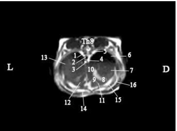
obtained byT2 sequence. Morphological interpretation of the gall bladder itemizedits three parts in alive aspect-fundus, body and neck and the beginning of the cystic duct (Figure 2).
Figure 2. Transversal MRI anatomical presenting of rabbit liver in Т2 sequence. (L-left, D-right) (abdominal recumbency). (1) left azygos vein; (2) abdominal aorta; (3) oesophagus; (4)caudal vena cava; (5) right azygos vein; (6) right lung; (7)right lobe of liver;(8) gall bladder fundus; (9) body of gall bladder; (10)neck of of gall bladder and cystic duct; (11) quadrate lobe; (12)left medial lobe of liver; (13) left lateral lobe of liver; (14) rectus abdominis muscle; (15) transverse abdominal muscle; (16)external and internal oblique muscles.
DISCUSSION
There is discussion about the number of rabbit liver lobes. Some authors (Barone, 1997, McCracken and Kainer, 2008) consider that the liver in rabbit is composed of five lobes and other find that the organ has six lobes (Bensley, 1948; Meredith and Raimond, 2000). There is a debate about topography and anatomical parts of gall bladder (Barone, 1997; Hristov et al., 2006). According to our imaging anatomical investigation rabbit liver is composed of five lobes, with sharply visible quadrate lobe on MRI. The right lobe of liver is not separated and the gall bladder does not reach the ventral liver border. Thus our results correspond to literature data of Barone (1997) and McCracken and Kainer (2008).
the liver, gall bladder, aorta, oesophagus, caudal vena cava, we find in details and in classic topographic aspect the parts of the gall bladder in the rabbit and the beginning of the cystic duct.
We give different protocol for MRI study of the rabbit liver, in comparison to that proposed by Samii et al. (1999)for feline abdominal organs. To obtain objective MRI anatomical data for rabbit liver’s lobes, organ’s topography and closeness, the studied biological object must be positioned in dorsoventral recumbency, and the applied sequence must be Т2. That corresponds to our previous published data (Stamatova-Yovcheva et al., 2012).
CONCLUSIONS
When is necessary to find topography of rabbit liver, its outlines, lobes, borders, position to close organs, transversal helical abnatomical CT study of the cranial abdominal region at the level of Th8 is a highly informative method. The data are with high resolution and content summarized information, obtained for one cycle of breathing.
The anatomical data for rabbit liver, obtained by MRI give accurate information about its lobes, topography of the big vessels, interlobar visualization of the gall bladder, beginning of cystic duct and topography of the studied organ to close structures, which are with similar and higher level of magnetization.
REFERENCES
Barone, R., 1997. Chapitre VII - Foie. In: Barone R, editor. Anatomie comparée des mammifères domestiques. Splanchnologie I. Tome troasième, Troisième edition. Paris: Vigot. p 507-560.
Bensley, B., 1948. Part III. Rabbit Dissection. IV. The Liver. In: Bensley B, editor. Practical Anatomy of the Rabbit: An Elementary Laboratory Textbook in Mammalian Anatomy. Toronto: The University Press. p. 120-121.
Boyd, S., Davison, P., Muller, R. & Gasser, J., 2006. Monitoring individual morphological changes over time in ovariectomized rats by in vivo micro-computed tomography. Bone 39, 854-862.
Catalano, O.,Singh, A., Uppot, R., Hahn, P., Ferrone, C. &Sahani,D., 2008. Vascular and Biliary Variants in the Liver: Implications for Liver Surgery. RadioGraphics, 28, 359 –378.
Champetier, J., Bas, J., Haouari, H. & Chaffanjon, P., 1992. Magnetic resonance imaging of the liver by oblique sections. Surg Radiol Anat, 14 (2), 159-167.
Champetier, J., Bas, J., Yver, R., Létoublon, C. & Hou, K., 1987. Magnetic resonance imaging of the liver by frontal (coronal) sections. Surg Radiol Anat, 9 (2), 107-121.
Dimitrov, R., Toneva, J., Yonkova, P. & Stamatova, K., 2010b. Comparative transversal imaging anatomic study on domestic rabbitbulbourethral glands. Journal of Mountain Agriculture on the Balkans 13, 59-70.
Dimitrov, R., Yonkova, P. & Stamatova, K., 2011a. Agreement between sagittal plane cross-sectional anatomy, sonoanatomy and computed tomography of rabbit prostate and bulbourethral glands. Bulgarian Journal of Veterinary Medicine 14, 11−16.
Dimitrov, R., Toneva, Y., Vladova, D., Stamatova, K. & Stefanov, M., 2011b. Computed Tomographic Imaging of Vesicular Glands in Rabbits. Journal of Animal and Veterinary Advances 10, 55-59.
Dimitrov, R., Stamatova, K. & Kostov, D., 2013. Comparative imaging of the vesicular glands in New Zealand white rabbits (Oryctolagus cuniculus). Turkish Journal of Veterinary and Animal Sciences 37, 97-101).
Dimitrov, R., 2013. Anatomical Imaging Analysis of the Prostate gland in Rabbit (Oryctolagus cuniculus) - Helical computed tomography study. Revue de Médecine Vétérinaire, 164 (5), 245-251.
Drake, R., Vogl, A., Mitchell, A., Tibitts, R. & Richardson, P., 2007. Grays atlas of anatomy. 8th edition. Churchill Livingston: Elsevier Publishers.
Elis, H., 1999. Human sectional anatomy: atlas of body sections, CT and MRI images. 2nd edition. Oxford: Butterworth-Heinemann.
Formica, D. & Silvestri, S.,2004. "Biological effects of exposure to magnetic resonance imaging: an overview". Biomed Eng Online 3: 11. doi:10.1186/1475-925X-3-11. PMC 419710. PMID 15104797.
Henninger, W., Frame, M. & William, M., 2003. CT features of alveolitis and sinusitis in horses. Veterinary Radiology and Ultrasound, 44, 269-276.
Hristov, H., Kostov, D. &Vladova, D., 2006. Topographical anatomy of some abdominal organs in rabbits. Trakia Journal of Sciences 4, 7-10.
Huda, W. & Vance A., 2007. Patient Radiation Doses from Adult and Pediatric CT. AJR 188, 540– 546.
Alonso-Farré, J. M.,Gonzalo-Orden,M., Barreiro-Vázquez, J. D.,Ajenjo, J. M.Barreiro-Lois, A.,Llarena-Reino, M. &Degollada,E., 2013. Cross-Sectional Anatomy, Computed Tomography and Magnetic Resonance Imaging of the Thoracic Region of Common Dolphin ( Delphinus delphis) and Striped Dolphin (Stenella coeruleoalba). Anantomia Histologia Embryologia, 44 (1), 221-229.
Lee, J., Lee, J.M., Kim, S., Kim, C. & Mun, W., 2001. MR Imaging-Histopathologic Correlation of Radiofrequency Thermal Ablation Lesion in a Rabbit Liver Model: Observation during Acute and Chronic Stages. Korean J Radiol, 2, (3), 151-158.
Lee, J., Liau, J., Murphy, P., Schroeder, M., Sirlin, C. & Bydder, M., 2012. Cross-sectional investigation of correlation between hepatic steatosis and IVIM perfusion on MR imaging. Magnetic Resonance Imaging, 30, 572-578.
Lenard, Z., 2008. New Advances In Small Animal Imaging – CT. Small Animal Medicine Chapter. In: ACVSc College Science Week. Murdoch University. p. 70-73.
McCracken, O. & Kainer, R., 2008. The Rabbit. In: McCracken O, Kainer R, editors. Color Atlas of Small Animal Anatomy: The Essentials. Oxford: Blackwell Publishing LTD. p 64-75.
Meredith, A. & Rayment, L., 2000. Liver Disease in Rabbits. Seminaris in Avian and Exotic Pet Medicine, 9, 146-152.
Novelline, R., Rhea, J. & Rao, P., 1999. Helical CT in emergency radiology. Radiology, 213, 321-339.
Ober, C. &Freeman, L., 2009. Computed tomographic magnetic resonance imaging and cross-sectional anatomic features of the manus in cadavers of dogs without forelimb disease. American Journal of Veterinary Research, 70, 1450-1458.
Paulus, M., Gleason, S., Easterly, M. & Foltz, C., 2001. A review of high-resolution X-ray computed tomography and other imaging modalities for small animal research. Lab Animal, 30, 36-45.
Penney, G., Blackall, J., Hamady, M., Sabharwal, T., Adam, A.&Hawkes, D., 2004. Registration of freehand 3D ultrasound and magnetic resonance liver images. Medical Image Analysis 8, 81-91.
Prather, A., Berry, C.& Thrall, D., 2005. Use of radiography in combination with computed tomography for the assessment of noncardiac thoracic disease in the dog and cat. Veterinary Radiology & Ultrasound, 46, 114-121.
Raes, E., Bergman, E., Van der Veen, H., Vanderperren, K., Van der Vekens, E. & Saunders, J., 2011. Comparison of cross-sectional anatomy and computed tomography of the tarsus in horses. Am J Vet Res, 72, 1209-1221.
Romans, L., 1995. Introduction to computed tomography. Philadelphia: Williams & Wilkins.
Sahani, D. &Kalva,S., 2004. Imaging the Liver. The Oncologist, 9, 385-397.
Samii, V., Biller, D. & Koblik, P., 1999. Magnetic resonance imaging of the normal feline abdomen: An anatomic reference. Veterinary Radiology & Ultrasound, 40 (4), 486-490.
Seeram, E., 2009. Single Slice Spiral/Helical Computed Tomography: Physical Principles and Instrumentation. Chapter 11. In: Wilke J, Held L, editors. Computed Tomography: Physical Principles, Clinical Applications, and Quality Control. St. Louis: Saunders ELSEVIER. p. 246-266.
Shojaei, B., Vosough, D. & Sharifi, F., 2012. Computed Tomographic Anatomy of the Thoracic Cavity Vessels in the Rayini Goat. IJVS, 7, 9-22.
Soler, M., Murciano, J., Latorre, R., Belda, E., Rodrıguez, M.J. & Agut, A., 2007. Ultrasonographic, computed tomographic and magnetic resonance imaging anatomy of the normal caninestifle joint. Vet J Journal, 174, 351–361.
Stamatova-Yovcheva, K., Dimitrov, R., Chaprazov, Zv., Russenov A. & Yovchev, D., 2012. Magnetic Resonance Imaging Application in Rabbit Liver Anatomy. Correlation with Cadaver Cross-sectional cuts. Book of Proceedings, Days of Veterinary Medicine, 3rd International Scientific Meeting, 239-242.
Van Caelenberg, A., De Rycke, L., Hermans, K., Verhaert, L., Van Bree, H. & Gielen, I., 2010. Computed tomography and cross-sectional anatomy of the head in healthy rabbits. Am J Vet Res, 71, 293-303.
Ziegler,A., Faber,C., Mueller, S.&Bartolomaeus,Th.,2008. Systematic comparison and reconstruction of sea urchin (Echinoidea) internal anatomy: a novel approach using magnetic resonance imaging. BMC Biology, 6, 33, doi:10.1186/1741-7007-6-33