Molecular docking and molecular dynamics (MD)
simulation of human anti complement factor H (CFH)
antibody Ab42 and CFH polypeptide (pCFH)
Bing Yang1,2†, Jiayi Ren3†, Tong Liu1,2, Shujian Lin1,2, Yueming Wang1,4, Chengming
Li1,4, Youwen He1,4, Wenwen Xu1,5, Weihong Zheng5, Xiaohui Yuan1,5*, Huaxin
Liao1,5*
1. Institute of Biomedicine, Jinan University, Guangzhou, 510632, China;
2. Guangdong Provincial Key Laboratory of Bioengineering Medicine, Guangzhou,
510632, China;
3. Zhuhai College of Jilin University, Zhuhai, 519041, China;
4. National Engineering Research Center of Genetic Medicine, Guangzhou, 510632,
China;
5. Zhuhai Trinomab Biotechnology Co., Ltd. Zhuhai, 519040, China;
†These authors contribute equally to this work.
*Corresponding to Yuan Xiaohui ([email protected]) and
Liao Huaxin ([email protected]).
Abstract
The details of antigen-antibody interactions and the identification of epitopes are
critical for the development of monoclonal antibody drugs. Ab42 is a native
human-derived anti-CFH monoclonal antibody. In this study, the interaction between
antigen pCFH and antibody (Ab42) was theoretically demonstrated by molecular
docking and MD simulation, combined with free energy calculation and
computational alanine scanning (CAS), and key amino acids and epitopes were
identified. Experimental alanine scanning (EAS) was then carried out to verify the
results of the calculation, and our results indicated that Ab42 antibody forms
hydrogen bonds and interacts hydrophobically with pCFH through the Tyr315, Ser100,
Gly33, and Tyr53 residues on its CDR, while the main pCFH epitopes are located at
the six sites of Pro441, Ile442, Asp443, Asn444, Ile447, and Thr448. In conclusion,
this study has explored the mechanism of antigen-antibody interaction from both
theoretical and experimental aspects, and our results have important theoretical
significance for the design and development of relevant antibody drugs.
Keywords: Complement factor H (CFH), Molecular docking, Molecular dynamics
(MD) simulation, Computational alanine scanning (CAS), Experimental Alanine
Introduction
CFH is a soluble glycoprotein expressed by normal epidermal cells with a
molecular weight of 155 kDa[1,2]. The main function of CFH is to protect the host
cells from complement system-mediated attack and destruction through inhibiting the
complement activation pathway, i.e., preventing the deposition of C3b on the surface
of target cells[3,4]. Certain malignant tumor cells escape the body’s tumor clearance through over-expressing CFH, which is one of the important mechanisms of
malignant tumors escaping the immunity[3,5].
The polypeptide domain SCR19 on the CFH protein (sequence:
GPPPPIDNGDITSFP) is an important region for its binding to the membrane of
C3b-expressing cells[2,5]. Previous literature also reported that anti-pCFH antibodies recognize and bind CFH on the surface of tumor cells and thereby facilitate the
activation of complement system, which in turn damages the tumor cells by both
complement-dependent cytotoxicity (CDC) and antibody-dependent
complement-mediated cytotoxicity (ADCC) and ultimately suppresses tumor growth
and preventing tumor metastasis[6,7].
Ab42 is an human-derived high-affinity anti-pCFH monoclonal antibody isolated
previously in our laboratory from the peripheral blood mononuclear cells of a
malignant glioma patient using a single memory B lymphocyte sorting and RT-PCR
method [8]. As a native human antibody, it can avoid the immune rejection of the body
and thus has an important application prospect for the development of anti-tumor
Information on the interaction of antibodies with their targets is critical for the
development of antibody drugs. Molecular docking[9,10] and MD simulation[11,12]
methods provide an advantageous means for studying the interaction between antigen
and antibody. Our previous studies have screened different solvent water models and
force field calculations in the MD simulation system and suggested that
amber99sb_spce is the best candidate model for studying antigen-antibody
interactions[13].
Therefore, the present work applied this amber99sb_spce model to study the
interaction between Ab42 antibody and pCFH by molecular docking, MD simulation,
and CAS[14,15], and successfully identified the binding epitope of Ab42. Finally, the
results of theoretical calculations were further verified by both EAS[1,16] and enzyme-linked immunosorbent assay (ELISA) antigen-antibody binding experiments.
Results and Discussion
The three-dimensional (3D) structure of Ab42
Ab42 was originally an intact antibody containing FC segment. However, in the
calculation part of this work, in order to more accurately describe the interaction
between the antigen and the antibody and simplify the calculation, only the Fab
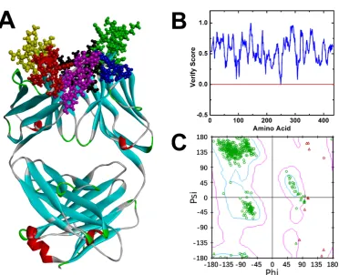
segment of Ab42 antibody was established (Figure 1A). We used the Verify Protein
(Profiles-3D) program under the Homology Modeling module to examine the
compatibility scores of each amino acid residue in its primary sequence on both the
Figure 1B shows that the Verify Score of Ab42 antibody structure is greater than
zero, indicating that the amino acid sequence of Ab42 antibody is compatible with its
corresponding 3D structure. The stereo-chemical accuracy of the model, including the
rationality of structural parameters, such as bond length, bond angle, and dihedral
angle, was evaluated by the Ramachandran plot analysis method, which shows that
most of the atoms are located in the first quadrant, second quadrant, and third
quadrant, demonstrating that the bond lengths, bond angles, and dihedral angles of the
entire molecule are reasonable (Figure 1C). These results are similar to the template
for homology modeling and are also consistent with the Profiles-3D predictions,
suggesting that the structure of Ab42 antibody was precisely optimized by energy
minimization with correct stereo-structure accuracy.
Molecular docking
The ZDOCK program was used to perform the molecular docking between pCFH
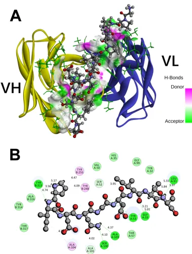
and Ab42 proteins to achieve the highest docking pose (Figure 2). Our results showed
that the entire pCFH molecule spans around the CDR of Ab42 molecule (Figure 2A),
and subsequent analysis further revealed that pCFH interacts strongly with the surface
amino acids of Ab42, mainly by hydrogen bonding and hydrophobic interaction
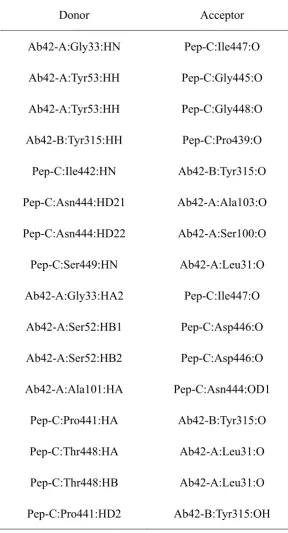
(Figure 2B), which play the major roles in the binding of pCFH to Ab42 antibody. As
shown in Figure 2B, pCFH forms hydrogen bonds with the sites of Tyr315, Ala103,
Ser100, Gly33, Ser52, and Leu31 on the CDR of Ab42 antibody, and interacts
hydrophobically with the sites of Tyr248, Tyr255, and Ala104 too.
Ab42-pCFH complex (Table 2) with an average length of approximately 2.0
angstroms, indicating that they play the most important roles in the binding between
Ab42 and pCFH during the complex formation, followed by the hydrophobic
interactions.
Stability analysis of Ab42-pCFH complex
MD simulation can be used to solve the energy barrier problem that EM
calculation cannot overcome, and the equilibrium trajectory of MD can be used for
the conformation sampling of MM-PBSA combined with free energy calculation. In
this study, the Ab42-pCFH complex was subjected to three MD simulations under the
force field of Amber99sb-spce with each run lasting 100 ns (designated as MD-1,
MD-2, and MD-3, respectively), and the root mean square deviations (RMSDs) were
analyzed subsequently to evaluate the balance of trajectory.
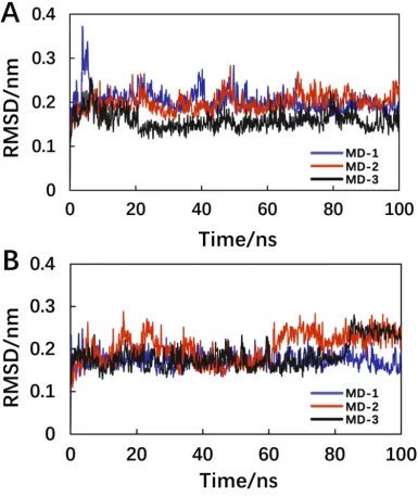
As shown in Figure 3A, the three MD repeats obtained similar MD trajectories,
and the system reached equilibrium at approximately 40 ns. Since the interaction of
antigen and antibody is mainly between the epitope of the antigen and the CDR of the
antibody, in the RMSD analysis, we used the index tool of gromacs (Make_ndx) to
define the CDR of Ab42 and the pCFH as a molecular group. In the 100 ns MD
process, in addition to analyze the RMSD of the total backbone, the variation curve of
CDR-pCFH was also analyzed (Figure 3B). The antibody CDR interacted with the
pCFH, and the results of RMSD analysis of the CDR-pCFH group showed that the
RMSD values remained within the range of 0.1 nm, indicating that the CDR regions
MM-PBSA and energy decomposition
In order to further explore the interaction mechanism between pCFH and its
antibody Ab42, the equilibrium phase of the three kinetic simulation of the
Ab42-pCFH complex structure was selected and sampled. Binding free energy and
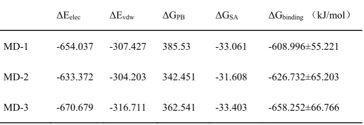
residue decomposition were then calculated using MM-PBSA. As shown in Table 3,
the binding free energies MD-1, MD-2, and MD-3 obtained from the three MD
simulations were -608.996±55.221 KJ/mol, -626.732±65.203 KJ/mol, and -658.252±
66.766 KJ/mol, respectively. During the interaction between pCFH antigen and Ab42
antibody, the electrostatic interaction, van der Waals force, and non-polar solvation
play important roles combinatorially.
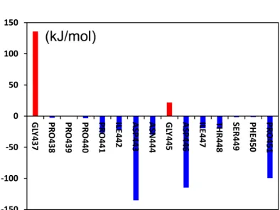
Taking average of the residual energy contribution values obtained from the
three simulations, we further analyzed the energy contributions of pCFH and the key
amino acid sites on the CDR of Ab42 separately, and the results showed that almost
all the amino acids in pCFH sequence (except for GLY437 and GLY445, which did
not interact with the antibody), displayed negative energy values (i.e., PRO441,
ILE442, ASP-443, ASP-446, ILE-447, THR448, and PRO451), strongly
demonstrating that these amino acids on the polypeptide facilitate the binding of
Ab42-pCFH, whereas the two sites of GLY437 and GLY445 may exert an adverse
effect on the antigen-antibody interaction.
Comparison of CAS and EAS
Alanine mutation of an amino acid replaces its reactive group with a small
structure, and hence is commonly used in observing the functional impact of amino
acids on proteins. Therefore, this study used alanine mutation to further verify the
results of CAS by performing EAS on the interaction of Ab42 with pCFH.
The free energy changes after amino acid mutation into alanine were mainly
considered in the CAS. An energy value of >0.5 Kcal/mol after the mutation indicates
that the structure is unstable and that the particular amino acid in situ plays an
important role in stabilizing the structure, whereas an post-mutational energy value of
<-0.5 Kcal/mol means that the structure remains stable and that the original amino
acid in situ is not conducive to the structural stability; similarly, a mutation energy
between 0.5 and -0.5 Kcal/mol indicates that there is no significant effect on the
structural stability before and after the mutation, i.e., these amino acids play little
roles on the structural stability.
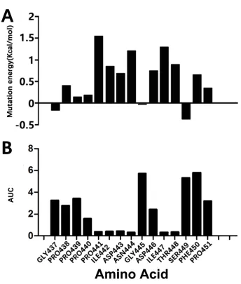
During the EAS, our CAS data showed that the sites at Pro441, Ile442, Asp443,
Asn444, Asp446, Ile447, Thr448, and Phe450 had an energy of >0.5 Kcal/mol after
mutation to Ala (Figure 5A), suggesting that when these amino acids were mutated,
conformational changes of the entire molecule occurred and thereby that the
antibody-polypeptide interaction mechanism changed, implying that the amino acids
at these sites play key roles in stabilizing the Ab42-pCFH complex.
As shown in Figure 5B, the area under the curve (AUC), as an important
pharmacokinetic parameter, represents the bioavailability of the drug (the extent to
which the drug is absorbed and utilized in the human body); the larger the AUC, the
Figure 5B shows that the AUCs after the alanine mutations of Pro441, Ile442,
Asp443, Asn444, Ile447, and Thr448 decreased, respectively, indicating that the
amino acids at these sites play important roles in the bioavailability of the protein.
Compared with the original CAS data, the results of experimental Ala mutations of
PRO441, ILE442, ASP-443, ASN444, ILE447, and THR448 were confirmatory.
However, we also noticed that the results of alanine scanning are not completely
consistent with the results of residue decomposition calculated by MM-PBSA,
especially for the ASP446, and the main reason may be that the analysis of the residue
energy decomposition was obtained in a single run, and it did not change the
molecular structure. The contribution of an amino acid to the binding free energy
reflects its importance in the complex structure during the alanine scanning process in
both CAS and EAS, and when mutated, it not only has a structurally complex effect
on the original site but also influence the amino acids around it. In general, our data
indicated that the six amino acids PRO441, ILE442, ASP-443, Asn444, Ile447, and
Thr448 are the key amino acids for the formation of a stable complex.
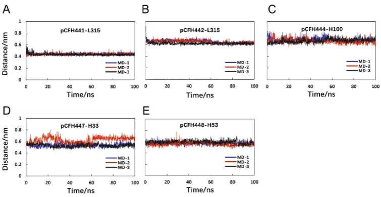
Distance monitoring of key amino acids
In order to find out the specific bonds between the key amino acids in the
structure of Ab42-pCFH complex, this study also investigated the distance changes
between several pairs of amino acids, through which pCFH interacts with Ab42 by
hydrogen bond formation, and the amino acid distance changing with time were
calculated.
in the CDR of the antibody, while the key amino acids on the polypeptide (Figure 6B)
are Pro441, Ile442, Asp443, Asn444, Ile447, and Thr448, which form hydrogen bonds
with the amino acids Tyr315, Ser100, Gly33, and Tyr53 on the CDR of Ab42
respectively. The relevant distance for pCFH is also very stable (Figure 6), implying
the stability of the CDR structure. As shown in Figure 6B, the decomposition energy
of these amino acid residues in the CDR are negative too, suggesting that the amino
acids at these sites contribute significantly to the antigen-antibody binding.
Conclusions
Revealing the details of antigen-antibody interactions and identifying the
important amino acid sites (epitopes) through which an antibody interacts with an
antigen are critical for the development of an antibody drug. In order to understand
the mechanism of interaction between human anti-CFH antibody Ab42 and its target
pCFH, the present study has used computational simulation methods, such as
homology modeling, molecular docking, MD simulation, and CAS, to study the
interaction between Ab42 and pCFH.
The amino acid energy contributions obtained from both the MM-PBSA
calculation and the CAS were used in our study to describe the key amino acids of the
antigen-antibody interaction, and the results indicated that the mechanisms of
interaction between Ab42 and pCFH are mainly hydrogen bond formation and
hydrophobic interaction between the amino acids Tyr315, Ser100, Gly33, and Tyr53
and Thr448 on the pCFH. Finally, all the computational results were verified by the
EAS method. As a fully natural human antibody, Ab42 can be further developed into
anti-tumor drugs without the concern of invoking immune rejections.
In summary, the present study has important guiding significance not only for the
development of Ab4 into anti-tumor drugs but also for the investigation of other
relevant antibody-antigen interactions.
Materials and Methods
Structural optimization
The structure of CFH polypeptide was retrieved from the PDB database
(PDB_ID: 5EA0)[1]. The Dmol3 quantum chemistry calculation in Discovery Studio software[17] was then applied, with the quantum chemical optimization setting at the density functional theory B3LYP method[12] for optimization, the maximum SCF
cycle setting at 300, and the calculated non-bond interaction value setting at 14
angstroms, respectively.
The human antibody Ab42, isolated from our laboratory, is a Fab antibody with
217 amino acids in the heavy chain and 219 amino acids in the light chain. According
to the IMGT numbering scheme, The CDR of Ab42 was defined as the following:
H-CDR1 (amino acids 26-33), H-CDR2 (amino acids 51-58), H-CDR3 (amino acids
97-108), L-CDR1 (amino acids 244-255), L-CDR2 (amino acids 273-275), and
L-CDR3 (amino acids 312-320).
method[17,18]. DS_Model Antibodies module in the Discovery Studio v4.5 (DS45) package was applied, and the antibody CDRs were optimized using the Model
Antibody Loops program. Finally, the charmm27 force field was used in the GBSW
solvent model, and the energy minimization (EM) was performed on the DS_Charmm
module in DS45. The final convergence was lower than 0.4184 kJ/(mol·nm), and the
output structure was the initial structure for molecular docking calculation.
Molecular docking
The molecular docking of pCFH with Ab42 was performed using the
ZDock[19,20] module under the Discovery Studio platform. The pCFH and Ab42
molecules were assigned to the all-atom Charmm27 force field[21], first using the
steepest descent (SD) method to optimize 1000 steps and then using the conjugate
gradient (CG) method, to optimize until the whole system convergence criterion
reaching 0.4184 kJ/(mol·nm), with pCFH as the ligand, Ab42 as the receptor, and the
antibody CDR defined as the binding region.
The RMSD Cutoff and the Interface Cutoff were set to 6 and 9 angstroms
respectively. All the docking poses obtained were scored by ZRank[22], and the highest scored Poses docking conformation was selected for the RDock[23] program. The simulated annealing method was used to reconstruct the docking compound, and
the RDock optimization parameters were as follows: Generalized Born with a simple
Switching (GBSW), Charmm force field, and dielectric constant 4.0.
MD simulations
complex. The MD trace was also used to calculate the MM-PBSA[24,25] binding free energy and select the dominant conformation for further analysis of the virtual
mutation. The MD was run using the Gromacs 5.1.2 software package[26,27], and the
initial model was dissolved in a cube box containing SPC/E water molecules; the
system was assigned to the Amber 99sb force field[28]. The entire system was
balanced by addition of neutral ions under the Genion program in the Gromacs
package.
Before the final production of MD, a 500-step SD optimization of protein
molecules in the system was performed to eliminate the positional conflicts from
unreasonable van der Waals force, and then a 2 ns position-restricted MD simulation
was performed with NVT and NPT[11] ensemble separately.
Finally, a 100 ns unrestricted MD simulation was performed at 300K, with
atmospheric NPT ensemble, and under periodic boundary conditions. All the bond
lengths were limited by the LINCS algorithm[18], and the electrostatic interactions
were calculated using the Particle Mesh Ewald (PME) summation scheme[29]. The
time step was set at 2 fs, and the conformations were stored every 20 ps, with each
MD trajectory file containing 1,000 conformations.
In the MD simulations, we mainly used the RMSD of backbone to examine
whether the system reaches its equilibrium and used the cluster analysis to obtain the
representative conformation after the system achieving balance. All of the Gromacs
MD simulation jobs were performed on the high performance computing platform of
Binding energy calculation by MM-PBSA
A total of 100 conformations were extracted from the equilibrium phase of each
MD locus (wild type and mutant), and the g_mmpbsa software package[30] was
executed; the binding free energy and residue decomposition of Ab42 and pCFH
complexes were calculated using the MM-PBSA method[13,31].
In MM-PBSA, the enthalpy of the system was calculated using the molecular
mechanics (MM) method; the contribution of the polar part and non-polar part of the
solvent effect to the free energy were determined by solving the Poisson-Boltzmann
(PB) equation and calculating the molecular surface area (SA) respectively. The basic
principle is shown in a formula as follows:
ΔGbind=ΔEMM+ΔΔGsol-TΔS
=ΔEMM+ΔGGB+ΔGSA-TΔS
=ΔEvdw+ΔEele+ΔGGB+ΔGSA-TΔS,
where ΔGbind is the binding free energy; ΔEMM is the difference in intramolecular
energy under vacuum; ΔΔGsol is the solvation free energy difference; T is the
absolute temperature, and ΔS is the entropy change. The ΔEMM can be calculated by
MM method; ΔΔGsol is composed of polar solvation free energy difference and
non-polar solvation free energy difference. While the polar part was obtained by
solving the finite difference PB equation, the non-polar part was fitted by estimating
the solvent accessible SA; finally, the TΔS was calculated using the normal mode
method.
Alanine scanning is to mutate an amino acid into alanine, thereby replacing any
functional groups on its side chain with a small neutral methyl group that exerts little
effect on the protein structure[14]. In this study, the importance of key amino acids
was determined by alanine scanning and calculation of the changes in binding free
energy before and after the mutation, in order to find the mechanism of interaction
between Ab42 and pCFH. The CAS was performed with the Calculate Mutation
Energy (Binding) module under the DS platform; the amino acids on the pCFH were
mutated into alanine one by one, and the energy differences between the wild type and
the mutants in the antigen-antibody complex were determined subsequently.
The GBIM (Generalized Born with implicit membrane) approximation[32] was
used to detect the effect of the solvent, and the electrostatic terms were approximated
by the sum of Coulombic interactions and polar contributions to solvation energy. The
energy function also contains the van der Waals interaction energy, a side-chain
entropy term, and a non-polar surface dependent term.
EAS
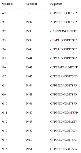
Each amino acid in pCFH was mutated into alanine one by one, and, including
the wild-type pCFH, a total of 16 polypeptides were synthesizeds for further
examination. As shown in Table 1, the peptide purity achieved 99%, and the biotin
was coupled for labeling. The ELISA was performed as follows: an ELISA plate was
coated with 20 μg/ml streptavidin coating solution (100 μl/well). The plate was
incubated overnight at 4 °C, washed with PBST (PBS containing 0.1% Tween 20),
washing with PBST, 20 μg/ml of each polypeptide was added and incubated at 37 °C
for 1 hour. After washing with PBST, the antibody was added and incubated at 37 °C
for 1 hour, and the secondary antibody, horseradish peroxidase-labeled goat
anti-human IgG (H+L), was applied. After incubation for another 30 minutes, 100 μl
of TMB color developer was added, and the OD values were read at the wavelength
of 450 nm.
Acknowledgements
The work was supported by the Guangdong Innovative and Entrepreneurial Research
Team Program (No. 2013Y113), National Natural Science Foundations (No.
31400795), Pearl River S&T Nova Program of Guangzhou (No. 201506010097),
Opening Fund of Guangdong Provincial Key Laboratory of Computational Science
(No. 2016004), Operating Fund of Guangdong Provincial Key Laboratory of
Bioengineering Medicine (NO.2014B030301050), Zhuhai Innovative and
Entrepreneurial Research Team Program (No. ZH01110405160015PWC), National
Basic Research Program of China (973 Program) (No. 2015CB553706).
Conflicts of Interest
The authors declare there is no conflicts of interest regarding the publication of this
paper.
1. Bushey, R.T.; Moody, M.A.; Nicely, N.L.; Haynes, B.F.; Alam, S.M.; Keir, S.T.; Bentley, R.C.; Roy Choudhury, K.; Gottlin, E.B.; Campa, M.J., et al. A Therapeutic Antibody for Cancer, Derived from Single Human B Cells. Cell Rep 2016, 15, 1505-1513, doi:10.1016/j.celrep.2016.04.038.
2. Herbert, A.P.; Kavanagh, D.; Johansson, C.; Morgan, H.P.; Blaum, B.S.; Hannan, J.P.; Barlow, P.N.; Uhrin, D. Structural and functional characterization of the product of disease-related factor H gene conversion. Biochemistry 2012, 51, 1874-1884, doi:10.1021/bi201689j.
3. Amornsiripanitch, N.; Hong, S.; Campa, M.J.; Frank, M.M.; Gottlin, E.B.; Patz, E.F., Jr. Complement factor H autoantibodies are associated with early stage NSCLC. Clinical cancer research : an official journal of the American Association for Cancer Research 2010, 16, 3226-3231, doi:10.1158/1078-0432.CCR-10-0321.
4. Kajander, T.; Lehtinen, M.J.; Hyvarinen, S.; Bhattacharjee, A.; Leung, E.; Isenman, D.E.; Meri, S.; Goldman, A.; Jokiranta, T.S. Dual interaction of factor H with C3d and glycosaminoglycans in host-nonhost discrimination by complement. Proc Natl Acad Sci U S A 2011, 108, 2897-2902, doi:10.1073/pnas.1017087108.
5. Ferreira, V.P.; Pangburn, M.K.; Cortes, C. Complement control protein factor H: the good, the bad, and the inadequate. Mol Immunol 2010, 47, 2187-2197, doi:10.1016/j.molimm.2010.05.007.
6. Campa, M.J.; Gottlin, E.B.; Bushey, R.T.; Patz, E.F., Jr. Complement Factor H Antibodies from Lung Cancer Patients Induce Complement-Dependent Lysis of Tumor Cells, Suggesting a Novel Immunotherapeutic Strategy. Cancer Immunol Res 2015, 3, 1325-1332, doi:10.1158/2326-6066.CIR-15-0122.
7. Hofer, J.; Giner, T.; Jozsi, M. Complement factor H-antibody-associated hemolytic uremic syndrome: pathogenesis, clinical presentation, and treatment. Semin Thromb Hemost 2014, 40, 431-443, doi:10.1055/s-0034-1375297.
8. Liao, H.X.; Levesque, M.C.; Nagel, A.; Dixon, A.; Zhang, R.; Walter, E.; Parks, R.; Whitesides, J.; Marshall, D.J.; Hwang, K.K., et al. High-throughput isolation of immunoglobulin genes from single human B cells and expression as monoclonal antibodies. J Virol Methods 2009, 158, 171-179, doi:10.1016/j.jviromet.2009.02.014.
9. Tonelli, M.; Boido, V.; Colla, P.; Loddo, R.; Posocco, P.; Paneni, M.S.; Fermeglia, M.; Pricl, S. Pharmacophore modeling, resistant mutant isolation, docking, and MM-PBSA analysis: Combined experimental/computer-assisted approaches to identify new inhibitors of the bovine viral diarrhea virus (BVDV). Bioorganic & medicinal chemistry 2010, 18, 2304-2316, doi:10.1016/j.bmc.2010.01.058.
10. Luo, Q.; Zhang, C.; Miao, L.; Zhang, D.; Bai, Y.; Hou, C.; Liu, J.; Yan, F.; Mu, Y.; Luo, G. Triple mutated antibody scFv2F3 with high GPx activity: insights from MD, docking, MDFE, and MM-PBSA simulation. Amino Acids 2013, 44, 1009-1019, doi:10.1007/s00726-012-1435-3.
11. Zhang, D.; Chen, C.F.; Zhao, B.B.; Gong, L.L.; Jin, W.J.; Liu, J.J.; Wang, J.F.; Wang, T.T.; Yuan, X.H.; He, Y.W. A novel antibody humanization method based on epitopes scanning and molecular dynamics simulation. PLoS One 2013, 8, e80636, doi:10.1371/journal.pone.0080636.
Journal of Quantum Chemistry 2011, 112, 909-921.
13. Jiayi, R.; Zhiwei, Y.; Nianjue, Z.; Junqi, L.; Chunlong, Y.; Shujian, L.; Bing, Y.; Junqing, H.; Huaxin, L.; Xiaohui, Y., et al. Effect of ForceFields and Water Models of EGFRvIII (scFv) Complex by Molecular Dynamics Simulation, MM-PBSA Calculation, and ITC Experiment.
Chem J Chinese Universities 2017, 38, 2070-2076.
14. Martins, S.A.; Perez, M.A.; Moreira, I.S.; Sousa, S.F.; Ramos, M.J.; Fernandes, P.A. Computational Alanine Scanning Mutagenesis: MM-PBSA vs TI. Journal of chemical theory and computation 2013, 9, 1311-1319, doi:10.1021/ct4000372.
15. Liu, X.; Peng, L.; Zhou, Y.; Zhang, Y.; Zhang, J.Z.H. Computational Alanine Scanning with Interaction Entropy for Protein-Ligand Binding Free Energies. Journal of chemical theory and computation 2018, 14, 1772-1780, doi:10.1021/acs.jctc.7b01295.
16. Gao, M.; Zhang, F.; Zhu, Y.; Gao, L.; Jiang, Y.; Luo, Y.; Zhuang, F.; Mao, Z.; Mao, J. Alanine scanning mutagenesis of SP70 epitope in characterizing speciesspecific antibodies induced by enterovirus 71based antigens. Mol Med Rep 2018, 17, 1006-1014, doi:10.3892/mmr.2017.7992.
17. Yuan, X.; Qu, Z.; Wu, X.; Wang, Y.; Wei, F.; Zhang, H.; Liu, L.; Yang, Z. Homology Modeling and Evolution Trace Analysis of Human Adenovirus Type 3 Hexon. Chem J Chinese Universities 2009, 30.
18. Yuan, X.H.; Wang, Y.C.; Jin, W.J.; Zhao, B.B.; Chen, C.F.; Yang, J.; Wang, J.F.; Guo, Y.Y.; Liu, J.J.; Zhang, D., et al. Structure-based high-throughput epitope analysis of hexon proteins in B and C species human adenoviruses (HAdVs). PLoS One 2012, 7, e32938, doi:10.1371/journal.pone.0032938.
19. Yang, Z.; Wu, F.; Yuan, X.; Zhang, S. Molecular interactions of GABA analogues against the
α+β-interface of GABAA receptor: Docking and molecular dynamics studies; 2015; Vol. 10, pp. 811-822.
20. Wiehe, K.; Pierce, B.; Tong, W.W.; Hwang, H.; Mintseris, J.; Weng, Z. The performance of ZDOCK and ZRANK in rounds 6-11 of CAPRI. Proteins 2007, 69, 719-725, doi:10.1002/prot.21747.
21. Sapay, N.; Tieleman, D.P. Combination of the CHARMM27 force field with united-atom lipid force fields. Journal of computational chemistry 2011, 32, 1400-1410, doi:10.1002/jcc.21726. 22. Pierce, B.; Weng, Z. ZRANK: reranking protein docking predictions with an optimized energy
function. Proteins 2007, 67, 1078-1086, doi:10.1002/prot.21373.
23. Li, L.; Chen, R.; Weng, Z. RDOCK: refinement of rigid-body protein docking predictions.
Proteins 2003, 53, 693-707, doi:10.1002/prot.10460.
24. Kuhn, B.; Gerber, P.; Schulz-Gasch, T.; Stahl, M. Validation and use of the MM-PBSA approach for drug discovery. Journal of medicinal chemistry 2005, 48, 4040-4048, doi:10.1021/jm049081q.
25. Wang, J.; Morin, P.; Wang, W.; Kollman, P.A. Use of MM-PBSA in reproducing the binding free energies to HIV-1 RT of TIBO derivatives and predicting the binding mode to HIV-1 RT of efavirenz by docking and MM-PBSA. J Am Chem Soc 2001, 123, 5221-5230.
26. Van Der Spoel, D.; Lindahl, E.; Hess, B.; Groenhof, G.; Mark, A.E.; Berendsen, H.J. GROMACS: fast, flexible, and free. Journal of computational chemistry 2005, 26, 1701-1718, doi:10.1002/jcc.20291.
GROMACS: High performance molecular simulations through multi-level parallelism from laptops to supercomputers. SoftwareX 2015, 1–2, 19-25, doi:http://dx.doi.org/10.1016/j.softx.2015.06.001.
28. Showalter, S.A.; Bruschweiler, R. Validation of Molecular Dynamics Simulations of Biomolecules Using NMR Spin Relaxation as Benchmarks: Application to the AMBER99SB Force Field. Journal of chemical theory and computation 2007, 3, 961-975, doi:10.1021/ct7000045.
29. Yuan, X.H.; Wang, Y.C.; Qu, Z.Y.; Ren, J.Y.; Wu, X.M.; Wang, J.F. Phylogenetic and structural analysis of major surface proteins hemagglutinin and neuraminidase of novel avian influenza virus A H7N9 from chinese patient. Chem Res Chinese U 2013, 29, 934-940, doi:10.1007/s40242-013-3200-x.
30. Kumari, R.; Kumar, R.; Lynn, A. g_mmpbsa--a GROMACS tool for high-throughput MM-PBSA calculations. Journal of chemical information and modeling 2014, 54, 1951-1962, doi:10.1021/ci500020m.
31. Thompson, D.C.; Humblet, C.; Joseph-McCarthy, D. Investigation of MM-PBSA rescoring of docking poses. Journal of chemical information and modeling 2008, 48, 1081-1091, doi:10.1021/ci700470c.
Figure Legends
Figure 1. The crystal structure and the structure evaluation of Ab42. (A) the crystal
structure of Ab42 with the six CDRs displayed by red, yellow, purple, black, blue, and
green spheres respectively; (B) Profile_3D verification result of the Ab42 model with
residues exhibiting reasonable folding; (C) the Ramachandran plot analysis shows
phi-psi torsion angles of all residues in the structure.
Figure 2. The interaction of Ab 42 with pCFH. (A) atomic surface contact of pCFH
with Ab42 CDR; (B) two dimensional diagram of the interaction between pCFH and
Ab42, with pCFH displayed by ball and stick and the key residues in CDR displayed
by disc presentation.
Figure 3. The RMSD as functions of the Ab42-pCFH MD simulation time. (A) the
backbone-atom RMSD of whole Ab42-pCFH structure;(B) the CDR-pCFH RMSD of
whole Ab42-pCFH structure
Figure 4. Amino acid energy decomposition of pCFH. The red bar or the blue column
indicates the binding energies positive or negative respectively after the mutation.
Figure 5. Results of alanine scanning and experimental alanine mutation. (A) CAS;
(B) experimental alanine mutation.
Figure 6. The Key amino acid distance between pCFH and Ab42. (A)
pCFH441-L315; (B) pCFH442-L315; (C) pCFH444-H100; (D) pCFH447-H33; (E)
Tables
Table 1. The amino acid sequences for peptide synthesis.
Mutation Location Sequence
Table 2. The hydrogen bonds formed in the docking complex.
Donor Acceptor
Ab42-A:Gly33:HN Pep-C:Ile447:O
Ab42-A:Tyr53:HH Pep-C:Gly445:O
Ab42-A:Tyr53:HH Pep-C:Gly448:O
Ab42-B:Tyr315:HH Pep-C:Pro439:O
Pep-C:Ile442:HN Ab42-B:Tyr315:O
Pep-C:Asn444:HD21 Ab42-A:Ala103:O
Pep-C:Asn444:HD22 Ab42-A:Ser100:O
Pep-C:Ser449:HN Ab42-A:Leu31:O
Ab42-A:Gly33:HA2 Pep-C:Ile447:O
Ab42-A:Ser52:HB1 Pep-C:Asp446:O
Ab42-A:Ser52:HB2 Pep-C:Asp446:O
Ab42-A:Ala101:HA Pep-C:Asn444:OD1
Pep-C:Pro441:HA Ab42-B:Tyr315:O
Pep-C:Thr448:HA Ab42-A:Leu31:O
Pep-C:Thr448:HB Ab42-A:Leu31:O
Table 3. The binding free energy of Ab42-pCFH complexes.
ΔEelec ΔEvdw ΔGPB ΔGSA ΔGbinding (kJ/mol)
Figures
![1 [4,5 Bis(benzyloxy) 2 methylphenyl]ethanone](data:image/gif;base64,R0lGODlhAQABAIAAAP///wAAACH5BAEAAAAALAAAAAABAAEAAAICRAEAOw==)