A Survey on Thermographic Segmentation
V.S.Narayana Tinnaluri
1, Proof. Dr. Anil Kumar
2Dept. of Computing sciences and engineering, School of Engineering, Sri Satya Sai University Of Technology And
Medical Sciences, India 1,2
ABSTRACT: Image segmentation is used in many application areas to solve the issues in different form of objects. Thermographic segmentation is used to identify the issues of Surface Temperature. Early detection of diseases helps to reduce the growth of diseases which results in healthy fruit growth. Thermal images of leaves used for early detection of diseases. The Plant leaves are affected by the diseases such as powdery mildew, Downy mildew and Anthracnose. In this paper we contemplated thermal image segmentation for detecting the thrips and anthracnose. Image segmentation algorithm is used to identify the diseased part over the leaves. The pests and anthracnose affect the part of the leaves. Then it spread over the plant and infect the berries which debase the quality of Plants. The thresholding technique used for the segmentation of thermal images. Thrips are sucking pests and anthracnose is fungal disease. To identify thrips and anthracnose on the leaves thermal images are used.
KEYWORDS: Thermal image, Image segmentation, Thresholding.
I. LITERATURE REVIEW
Thermal image segmentation is used in agriculture for disease detection on leaves of fruits and vegtables. Thermal images are useful for early disease detection. It will help to identify the diseases over the leaves. Diseases may harm fruits or vegetables. To reduce growth of diseases on plants there is need for segmentation of thermal images. Thermal images are used to identify the “region of interest’. Thermal images detect the amount heat emitted from object. The heat emitted from object help to identify the objects or region of interest. Thermal images are conventionally used in biomedical areas. Recently, it has been used in electrical field and agricultural field.
In India, Andhra Pradesh is one of the big export state. In Andhra Pradesh state Hyderabad and Sangali districts are the prominent for cultivation of Plants. Hyderabad is also known for “Plant capital of India”. Plants are cultivated in hot and dry climate. Vegetative growth and fruiting of Plants requires high humidity. The high humidity affects the vines fruit size and quality. But after forward pruning of vines it may increase the fungal disease.
The Plants are affected by pests or Plant diseases. The disease and pests are get differentiated at every stage of growth of Plants. Plants diseases and pests are varies based on the regions of cultivation. As atmosphere is really affects the growth of Plants. The pests are mealay bugs, flea beetle, girdle bittle, thrips, hoppers, stem borers, leaf eating caterpillar, and Plant leaf folder. The diseases like downy mildew, powdery mildew, anthracnose, greenaria bitter rot, bacterial leaf spot, alternia blight, black rot. The image segmentation help to identify the part of image useful for disease detection. The thermal image segmentation will reduce the task of image pre-processing. This reduces the wastage of time in pre-processing of the image. Thermal image overcome the timestamp for processing of thermal image segmentation. Thermal image segmentation recognize the area of interest. The goal of the image segmentation is to remove the noise at the background, by separating the object from the background the Region of Interest is get highlighted. distinct segmentation techniques are described in this section.
and V channel are separated for clear view of the leaf and disease over leaf respectively. After that thresholding is using Otsu adaptive method applied to differentiate between healthy and unhealthy leaves. After that some post-processing tools are applied and leaf disease get distracted.
B. Otsu thresholding
Otsu thresholding is a kind global thresholding and also known for. The threshold value of each pixel is set either 0 or 1 i.e. background or foreground. It reduces the conversion of grayscale image. Salvador et al. [10] propound the most popular thresholding techniques rates through all possible threshold values, after that it will calculate a measure extent for pixel levels each side of the threshold value by manipulating the sum of foreground and background. Otsu method can be suitable for any application.
II. THRIPS AND ANTHRACNOSE
A. Thrips
Brown spot appeared on leaf
Infected Plant berries
B. Anthracnose
Fungal black spots
III.THERMAL IMAGE SEGMENTATON
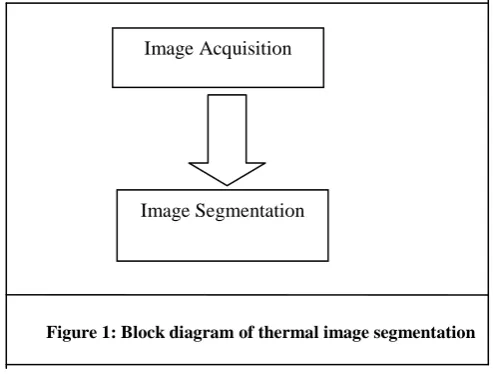
Thermal image segmentation that consumes less time for detection of region of interest nothing but the disease part infected by anthracnose and thrips. Simple structure for thermal image segmentation is shown in figure1. The diagram sows only two parts Image acquisition and Image Segmentation. The general image segmentation has three stage one is image acquisition, then pre-processing of the image and then segmentation which consumes time for image segmentation part. By reducing the time for pre-processing of the image it is possible to reduce the time consumption for image segmentation.
A. Image Acquisition
B. Image Segmentation
Testo IR soft uses six types of thermal color paletes, these formats are useful to focus on the region of interest on the leaves part. The region interest is diseased part. The IR soft is a helpful tool for thermal images.
Figure 1: Block diagram of thermal image segmentation
After this part it’s being easy for segmentation of image. The simple thresholding technique is able segment the image. For segmentation of the image matlab R2014a software is used. The thresholding technique is applied for segmenting an image.
Image acquisition
Image Acquisition
Captured Thermal Image
Segmented Image
Figure 2: The structure of Thermal Image Segmentation
IV. RESULT
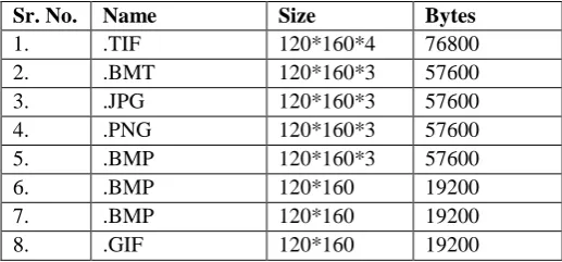
Comparative result of image extensions
This result is similar for some image extensions. It is possible only if the image is thermal image in .BMT format. The result analysis shows that thermal image segmentation is the simple and less time consuming way for image segmentation. Also the thermal image shows the resultant data in the following way. The thermal report of this Plant leaves conclude with the emmisivity of 0.95 and Refl. Temp [oC] of 20.0
The IR soft gives the some information regarding thermal images like histogram, temperature details table and color bar which shows the temperature. Here the result of the image transformation with information.
Digital image
Thermal Image
No Temp. [°F] Emiss. Refl.Temp. [°F]
M1 85.9 0.95 68.0
M2 87.4 0.95 68.0
M3 84.8 0.95 68.0
Color bar with isotherm indication
Histogram based on thermal image
V. CONCLUSION
[9]Carlos M.Travieso, Jesús B. Alons, Varun Gupta, Namita Sengar, Malay Kishore Dutta, o“Automated Segmentation of Powdery Mildew disease from Cherry Leaves using Image Processing,” IEEE, 2017, p. 301, 1982].
[10]Salvador Hinojosa, Gonzalo Pajares, Erik Cuevasand Noé Ortega-Sanchez” Thermal Image Segmentation Using Evolutionary Computation Techniques”,Researchgate, January 2018, p.65-68