Scholarship@Western
Scholarship@Western
Electronic Thesis and Dissertation Repository
8-27-2013 12:00 AM
Characterization of Staphylococcus aureus Lipase
Characterization of Staphylococcus aureus Lipase
Vithooshan Vijayakumaran The University of Western Ontario
Supervisor
Dr. David Heinrichs
The University of Western Ontario Joint Supervisor Dr. Martin McGavin
The University of Western Ontario
Graduate Program in Microbiology and Immunology
A thesis submitted in partial fulfillment of the requirements for the degree in Master of Science © Vithooshan Vijayakumaran 2013
Follow this and additional works at: https://ir.lib.uwo.ca/etd
Part of the Bacteria Commons, Bacteriology Commons, Biology Commons, Genetics Commons,
Immunology of Infectious Disease Commons, Molecular Genetics Commons, and the Pathogenic Microbiology Commons
Recommended Citation Recommended Citation
Vijayakumaran, Vithooshan, "Characterization of Staphylococcus aureus Lipase" (2013). Electronic Thesis and Dissertation Repository. 1584.
https://ir.lib.uwo.ca/etd/1584
This Dissertation/Thesis is brought to you for free and open access by Scholarship@Western. It has been accepted for inclusion in Electronic Thesis and Dissertation Repository by an authorized administrator of
i
Characterization of Staphylococcus aureus Lipase
Thesis format: Monograph
By
Vithooshan Vijayakumaran
Graduate Program in Microbiology and Immunology
A thesis submitted in partial fulfillment of the requirements for the degree of
Master of Science
The School of Graduate and Postdoctoral Studies The University of Western Ontario
London, Ontario, Canada
ii
ABSTRACT
USA300, a strain of community-associated methicillin resistant Staphylococcus
aureus (CA-MRSA), has become prevalent in the community. Colonization of human skin
requires mechanisms that allow this bacterium to overcome the innate immune defenses on
the skin, including secretion of antimicrobial lipids. Antimicrobial lipids inhibit S. aureus
growth and induce the staphylococcal proteolytic cascade, producing aureolysin (Aur) which
processes the lipase glycerol ester hydrolase (Geh). Nearly all S. aureus strains secrete Geh,
yet little information exists concerning its function. Using purified Aur and Geh we confirm
that aureolysin processes proGeh to Geh. We then confirmed that geh was required for lipase
activity and both forms of the purified enzyme had lipase activity. Finally we showed that
optimal growth in trilinolein requires Aur, and might reflect a requirement for the proGeh
form of the enzyme to convert trilinolein into toxic linoleic acid, despite that fact that both
unprocessed and processed Geh catalyze the hydrolysis of trilinolein.
iii
ACKNOWLEDGEMENTS
I would like to begin by thanking both of my supervisors, Dr. David Heinrichs and
Dr. Martin McGavin for providing me the opportunity to complete my thesis in their
laboratories. Their mentorship, patience and support through all the challenges that I have
faced during the completion of my Master’s project have been crucial to my success.
I would also like to thank Dr. Mark Bernards and Dr. Sung Kim for their advice and
guidance as members of my advisory committee and Dr. Bernards for assistance with
GC-MS and lipid analysis within a short time-frame.
Finally, there are no words I can use to describe my gratitude towards my parents for
iv
TABLE OF CONTENTS
Page
Abstract ii
Acknowledgements iii
Table of Contents iv
List of Tables vi
List of Figures vii
List Abbreviations viii
Chapter 1 – INTRODUCTION 1
1.1 Staphylococcus aureus 2
1.1.1 Description 2
1.1.2 S. aureus and Virulence 2
1.1.3 MRSA 4
1.1.4 USA300 5
1.2 Lipids 9
1.2.1 Chemical and Physical Properties of Lipids 9
1.2.2 Biological Role of Lipids 12
1.2.3 Lipids in Infection and Immunity 12
1.2.4 Resistance to Antimicrobial Fatty Acids 13
1.3 Staphylococcal Proteases 15
1.3.1 Proteases 15
1.3.2 The Staphylococcal Proteolytic Cascade 15
1.4 Lipases 19
1.4.1 Microbial Lipases 19
1.4.2 S. aureus Lipases 20
1.5 Rationale and Hypothesis 21
Chapter 2 – MATERIALS AND METHODS 22
2.1 Bacterial Strains and Growth Conditions 23
2.2 DNA Methodology 25
2.2.1 Plasmid Isolation from E. coli 25
2.2.2 Plasmid Isolation from S. aureus 27
2.2.3 Isolation of Chromosomal DNA from S. aureus 27
2.2.4 DNA Ligation 28
2.2.5 Recombinant DNA Methodology 28
2.2.6 Agarose Gel Electrophoresis 28
2.2.7 Isolation of DNA Fragment from Agarose Gels 29
v
2.2.9 DNA Sequencing 29
2.2.10 Computer Analysis 29
2.3 Transformation Methodologies 30
2.3.1 Preparation of Transformation Competent E. coli 30 2.3.2 Transformation of CaCl2 Competent E. coli 30
2.3.3 Preparation of Transformation Competent S. aureus 31 2.3.4 Transformation of Electrocompetent S. aureus 31
2.4 Mutagenesis and DNA Cloning Methods - Mutagenesis of geh 31
2.5 Protein Methodology 33
2.5.1 TCA Precipitation, and Visualization of Secreted Proteins 33
2.5.2 Purification of Aur and Geh 34
2.6 Assays 35
2.6.1 Lipase Assays 35
2.6.2 Protease Assays 36
2.6.3 Aureolysin-Geh Processing 36
2.7 Influence of triglycerides on S. aureus growth 37
2.8 Lipid Extraction and Analysis by GC/MS 37
Chapter 3 – RESULTS 39
3.1 Deletion of geh locus 40
3.2 Growth in Fatty Acid Results in Processing of Geh 42
3.3 Aureolysin is required for Processing of Geh 42
3.4 The USA300 geh mutant has no detectable lipase activity 47 3.5 Growth in Trilinolein Results in Inhibition of Growth 49 3.6 Aureolysin is required for optimal growth in trilinolein 51 3.7 GC/MS confirms that trilinolein is a substrate for proGeh and Geh 57
Chapter 4 – DISCUSSION 60
REFERENCES 65
vi
List of Tables
Table Page
Table 1. Lipid categories 10
Table 2. Bacterial strains used in this study 24
Table 3. Plasmids used in this study 26
Table 4. Oligonucleotides and their sequences 32
vii
List of Figures
Figure Page
Figure 1. The arginine mobile genetic element 7
Figure 2. Structures of lipids 11
Figure 3. The staphylococcal proteolytic cascade 16
Figure 4. Generation of a Δgeh mutant in S. aureus strain USA300 41
Figure 5. Induction of Aur by linoleic acid results in processing of
proGeh to Geh in S. aureus culture supernatants 43
Figure 6. Purification of Aur and proGeh 45
Figure 7. Visualization of proGeh processing by aureolysin 46
Figure 8. pNPP lipase assay of culture supernatants and purified Geh 48
Figure 9. Grow curve analysis of USA300 wild-type, Δgeh and Δaur
with trilinolein 50
Figure 10. Secreted protein profile of cells grown in 50 M trilinolein 52
Figure 11. Comparison of cell-surface proteins of washed and
unwashed cells and growth of washed cells in trilinolein 53
Figure 12. Delayed growth of Δgeh strain in trilinolein after
re-suspension in supernatant from wild-type and Δaur 55
Figure 13. Impaired growth of Δgeh in trilinolein with addition of
purified proGeh or Geh at inoculation 56
Figure 14. Gas chromatography-mass spectrometry confirms release of
viii
List of Abbreviations
Bp Base pair
CA-MRSA Community associated methicillin resistant S. aureus
CoNS Coagulase negative staphylococci
DNA Deoxyribonucleic acid
Em Erythromycin
HA-MRSA Health-care acquired methicillin resistant S. aureus
Km Kanamycin
LB Luria-Bertani broth
M Molar
mg Milligram
ml Milliliter
mM Millimolar
nm Nanometer
OD optical density
PCR Polymerase chain reaction
SDS Sodium dodecyl sulphate
TAE Tris-acetate EDTA
TBE Tris-borate EDTA
TSB Tryptic soy broth
μg Microgram
μl Microliter
INTRODUCTION
1.1 Staphylococcus aureus
1.1.1 Description
Staphylococcus aureus is a Gram-positive bacterium where the individual cocci
divide in two planes resulting in the formation of grape like clusters, and for this reason the
word Staphylococcus is derived from the Greek term staphylé (“bunches of grapes”). S.
aureus is the most pathogenic of the staphylococci and was identified in the 19th century. S.
aureus strains are characterized by a golden-yellow pigment, staphyloxanthin (aureus:
“golden”). Additionally, S. aureus are the only staphylococci with the ability to produce the
protein coagulase which allows for the conversion of fibrinogen to fibrin, allowing fibrin to
coat the bacteria and thus protecting them from attacking host cells (59). S. aureus is a highly
persistent organism which can survive in high salt conditions (up to 15 % NaCl), dry
environments and can survive over a wide pH range from 4.8 to 9.4 (15, 58). These survival
mechanisms allow it to thrive in many harsh environments, ranging from the surfaces of
medical equipment to the epidermal and mucosal layer of mammals.
1.1.2 S. aureus and Virulence
Staphylococcus aureus is a human commensal bacterium found to persistently
colonize about 30% of the human population (73). While the organism is generally harmless
to healthy individuals, S. aureus is a significant opportunistic pathogen and is the causative
agent in over 10% of all nosocomial bacterial infections in the United States (25).
severe conditions such as necrotizing pneumonia, osteomyelitis, toxic shock syndrome,
bacteremia and infective endocarditis (56).
Virulence factors provide bacteria with mechanisms that aid in their in vivo growth
and enhance their ability to cause disease. S. aureus possesses a vast array of virulence
factors which contribute to its success as a pathogen. S. aureus possesses a plethora of
surface-associated virulence factors which aid in adherence followed by the establishment of
infection (15). Furthermore, some of these surface-associated proteins also provide an
immune evasion mechanism by modulating the host immune response. Cell surface virulence
factors can include cell wall anchored proteins or polysaccharides. Polysaccharides such as
teichoic acid on the S. aureus cell wall aid in adhesion to endothelial surfaces while other cell
surface components such as peptidoglycan and lipoteichoic acids can cause neutrophil
infiltration and septic shock (45, 50, 95, 96). S. aureus also produces several pore forming
toxins which create pores in the cell membrane resulting in lysis through osmotic imbalance.
Two of these pore forming toxins are known as α-toxin and β-toxin which are capable of
destroying erythrocytes (15). Destruction of erythrocytes is a key nutritional strategy that
provides S. aureus with a source of heme-iron for uptake. Additionally S. aureus secretes a
variety of toxins and exoenzymes capable of causing damage to host tissue. For example, S.
aureus produces the toxic shock syndrome toxin-1 (TSST-1), a known superantigen, which
through the activation of T cells induce a massive cytokine storm resulting in fevers, rashes,
hypotension, multi-organ failure and death (15, 92). Exoenzymes such as proteases, lipases
and nucleases are some of the less well-understood virulence factors of S. aureus, and the
1.1.3 Methicillin Resistant Staphylococcus aureus
Administration of penicillin was the effective traditional treatment for S. aureus
infections and could significantly reduce mortality rates in patients (27). However, due to the
use of penicillin, antibiotic-resistant strains of S. aureus emerged and widespread penicillin
resistance around the world had occurred by the 1950s (15). Even with the development of
next generation antibiotics, both resistant hospital-acquired and community-acquired strains
emerged. The emergence of resistance to beta-lactam antibiotics such as cephalosporins,
carbapenems, cephamycins and monobactams prompted development of methicillin.
Unfortunately, by 1961, just 2 years since the introduction of the drug to penicillin resistant
S. aureus, the first methicillin resistant S. aureus (MRSA) were reported. Over the next
several decades MRSA spread around the world, and is now endemic in most healthcare
facilities in developed countries. Methicillin and beta-lactam class antibiotic resistance is
encoded on a genomic island called the staphylococcal cassette chromosome mec (SCCmec),
which also encodes tetracycline resistance (38). A major concern is the increase of multidrug
resistant strains of MRSA, which have evolved resistance to other classes of antibiotics
including rifampin, fluoroquinolones, aminoglycosides, tetracyclines and chloramphenicol.
Additionally, the emergence of MRSA strains resistant to vancomycin, the treatment of last
resort for some MRSA strains, leaves the possibility of emerging S. aureus infections that
would be untreatable with current antibiotics (16, 27).
In contrast to healthcare-associated MRSA (HA-MRSA), which often infect
individuals who are at high risk or already ill, community-associated MRSA (CA-MRSA)
strains can infect healthy individuals. This, in combination with the unusually severe disease
caused by these strains, suggests that CA-MRSA possess greater virulence potential than
an infected individual and this often occurs among military settings, locker rooms and among
children in daycare centers where there is a combination of close body contact and low
personal hygiene. In particular the CA-MRSA strain USA300 has become a leading cause of
visits to hospital emergency departments as it is easily transferred to household contacts
compared to other S. aureus strains (24, 63).
1.1.4 USA300
The USA300 strain is a particularly hypervirulent strain of CA-MRSA which has
become endemic within the community. USA300 clones show a high level of genetic
similarity, with different isolates being distinguished only by a few single nucleotide
polymorphisms (71). In a brief time span this strain has become the most frequent cause of
skin and soft tissue infections in emergency rooms in the United States, being responsible for
97% of MRSA and 58% of all reported skin infections (67). Since the appearance of USA300
the percentage of MRSA carriers has doubled (31). It can be inferred that the increased
incidence of USA300 infections can, in part, be attributed to an increased frequency of
colonization.
In addition to its exceptional transmission and colonization from host to host, the
USA300 strain has been associated with highly invasive diseases such as necrotizing
pneumonia, severe septicaemia and necrotizing fasciitis (28, 30, 64). USA300 was found to
be more resistant to killing by human polymorphononuclear leukocytes and more effective at
causing host cell lysis. Mouse bacteraemia models show that it is significantly more virulent
than other MRSA strains and is more highly invasive of major organs (94).
Like other CA-MRSA, USA300 carries the type IV SCCmec cassette, which lacks
this cassette when there is a lack of antibiotic stress (19). However, the extraordinary success
of USA300 as a pathogen and its ability to overtake other CA-MRSA clones can be
attributed to the presence of unique genetic elements that enhance colonization and
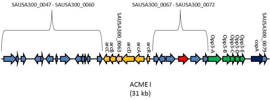
transmission. Exclusive to the USA300 strain is the 31-kb arginine catabolic mobile element
(ACME) (Figure 1) which is a putative pathogenicity island found adjacent to SCCmec (19).
In USA300, ACME is thought have been acquired from S. epidermidis, likely through
horizontal gene transfer. However, ACME has also been identified among other
coagulase-negative staphylococci (CoNS) such as S. haemolyticus and S. capitis in which it has a more
genetically diverse organization than in S. aureus (83). ACME encodes two major gene
clusters which include an arginine deiminase (arc), a spermine/spermidine acetyl-transferase
(speG) and oligopeptide permease (opp) operon, both of which are homologs of genes that
are accepted virulence factors (18).
Most pathogens often have difficulty colonizing the harsh environment of the human
skin, yet USA300 is able to colonize this niche at a higher rate than other strains. One
attribute of skin that deters infections is the acidic pH (4.2-5.9). However, the presence of
Arc encoded by ACME has been found to enhance the acid tolerance of USA300, including
to, for example, exogenous lactic acid, the major organic acid present on human skin (1, 62,
93). The ACME Arc encoded arginine deiminase pathway converts L-arginine to carbon
dioxide, ATP and ammonia, which counteracts the acidity of the environment (18, 19, 66, 83,
93).
Polyamines are aliphatic compounds produced by many organisms that exert a variety
Figure 1. The Arginine mobile genetic element. The 31-kb ACME is unique to the
USA300 strain of S. aureus and is thought to confer upon this strain an enhanced ability to
and are all compounds synthesized from L-arginine. However, S. aureus is unable to produce
polyamines and exogenous polyamines actually inhibit S. aureus growth, and are bactericidal
at concentrations found in humans. USA300 is a notable exception in that it exhibits
complete resistance to high levels of exogenous polyamines due to the presence of a
spermine/spermidine acetyl-transferase system encoded on ACME (41).
Additionally ACME encodes a putative oligopeptide permease operon, Opp-3. Opp
operons in Gram-positive and Gram-negative bacteria possess a range of functions including
peptide uptake, quorum sensing, chemotaxis, binding of serum components and expression of
virulence determinants (74). In vivo models have shown that disruption of Opp-1 and Opp-2,
which are found on the core genome have been shown to result in major growth defects and
attenuated virulence (12).
USA300 also possesses several other newly acquired genes such as the enterotoxin K
and Q (sek2 and seq2) in the unique SaPI5 pathogenicity island, and these contribute to
pathogenesis by binding T-cells (18). USA300 also possesses Panton-Valentine leukocidin
(PVL), a pore forming toxin which causes necrosis and apoptosis in leukocytes. PVL is a
bi-component toxin encoded by the lukS-PV and lukF-PV genes (12). Furthermore, research
indicates a high level of correlation between the production of PVL and severe skin and soft
tissue infections, necrotizing pneumonia and fasciitis (14, 29). Levels of PVL sufficient to
result in rapid neutrophil lysis can be directly detected in abscesses on human skin (4, 5). In
rabbit models, USA300 strains lacking PVL displayed attenuated virulence in pneumonia,
osteomyelitis and skin abscess models (13, 17, 48). Additionally, the expression of several
virulence genes such as α-toxin (Hla), a potent pore forming toxin, δ-toxin (hld) and α-type
Consequently, the success of USA300 as a pathogen can be attributed to the amalgamation of
an array of factors.
1.2 Lipids
1.2.1 Chemical and Physical Properties of Lipids
Lipids are an integral part of the physiology and pathophysiology of biological
systems. Although there is no widely accepted definition of the term lipid, a broad definition
would be that lipids are hydrophobic or amphipathic molecules that are created either entirely
or partly from carbanion-based condensations of thioesters or by carbocation-based
condensations of isoprene units (26). Although there are several categorization systems for
lipids (Table 1), they can broadly be classified as “simple” and “complex” lipids. Simple
lipids yield two products upon hydrolysis while complex lipids yield three or more products.
A lipid is saturated if all available spaces where hydrogen bonding to carbon can occur are
occupied, while an unsaturated lipid has at least one double bond between carbon atoms. As
a result, unsaturated lipids tend to have lower melting points and are more vulnerable to lipid
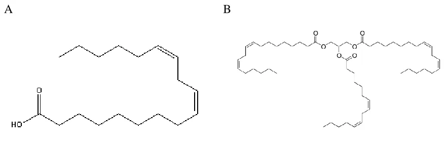
peroxidation (51). In this study we will focus on free fatty acids and triglycerides such as
Table 1. Lipid categories
Category Example
Fatty acyls dodecanoic acid
Glycerolipids 1-hexadecanoyl-2-(9Z-octadecenoyl)-sn-glycerol
Glycerophospholipids
1-hexadecanoyl-2-(9Z-octadecenoyl)-sn-glycero-3-phosphocholine
Sphingolipids N-(tetradecanoyl)-sphing-4-enine Sterol lipids cholest-5-en-3β-ol
Prenol lipids 2E,6E-farnesol
Saccharolipids
UDP-3-O-(3R-hydroxy-tetradecanoyl)-αD-N -acetylglucosamine
A B
Figure 2. Structures of lipids. A) Linoleic acid and B) trilinolein which is composed of
1.2.2 Biological Role of Lipids
Lipids have several important roles in biological systems, which include energy
storage, signaling, and as one of the structural components of the membrane (26, 90). The
biological membrane is essentially a lipid bilayer, the formation of which is energetically
favoured when glycerophospholipids are put in an aqueous environment, arranging
themselves so that the polar head groups face the aqueous side and the hydrophobic fatty acid
tails face each other (97). Recently, it has been shown that lipid signaling is important for
communication in cells. Some of these signaling lipids are involved in regulation of cell
growth, apoptosis, protein activation, and calcium mobilization (36, 80). One of the most
important and well understood functions of lipids is for energy storage in animals and plants,
especially in the form of triglycerides (8).
1.2.3 Lipids in Infection and Immunity
The sebaceous glands on the skin secrete several factors that are growth inhibitory to
many invading pathogens. An innate immune mechanism used to hinder bacterial persistence
is the secretion of sebum (65, 98), a liquid concoction of lipids composed of 28% free fatty
acids, 32% triglycerides, 25% wax esters and 11% squalene (88). Sapienic acid (C16:1Δ6), is
the major constituent of sebum triglycerides and fatty acids (98), and S. aureus is also
significantly exposed to linoleic acid (C18:2) from nasal secretions, during colonization of
the anterior nares (20). Previous studies have shown that at physiological concentrations,
linoleic acid (50 μM) is inhibitory to the growth of USA300 and results in an extended 12
hour lag phase in vitro (2). Fatty acids have been reported to interfere with cell growth by
altering cell permeability, uncoupling oxidative phosphorylation or by blocking electron
Generally lipid extracts from animal and microbial sources contain roughly 60 to 80%
phosphates and glycolipids with the remainder consisting of neutral lipids (44). However
lipid extracts from staphylococcal abscess homogenates revealed the composition to be 90%
neutral lipids and 10% glycolipids and phosphatides, and of the neutral lipids about 40%
were free fatty acids, including linoleic acid which is found to accumulate to high levels in
tissue abscesses which are characteristic of S. aureus skin and soft tissue infection (22). This
raises the question of how S. aureus can colonize and persist on human skin, mucosal
membranes, and within abscesses, despite the abundance of antimicrobial lipids such as free
fatty acids, monoglycerides and triglycerides, found in these environments (55).
1.2.4 Resistance to Antimicrobial Fatty Acids
While some bacteria can use fatty acids as a means of energy, this only occurs when the
fatty acid is broken down into two-carbon acetyl-CoA molecules through the β-oxidation
process so that they can enter the TCA cycle. However, S. aureus lacks the enzyme required
for β-oxidation (18, 72) and consequently is unlikely to have the ability to break down fatty
acids in this way. In addition to using fatty acid for energy, some bacteria secrete fatty acids
such as linoleic acid. For example, Lactobacilli which are a part of the nasal microbiota,
secrete linoleic acid, which has been shown to inhibit S. aureus growth (2). Since S. aureus is
also part of this niche, it competes with the nasal microbiota for colonization, including
several bacterial phyla such as Firmicutes, Proteobacteria, Bacteroidetes, and Actinobacteria.
Evidence of bacterial competition was seen from studies indicating an inverse correlation
between the members of Actinobacteria family and the amount of Staphylococcacaece
present (52). Differences in the fatty acid profile of nasal secretions may be dependent on the
One proposed mechanism of fatty acid resistance is the Fatty Acid Modifying
Enzyme (FAME). It has been shown that S. aureus uses FAME, an extracellular enzyme, to
counteract the staphylocidal activity of free fatty acids and monoglycerides and has been
detected in 80% of S. aureus strains (55). Contrary to typical lipases, FAME has been shown
to have optimal activity at a pH ranging from 5.5 to 6.0, which is similar to the acidic pH of
human skin, which can range from pH 4.2 to 5.9 (18, 42). When fatty acids were incubated
with FAME in the presence of ethanol or cholesterol, fatty acid esters were produced. Thus
FAME acts by esterifying free fatty acids directly or transferring the acyl group of
monoglycerides to short chain alcohols and cholesterol (57). While FAME has been detected
experimentally, neither the FAME protein nor its corresponding gene has yet been identified
(18, 42, 68). Interestingly, staphylococcal FAME is strongly inhibited by triglycerides, which
are found in large pools in abscesses. However, studies show that all strains with FAME
activity also exhibit lipase activity. In order to evade the issue of FAME inhibition, it is
believed that S. aureus employs a lipase which releases the fatty acids from their glyceride
backbone so that FAME can esterify the free fatty acids (55). In murine models, strains
possessing FAME activity exhibit greater virulence than strains in which FAME has not been
detected (68).
Other suggested mechanisms of fatty acid resistance implicate a cell wall component
as a requirement for fatty acid resistance. This hypothesis is based on literature suggesting
that the lack of a cell wall anchored component causes increasing sensitivity to fatty acids.
Some of the components implicated include teichoic acids, surface protein G (SasG) and the
elements confer resistance has yet to be established, the implication is that they prevent fatty
acids from seeping into the cell by acting as a barrier.
1.3 Staphylococcal Proteases
1.3.1 Proteases
Proteases are enzymes, which induce protein catabolism by hydrolyzing the peptide
bonds that hold together the amino acids that form a polypeptide. Studies have shown that
staphylococcal proteases are also key virulence determinants, as they have been known to
cleave and degrade a variety of important host proteins including elastin, plasma proteinase
inhibitor and the heavy chains of all human immunoglobulin classes (75-78). Furthermore,
more recent studies suggest that proteases may be involved in the conversion of S. aureus
from an adhesive to invasive state through the degradation of cell surface proteins such as
fibronectin binding protein (43, 61, 79, 91).
1.3.2 The Staphylococcal Proteolytic Cascade
S. aureus encodes four major extracellular proteases, including a metalloproteinase
(aureolysin, Aur), a serine glutamyl endopeptidase (serine protease, SspA) and two cysteine
proteases, staphopain A (ScpA) and staphopain B (SspB) (3). SspA and SspB are
co-transcribed in the sspABC operon, which also includes a third open reading frame sspC,
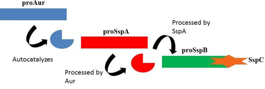
encoding a cytoplasmic inhibitor of SspB (60). The Aur, SspA, and SspB proteases are
expressed as proenzymes, which are subsequently activated in what is known as the
Aureolysin is a secreted protease of S. aureus belonging to the M4 metalloproteinase
family of enzymes, which include a number of enzymes which are acknowledged virulence
factors. Aureolysin cleaves several host proteins, including peptides involved in immune
escape. In vitro experiments have shown that aureolysin cleaves some plasma proteinase
inhibitors and activates prothrombin in human plasma (69).
Aureolysin is regulated by agr (accessory gene regulator) and sarA (Staphylococcal
accessory regulator), both of which are known virulence regulators in S. aureus despite the
lack of understanding on the importance of aureolysin in virulence. Transcription of
extracellular proteases is generally repressed by sarA and stimulated by agr (54). The agr
gene system results in production of RNA III, which stimulates the expression of several
exoprotein genes. While the exact mechanism of RNA III dependent regulation is unknown,
it is believed to interact with other regulatory proteins rather than direct binding to the
promoter of target genes. During regulation of agr, RNA III is believed to neutralize a
repressor of aur, the Rot protein expressed by the rot gene, a global gene regulator. However,
a direct repressor of aur is SarA, the product of sarA, which binds the aur promoter (54, 82).
When the gene encoding aureolysin was characterized, it was revealed that the
product would be about 509 amino acids in length, whereas the crystal structure of the
protein revealed only 301 amino acids, suggesting that aureolysin undergoes
post-translational processing. The enzyme in its unprocessed form is expressed as a 56 kDa
propeptide which undergoes autocatalysis resulting in the removal of a 23 kDa propeptide to
yield the 33 kDa active mature enzyme (69). As seen with other members of the M4 family
of metallopeptidases, aureolysin acts as a proprotein convertase and processes and activates
other enzymes. The activity of this protease depends on both a zinc ion in the active site, as
such as EDTA. Aureolysin is required for the progression of the Staphylococcal proteolytic
cascade, and inactivation of aureolysin results in a culture which lacks proteolytic activity
(69).
Activation of the serine glutamyl endopeptidase is dependent on aureolysin, although
it is necessary for proSspA to first undergo a series of initial autocatalytic steps in order for
efficient final activation by aureolysin to occur. Autocatalyis is enabled by the presence of a
glutamine rich region in the propeptide, and subsequent release of the mature enzyme occurs
from cleavage at Leu56 followed by Val69 by aureolysin. The mature form of SspA is
involved in the moderation of S. aureus adhesion to fibronectin, as it degrades cellsurface
fibronectin binding proteins and consequently contributes to invasive infection (70).
Furthermore, the mature SspA is also responsible for the activation of proSspB.
The final product of the staphylococcal proteolytic pathway is the mature form of the
Staphopain B cysteine protease. It has been established that SspC helps to maintain the SspB
precursor as an inactive zymogen (60). The activation of proSspB is dependent upon SspA
cleavage of the C-terminal end of the propeptide of SspB. The mature SspB protease has a
variety of roles, one of which is moderating the adhesive functions of the cell, and it has been
shown to preferentially cleave fibronectin to release the N-terminal portion.
The proteases of S. aureus staphylococcal cascade contribute to the virulence of this
pathogen by degrading plasma molecules, degrading molecules of the immune system
1.4 Lipases
1.4.1 Microbial Lipases
Lipases are enzymes found in animals, plants and microorganisms and have become
important for industrial usage. They are a class of hydrolases and, more specifically,
esterases which catalyze the hydrolysis of acylglycerol into glycerol and free fatty acids in a
lipid-water interface. Lipases can also catalyze the hydrolysis and transesterification of other
esters in addition to the synthesis of esters. Lipases are differentiated from esterases as they
are capable of degrading both emulsions and monomeric substrate where esterases only
degrade the latter (39). Thus lipase activity is dependent on the presence of an interface, such
that lipases were further defined as carboxylesterases acting on emulsified substrates (39).
Lipases can be classified into three groups, based on their substrate specificity. One
group of lipases hydrolyses only the primary ester bond of a glyceride, although recently
lipases with low activity towards hydrolyzing the second ester bond have also been
discovered. Another group of lipases exhibit high preference for certain fatty acids, for
example lipase B from Geotrichum candidum is specific for fatty acids with a double bond
between C9 and C10 (85). However no bacterial lipase belongs to this group. The final group
of lipases, which includes the lipases from S. aureus, have no positional or fatty acid
specificity (39). Bacterial lipases vary greatly in size, and although the overall homology of
lipases is relatively low, all share a similar three dimensional fold known as the α/β
hydrolase, and the region of highest conservation is the active site containing a Ser-His-Asp
catalytic triad (81). In most lipase structures, the active site is covered by surface loops and
helical structures, making it inaccessible. The active site becomes exposed upon interaction
1.4.2 S. aureus Lipase
Lipolytic activity in staphylococci was discovered as early as 1901. Lipolytic activity
by S. aureus is known to release large amounts of fatty acids, for example linoleic acid in
human plasma. The lipases responsible for these reactions are extracellular lipases which are
secreted into the medium (32). The Glycerol Ester Hydrolase lipase of S. aureus, is secreted
as a 72 kDa precursor enzyme (proGeh), which is then processed into mature 42 kDa form
(Geh). Its function, as the name suggests is to catalyze the hydrolysis of the ester bonds
between glycerol and fatty acids, which form triglycerides and this is believed to aid the
bacteria by contributing to the breakdown of host tissue, subsequently liberating nutrients.
The lipolytic activity of this lipase is sensitive to metal ion chelators such as EDTA, as the
enzyme requires calcium to function (32).
Geh has been implicated in virulence, and has been shown to interfere with the host
granulocyte function, and increase survival of the bacteria against the host defense by
inactivating bactericidal lipids (37). S. aureus strains recovered from deep infections show
significantly more lipase activity than those obtained from superficial abscesses.
Furthermore, during biofilm formation the lipase encoding genes were found to be induced
and were found to be up-regulated during biofilm formation in S. aureus. Furthermore, lipase
inhibitors have been shown to reduce biofilm formation in S. aureus. Additionally, mutation
of the lipase genes results in reduced peritoneal abscess formation (37). Other than this,
however, there has not been any significant research regarding the contribution of Geh to S.
1.5 Rationale and Hypothesis
The goal of this research was to elucidate a mechanism involving lipid metabolism in S.
aureus. Previous research indicates that when grown in the presence of linoleic acid, the lag
phase of growth of USA300 was severely prolonged. Additionally, robust induction of the
Staphylococcal proteolytic cascade was also observed, resulting in processing of proGeh to
Geh, due to the presumed proteolytic activity of aureolysin. As a result, we hypothesize that
lipid metabolism is a key factor contributing to the persistence of USA300 on human skin and
its dissemination within the community. The first objective of this study was to purify both
proGeh and aureolysin, and confirm that aureolysin was required for the processing of
proGeh. The second objective was to determine the effect of triglycerides on growth of
USA300 and mutants deficient for proGeh (and consequently the mature Geh) and
aureolysin. This was a follow-up on the multitude of events seen during growth in linoleic
acid, including the effects on the proteolytic cascade, lipase maturation and overall growth.
From these experiments it was determined that trilinolein resulted in an extended lag phase in
wild-type and aureolysin deficient strains, and the lag phase was especially prolonged in
aureolysin deficient strains. Conversely, trilinolein had no effect on Geh deficient strains.
Since the aureolysin mutant, which produces only proGeh, had a longer lag phase than the
wild-type, the third objective was to determine which factors caused the lag in growth and
MATERIALS AND METHODS
2.1 Bacterial strains and growth conditions
The bacterial strains and plasmids used in this study are described in Table 2. MRSA
isolate pulsed-field gel electrophoresis type USA300 LAC that had been cured of the
erythromycin resistance plasmid was used in all experiments as the wild-type (WT) strain.
Unless otherwise indicated, both E. coli and and S. aureus were cultured at 37°C and stored
at -80°C in 20% glycerol. E. coli strains were grown in Luria Bertani (LB) medium. S.
aureus strains were grown in tryptic soy broth (TSB). For strains carrying resistance genes,
antibiotics were used at the following concentrations: chloramphenicol (10 μg/mL) and
erythromycin (3 μg/mL) for growth of S. aureus strains; ampicillin (100 μg/mL) and
kanamycin (40 μg/mL) for growth of E. coli strains. Solid media were obtained by the
addition of 2% (w/v) Bactor agar (Difco). Water for preparation of growth media and
solutions was obtained by passage through a Milli-Q water filtration system (Millipore
Table 2. Strains used in this study
Strain or
plasmid Description
a
Source or reference
Strains
S. aureus
USA300 LAC Community-acquired MRSA; WT strain, cured
of resistance plasmid (2)
RN4220 rK- mK+ ; capable of accepting foreign DNA (49)
H2660 USA300Δgeh This Study
H2789 USA300Δaur::Erm; Ermr (2)
RN6390ΔsarA RN6390ΔsarA (70)
E. coli
DH5α
F-ϕ80dlacZΔM15 recA1 endA1 gyrA96 thi-1 hsdR17 (rK− mK+) supE44 relA1 deoR
Δ(lacZYA-argF)U169phoA
Promega
BL21 (DE3) E. coli B (DE3)[F- dcm ompT hsDa (rB- mB-)] (89)
a
2.2 DNA methodology
2.2.1 Plasmid isolation from E. coli
All plasmids used in this study are listed in Table 3. Plasmid DNA was prepared from
E. coli using the E.Z.N.A. Plasmid Miniprep Kit (Omega Biotek) according to the
manufacturer’s instructions. Briefly, approximately 5 mL of stationary phase culture of E.
coli were pelleted via centrifugation and resuspended in 250 μL of SolutionI/RNase (50mM
Tris, pH 8.0, 20 mM EDTA, 100 μg/mL of RNaseA). Cells were lysed by adding 250 μl of
Solution II (200 mM NaOH, 1% (w/v) SDS), gently inverting the tubes followed by
incubation at room temperature for 2-5 minutes. To neutralize the solution, 350 μl of
Solution III (guanidine hydrochloride with acetic acid) was added to the lysate and was
immediately inverted several times until a precipitate formed. The insoluble material was
subsequently centrifuged for 10 minutes at 13000 rpm to form a pellet. The resulting cleared
supernatant was aspirated into a HiBind DNA Miniprep Column (I) and was centrifuged at
13000 rpm for 1 minute. 500 μl of Buffer HB was added to wash the column and ensure that
residual protein contaminations are removed. 700 μl of DNA Wash Buffer diluted with
absolute ethanol was added to the column and centrifuged for 1 minute at 13000 rpm. The
column was subsequently centrifuged for 2 minutes at 13000 rpm to dry the column matrix.
Plasmid DNA was then eluted from the column into a fresh microcentrifuge tube by addition
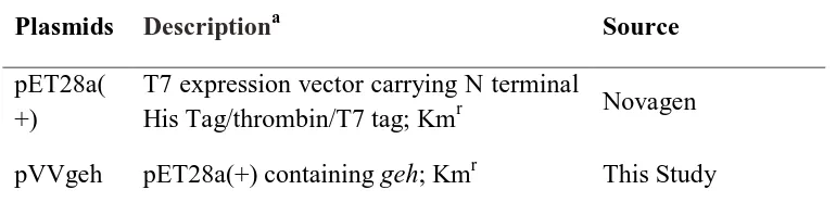
Table 3. Plasmids used in this study
Plasmids Descriptiona Source
pET28a( +)
T7 expression vector carrying N terminal
His Tag/thrombin/T7 tag; Kmr Novagen
pVVgeh pET28a(+) containing geh; Kmr This Study
a
2.2.2 Plasmid isolation from S. aureus
Plasmid DNA isolation from S. aureus followed the same protocol as described for E.
coli but with a few modifications. The harvested cells were incubated at 37°C for 30-60
minutes in a 250 mL mixture of Solution I containing lystostaphin (Sigma) (1 mL Solution I
added to 50 μg of lysostaphin) in lieu of RNase A solution, prior to addition of Solution II.
2.2.3 Isolation of chromosomal DNA from S. aureus
Chromosomal DNA was obtained from S. aureus by pelleting 500 μl of overnight
stationary phase cells grown in TSB culture. 200 μl of STE (75 mM NaCl, 25 mM EDTA, 20
mM Tris pH 7.5) was added to the cells along with 50 μg/ml of lysostaphin dissolved in 20
μL of STE in order to facilitate cell lysis. The cell suspension was incubated at 37°C for 1
hour. 20 μl of 10% SDS and 20 μl of Proteinase K (New England Biolabs) were added and
incubated overnight at 55°C. Subsequently, 80 μl of 5M NaCl was added and mixed by
inversion. 320 μl of a 25:24:1 phenol : chloroform : isoamyl alcohol (IAA) (Invitrogen) was
added and was allowed to sit at room temperature for 30 minutes. The aqueous layer was
removed after the mixture was spun at 12000 rpm for 10 minutes. Addition of 300 μl of 24:1
chloroform:IAA was added and then the mixture wasspun at 12000 rpm for 10 minutes. The
aqueous layer was subsequently removed, and 400 μl of isopropanol was added until the
DNA formed a visible mass while gently inverting. The mixture was allowed to sit for 10
minutes at room temperature, and was then spun at 12000 rpm for 5 minuts. The pellet was
2.2.4 Restriction Enzyme Digests
Restriction enzymes were purchased from Life Technologies, MBI Fermentas, New
England Biolabs, or Roche Diagnostics. Reactions were typically carried out in 30-40 μl
volumes over a 1-2 hour incubation at the appropriate temperature (typically 37°C). Digested
DNA was subsequently cleaned using a QIAquick PCR purification kit (QIAgen) as
described by the manufacturer.
2.2.5 DNA ligations
DNA fragments were ligated in a 20 μl reaction volume using a 10:1 ratio of insert to
vector DNA. Reactions were carried out using the T4 DNA ligase Rapid Ligation Kit (Roche
Diagnostics) in accordance with the manufacturer’s recommendations.
2.2.6 Agarose gel electrophoresis
Agarose gel electrophoresis was used for the separation and analysis of DNA
fragments. Agarose gels (0.8% w/v) were prepared using 1X TAE buffer (40 mM Tris
acetate, 1 mM EDTA) to which either 1.5 μg/ml of ethidium bromide of 2 μl of SYBR Safe
DNA gel stain (Invitrogen) was added. DNA samples to be run were mixed with loading
buffer (5% glycerol, 0.04% bromophenol blue, 0.04% xylene cyanol, 10 mM EDTA, pH 7.5)
prior to being loaded in the gel. Electrophoresis was typically carried out at 110 V for 20-25
minutes. The 1 kb-Plus ladder (Invitrogen) was used as a standard reference marker for
estimation of DNA fragment size. Following electrophoresis, DNA fragments were
2.2.7 Isolation of DNA fragments from agarose gels
Desired DNA fragments were visualized under long-wave UV light (365 nm) and
excised from agarose gels following electrophoresis. DNA was isolated using the QIAquick
Gel Extraction Kit (QIAGEN) using a protocol as described by the manufacturer.
2.2.8 Polymerase chain reaction (PCR)
PCR reactions were carried out in 50 μl reactions containing: DNA template, 1x PCR
buffer, 200 uM dNTP mix (Roche Diagnostics), 12 pM of forward and reverse primers, and
0.5 units of Taq DNA polymerase. PCR reactions were also carried using KAPA HiFi
HotStart PCR Kits (Kapa Biosystems). Briefly, 25 μl reactions containing template DNA, 1X
Kapa HF Buffer (2.0 mM Mg 2+ at 1X), 300 μM of dNTPs, 300 nM each of forward and
reverse primer and 0.5 units of KAPA HiFI HotStart DNA Polymerase. PCRs were
performed using the GeneAmp PCR system (Perkin Elmer), DNA engine Gradient Cycler
(Bio-rad) or the MJ Mini Personal Thermal Cycler (Bio-rad). Oligonucleotide primers were
obtained from Integrated DNA Technologies.
2.2.9 DNA sequencing
DNA sequencing was performed at the DNA Sequencing Facility of the Robarts
Research Institute (London, Ontario, Canada), with sequencing reactions prepared according
to their guidelines.
2.2.10 Computer Analyses
DNA sequence analysis, sequence alignments, and oligonucleotides primer design
Maryland). Blast searches were performed using tools available through the National Center
for Biotechnology Information (http://www.ncbi.nlm.nih.gob/blast/).
2.3 Transformation and transduction methodologies
2.3.1 Preparation of transformation competent E. coli
E. coli DH5a or E. coli BL21 DE3 CaCl2 competent cells were prepared as follows.
An overnight, stationary phase culture of DH5a was diluted 1:100 into 500 ml of fresh LB
and grown to an OD600 of approximately 0.5 and placed on ice for 30 minutes. The cells were
then harvested via centrifugation and resuspended in 100 ml of ice cold 100 mM CaCl2 plus
15% glycerol and incubated on ice for 30 minutes. Cells were again collected by
centrifugation, resuspended in 4 ml of CaCl2 plus 15% glycerol, and stored as 100 μl aliquots
at -80°C.
2.3.2 Transformation of CaCl2 competent E. coli
To transform CaCl2 competent E. coli DH5a or BL21, purified plasmid DNAor
ligation mixtures were added to an aliquot of competent cells and kept on ice for 45 minutes,
after which cells were subjected to a heat shock treatment at 42°C for 2 minutes immediately
followed by a 2 minute incubation on ice. An 800-ul aliquot of LB broth was added to the
tube, mixed, and the cells were permitted to recover for 1 hour at 37°C before being plated
2.3.3 Preparation of transformation competent S. aureus
Strains of S. aureus were made competent for transformation via electroporation as
follows. An overnight culture of S. aureus was diluted 1:100 into 100 mL of fresh TSB and
grown to an OD600 of approximately 0.3. Cells were then harvested via centrifugation and
resuspended in 500 mM sucrose. Cells were subsequently washed three times with ice cold
500 mM sucrose. After the final wash, cells were resuspended in 1 mL of 500 mM sucrose
and stored as 80 μl aliquots at -80°C.
2.3.4 Transformation of electrocompetent S. aureus
Electrocompetent S. aureus were transformed using purified plasmid DNA (typically
5 μl from an EZNA miniprep). DNA was added to a tube of competent cells and allowed to
incubate on ice for 30 minutes before being transferred to an ice-cold electroporation cuvette
(2mm, Bio-Rad) for electroporation. Following incubation, electroporation was performed
using a Bio-Rad Gene Pulser II with setting of 2.5 V, 200 mA, and 25 Ω. Ice cold TSB (800
μl) was immediately added to pulsed cells, which were then recovered for a minimum of 1
hours at 37°C before being plated on appropriate selective media.
2.4 Mutagenesis and DNA cloning methods - mutagenesis of geh
A S. aureus USA300 geh deletion mutant was constructed using the pKOR-1 plasmid
(6). Sequences flanking the geh locus of USA300 FPR3757 were PCR amplified using
specific primers (Table 4) to produce the upstream and downstream arms. A PCR amplicon
of the joined DNA fragments was recombined into the temperature sensitive pKOR1 vector,
resulting in the pKOR-1Δgeh plasmid. The resultant plasmid was first passaged through S.
Table 4.
Oligonucleotides Descriptiona
geh 5′F
GGGGACAAGTTTGTACAAAAAAGCAGGCTAACA TAGGGCATAAGTGGAC
geh 5′R CGCTAACACTGACACCACG
Generation of S. aureusgeh allele, 5’ arm
geh 3′F
/5Phos/GGTATCTGGCAAGTTAAACC
geh 3′R GGGGACCACTTTGTACAAGAAAGCTGGGTTTGCA
CAACTCACTTCACC
Generation of S. aureus geh allele, 3’ arm
geh 5′F-NdeI
TTTTCATATGTTAAGAGGACAAG
geh 3′R-EcoRI TTTGAATTCCAGCACGATTTACATAGC
Cloning of S. aureusgeh
a
allelic replacement of the geh locus was achieved using the method previously
described (6). The correct deletion of codons 25 to 631, of the geh gene, was confirmed by
PCR and DNA sequence analysis.
2.5 Protein methodology
2.5.1 TCA Precipitation, and visualization of secreted proteins
Proteins in the cell-free culture supernatant were precipitated by mixing supernatant
with an equal volume of ice-cold 20% trichloroacetic acid, washed in ice cold 70% ethanol,
then air dried and dissolved in SDS-PAGE reducing buffer (70). Density of the culture
(OD600) was determined before preparation of cell-free culture supernatant, and for the
analysis of secreted protein profiles, TCA precipitated protein derived from 5.0 OD600 units
of culture was loaded into the lanes of an 12% acrylamide gel. The gels were stained with
Coomassie Brilliant Blue R-250 to visualize the protein bands.
Protein bands were excised using an Ettan™ Spot Picker, and processed for mass
spectrometry using a Waters MASSPrep Automated Digestor. Processed protein samples
were spotted on MALDI plates and analysis was done using an Applied Biosystems 4700
Proteomics Analyzer. Data were acquired and processed using the 4000 Series Explorer and
Data Explorer (Applied Biosystems), and using the MASCOT search engine, the peptide
2.5.2 Purification of Aur and Geh
For purification of aureolysin, 1.5 liters of RN6390ΔsarA was grown overnight in
TSB at 37°C in an orbital shaker, set at 200 rpm and the cell-free supernatant was collected.
The supernatant was precipitated by adding ammonium sulphate up to 85% saturation. The
precipitate was then resuspended in binding buffer (20 mM Tris-HCl pH 7.4, 5 mM CaCl2)
and loaded on a sepharose packed 5 mL HiTrap column equilibrated with binding buffer. The
protein was eluted over a gradient of 0%-80% elution buffer (20 mM Tris pH 7.4, 500 mM
NaCl, 5 mM CaCl2). Protein fractions were collected and dialysed against 20 mM Tris pH
7.4 with 5 mM CaCl2. Protein purity was confirmed using SDS-PAGE, and protease activity
was confirmed with a protease assay (described above).
The full length of the gene encoding Geh was PCR-amplified from S. aureus strain
USA300 using forward and reverse primers containing NdeI and EcoRI restriction sites,
respectively. The amplicon was digested with NdeI and EcoRI and cloned into NdeI-Eco
RI-digested pET28a(+) (Novagen), which incorporates a thrombin-cleavable His6 tag at the
N-terminus of the encoded recombinant protein. The pVVgeh (pET28 with Geh) construct was
introduced into E. coli BL21 (DE3). Cells were grown in LB media containing 50 μg/mL
kanamycin at 37°C in an orbital shaker, set at 200 rpm. When the culture reached an OD600 of
0.6, isopropyl β-D-1-thiogalactopyranoside was added to a concentration of 0.5 mM. The
culture was further incubated for another 16 hours at 25°C, after which cells were collected
via centrifugation. The cell pellet was re-suspended in 30 mL Buffer A (10 mM Tris-HCl pH
8.0, 300 mM NaCl, 10 mM imidazole). The cells were lysed using a cell disrupter (Constant
Systems Inc.) at 30 psi after which 10 μM E64, a cysteine protease inhibitor, was added to
Any remaining cellular debris was removed by ultracentrifugation (50000 rpm for 60
minutes) and the soluble lysate was applied to a nickel-loaded 1-ml HisTrap column (GE
Healthcare) equilibrated with buffer A. The His6-tagged protein was eluted from the column
with a gradient of 0%–80% buffer B (10 mM Tris-HCl pH 8.0, 300 mM NaCl, 500 mM
imidazole) using an ÄKTA FPLC (GE Healthcare). Protein fractions were collected and
dialysed against 10 mM Tris pH 8.0. Protein purity was confirmed using SDS-PAGE. Lipase
activity was confirmed using the pNPP based assay as described previously (34) and the
protein was quantified using a Bradford Assay.
2.6 Assays
2.6.1 Lipase Assay
Lipase activity was assayed with para-nitrophenyl palmitate (pNPP) substrate
(Sigma). For assessment of lipase activity in cell-free supernatants, cultures were grown for
18 hours and filtered across a 0.22 micron filter. Supernatant was concentrated using Amicon
10 K centrifugal filters by centrifugation for 30 minutes 3000 rpm. Prior to assay, the
supernatant samples were normalized by dilution with sterile water as needed, to adjust for
minor differences in cell density of the stationary phase cultures at time of harvest. Solution
A (795 μM pNPP substrate in isopropanol) was added to solution B (0.005 % Triton X-100,
50 mM Tris-HCl pH 8.0, 1 mg/mL gum arabic) at a 1:9 ratio. Aliquots of the normalized
supernatant were added to the assay buffer (Solution A and B). For measurement of activity
of purified proGeh or mature Geh (as described previously), aliquots of enzyme were added
to the assay buffer. Samples were incubated for 30 minutes at 37°C in the dark and
2.6.2 Protease assay
Total protease activity of culture supernatants, as well as purified aureolysin, was
assayed with FITC-casein substrate (Sigma). Before the assay, the supernatant samples were
normalized by dilution with sterile water to adjust for minor differences in cell density of the
stationary phase cultures at time of harvest. For purified aureolysin, aliquots of the protease
were mixed in sterile water to a volume of 490 μL. These samples were mixed with 460 μL
of incubation buffer (40 mM Tris-HCl pH 7.4, 300 mM NaCl, 20 mM CaCl2, and 2 mM
L-cysteine) and 50 μL of 0.2% w/v FITC-casein. EDTA was used as an aureolysin inhibitor.
The samples were incubated at 37°C for 2 h in the dark. Trichloroacetic acid was then added
to 4% w/v to stop the reaction, and the samples were centrifuged at 15000 rpm for 15
minutes to pellet undigested casein. The supernatant was then mixed with an equal volume of
0.5 M Tris-HCl, pH 8.5, and after transfer to Optilux black clear bottom microtitre plates
(BD Falcon), fluorescence was quantified on a Biotech plate reader using excitation at 485
nm and emission at 535 nm.
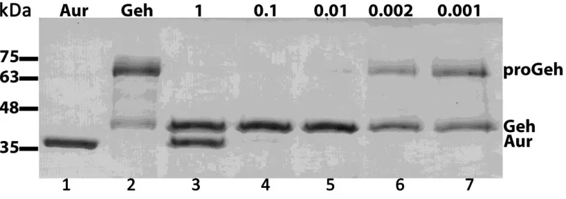
2.6.3 Aureolysin-Geh processing
The rate of Geh processing by aureolysin was determined to optimize the minimum
quantity of aureolysin required to produce mature Geh. Briefly, 3 μg of Geh was first
incubated at 37°C for 3 hours in lipase buffer (50 mM Tris HCl pH 8.0) containing varying
amounts (1 - 0.001 μg) of aureolysin. Additionally, a suitable time frame for the reaction was
determined by incubation of 3 μg of Geh at 37°C in lipase buffer (50 mM Tris HCl pH 8.0)
with 0.01 μg of aureolysin. Samples were removed from the incubator at varying time points
followed by heating for 5 minutes at 95°C. Assessments of the levels of proGeh and mature
Geh were made visually following SDS-PAGE.
2.7 Influence of triglycerides on S. aureus growth
Trilinolein was purchased from TCI America. Prior to supplementing TSB media, the
triglyceride was first mixed with an equal volume of DMSO, and then diluted in TSB to a
working stock concentration of 5 mM. Cell-free supernatant was obtained from cultures
grown for 18 hours in 25 mL volume TSB at 37°C on an orbital shaker incubator. Purified
proGeh was prepared by incubation at 37°C in lipase buffer (50 mM Tris HCl pH 8.0)
whereas mature Geh was prepared by incubation of proGeh with aureolysin (0.002 μg) in
lipase buffer for 3 hours at 37°C. Washed cells were prepared by removing the cell free
supernatant from precultured cells, and washed in saline twice before re-suspension in fresh
TSB media.
For growth analyses, bacteria from single colonies on TSB agar were inoculated into
culture tubes containing 5 mL of antibiotic free TSB, and grown overnight at 37°C on an
orbital shaker, followed by measurement of OD600. A 25-mL volume of TSB, supplemented
with triglyceride, supernatant (100 μL) and/or purified protein (2.7 μg) , was then inoculated
to achieve a starting OD600 of 0.01, and the cultures were grown at 37°C on an orbital shaker
incubator, set at 200 rpm. Measurements of OD600 were taken at set time points. All growth
analyses were conducted in at least triplicate, from individual cultures.
2.8 Lipid extraction and analysis by GC/MS
The substrate trilinolein was tested against the two forms of Geh, proGeh and mature
Tris-HCL pH 8.0) for 3 hours at 37°C. To obtain mature Geh, 3 μg of proGeh was incubated in
lipase buffer (50 mM Tris-HCL pH 8.0) with 0.002 μg of aureolysin for 3 hours at 37°C. Post
incubation, trilinolein was added to a final concentration of 5 μM in a total 50 μL final
reaction volume and incubated at 37°C for 60 minutes.
Fatty acids were then extracted two consecutive times with 500 L of hexane, and the
hexane extracts were then pooled together and dried under a constant stream of nitrogen gas.
Dried samples were trimethylsilylated with 50 L of pyridine and 50 L of,
Obis(trimethylsilyl)trifluoroacetamide (BSFTA) + 1% Trimethylchlorosilane (TMS)
(Sigma), and incubated at 70°C for 40 minutes.
Fatty acids were chromatographed as their TMS-esters on an Agilent 7890 GC
equipped with a CP-Sil 5 column (0.25 mm x 30 m) and a flame ionization detector (FID).
Samples (1 µL) were injected in splitless mode onto the column and eluted with a
temperature gradient as follows: initial temp. 80°C held for 2 min, followed by a ramp up to
220°C (40°C/min) and then a ramp up to 300°C (15°C/min). The final temperature was held
for 4.2 min for a total run time of 15 min. Nitrogen (N2) was used as a carrier gas at 2.0
RESULTS
3.1 Deletion of the geh locus
As stated previously, our laboratories' previous work showed that unsaturated fatty
acids, including linoleic acid, induced robust expression of the proteolytic cascade. One
result of this was the processing of proGeh (pro glycerol ester hydrolase; lipase;
SAUSA300_0320) to its mature form. One of the main goals of this study was to determine
if this processing had any role to play in the biological response of S. aureus to the presence
of fatty acids. The geh gene is 2, 073 bp and encodes a protein containing 690 amino acids
with a molecular mass of 72 kDa. In order to assess the importance of Geh during growth in
the presence of antimicrobial lipids, a geh in-frame deletion was constructed as described in
the Materials and Methods section. PCR amplification across the geh locus of wildtype and
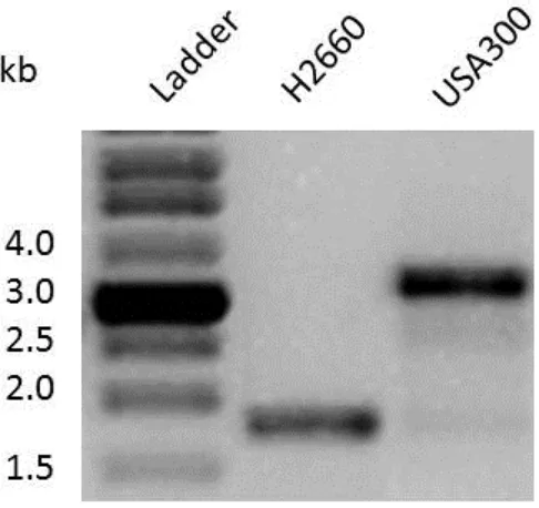
Figure 4. Generation of a Δgeh mutant in S. aureus strain USA300. PCR amplification
across the geh open reading frame confirms the deletion in the mutant strain. The PCR
amplicon was sequenced for final confirmation. Strain H2660 is the number given to the
3.2 Growth in fatty acid results in processing of Geh
As stated above, previously, our laboratories showed that when grown in 50 μM
linoleic acid, USA300 exhibited an extended lag phase in addition to the robust expression of
the staphylococcal proteolytic cascade (2). This proteolytic cascade begins with the
expression of the metalloprotease aureolysin and culminates with the activation of the
cysteine protease SspB. Additionally under these conditions, the 72 kDa precursor form of
Geh is processed into its mature 42 kDa form, but this processing is absent in an aureolysin
mutant (2). The previous data was shown using MS to identify proteins of interest. We
confirmed this phenotype and, using the geh and aur mutants, proved that indeed Geh was
the protein being processed in an Aur-dependent fashion (Figure 5). Moreover, when we
followed the kinetics of growth in the presence of linoleic acid, both the USA300Δgeh
mutant and the USA300Δaur mutant displayed the same 12 hour lag period as wild type
USA300 (data not shown), indicating that neither geh nor aur are required for growth in
inhibitory concentrations of linoleic acid, nor are they directly responsible for the delay in
growth in response to linoleic acid.
3.3 Purification of Aur and Geh, and confirmation of their activity
In a mutant lacking the aur gene, proGeh is among several proteins which remain
unprocessed, as they require the proteolytic activity of the metalloprotease aureolysin.
aureolysin is a zinc and calcium ion-dependent metalloprotease which initiates the
staphylococcal proteolytic cascade. Therefore, to conduct a detailed analyses of the role of
Aureolysin in processing of proGeh, we first needed to purify the two enzymes. As
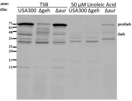
Figure 5. Induction of Aur by linoleic acid results in processing of proGeh to Geh in S.
aureus culture supernatants. SDS-PAGE of the culture supernatants of wild type and
mutant USA300 strains grown in TSB or TSB supplemented with 50 μM linoleic acid for 18
hours. Proteins in the cell free culture supernatant were precipitated in ice-cold TCA, and
after solubilization in SDS-PAGE reducing buffer, protein equivalent to 3.5 OD600 units of
culture supernatant was loaded in each lane. Note that while the cultures grown in the
presence of LA took longer to reach stationary phase, the cultures eventually reach the