Goldsmiths, University of London
Department of Psychology
PhD in Psychology
2012
Traumatic brain injury:
Relationships between brain structural abnormalities and
cognitive function
Kirsi M. Kinnunen
Goldsmiths, University of London
Submitted in fulfilment of the requirements for completing a PhD in Psychology © Department of Psychology, Goldsmiths, University of London
Acknowledgements
Firstly, I would like to thank my supervisors, Professor Jane Powell of Goldsmiths, University of London and Dr David Sharp of Imperial College London, who provided me with their intellectual mentorship over the course of this doctorate. I especially owe many thanks to Jane for her thorough approach, advice and patience during the write-up and to David for hosting me in his research group. The contribution made by the patients and healthy volunteers who took part in the studies is also invaluable – I cannot thank them enough.
For collaborations, I owe gratitude to the Traumatic Brain Injury team based at the Computational, Cognitive and Clinical Neuroimaging Laboratory (C3NL) of Imperial College London, and the wider Cognitive Neuroimaging Group there, especially Dr Robert Leech. I should also thank Group Leader Professor Richard Wise. Further, I would like to thank Dr Richard Greenwood of the University College London/Institute of Neurology, the National Hospital for Neurology and Neurosurgery, and the Regional Neurological Rehabilitation Unit of the Homerton University Hospital, as well as Professor Alan Pickering of Goldsmiths. Importantly, I would like to express my gratitude to the Department of Psychology of Goldsmiths – many thanks for the support.
This doctorate has been an eventful journey with its highlights and challenges. I am truly grateful to everyone who has been there for me along the way in one capacity or another. Most importantly, it is hard for me to imagine having successfully completed this thesis without my family and their unconditional support and encouragement. I also cannot imagine what these past few years would have been like without my friends. For their endless patience, generosity and numerous other reasons – including just being there and being great company – I owe my nearest and dearest the most heartfelt thanks.
Many thanks also to:
Internal Examiner: Professor Paul Burgess, Institute of Cognitive Neuroscience (ICN) and the Department of Psychology, University College London
External Examiner: Dr Michael O’Sullivan, Cardiff University Brain Research and Imaging Centre (CUBRIC) and Schools of Psychology and Medicine, Cardiff University
Statement of Contributions
Whilst the overall research project that the studies reported in this thesis are part of was an effort by the entire Traumatic Brain Injury Team at Imperial College London, all material presented here is based on my cental role in all aspects of the studies: their design, participant recruitment, the general running of the studies, data collection and analysis, and the interpretation of results and their preparation for publication in journals and presentation at scientific meetings and conferences. As regards data collection, at the same time as I was carrying out neuropsychological testing, other members of our team, including team leader Dr David Sharp, PhD students Ms Valerie Bonnelle and Dr Tim Ham and visiting researcher Dr Xavier De Boissezon, with the help of Ms Emer Hughes and Ms Amy McGuinness, assumed primary responsibility over the collection of the neuroimaging data. It was my responsibility to collect all patient neuropsychological data as well as to oversee the collection of healthy volunteer data, including the training of MSc students Ms Shezmin Kassam and Mr Peter Hawkins of Goldsmiths to carry out the healthy volunteer assessments.
Signed
Abstract
Traumatic brain injury (TBI) is the leading cause of disability in young adults and a major public health problem. Persistent cognitive impairments are common, and constitute a significant source of long-term disability. The specific pathophysiological mechanisms underlying these impairments remain poorly understood. As it disconnects brain networks, white matter damage can be a key determinant of cognitive impairment after TBI. Neuroimaging and neuropsychological methods were employed to explore the relationships between indices of brain structure and cognitive function. The participants were 40 TBI patients and 40 healthy controls. First, relationships between focal lesions and cognitive performance were investigated using structural magnetic resonance imaging (MRI) and a battery of neuropsychological tests. The results demonstrated that lesion location and load are not good indices of the cognitive deficits - probably because diffuse axonal injury is poorly assessed by standard MRI. By contrast, diffusion tensor imaging (DTI) can be used to quantify the microstructure of white matter. A ‘whole-brain’ technique, tract-based spatial statistics (TBSS), was used to flexibly analyse the structure of white matter tracts. Despite only small amounts of focal damage observed using standard MRI, TBSS revealed widespread white matter abnormalities after TBI. White matter damage was found in patients with no evidence of focal damage, and in patients classified as ‘mild’ clinically. Relationships between white matter tract structure and specific cognitive functions were then explored. The structure of the fornix, an important white matter pathway of the hippocampus, correlated with verbal associative memory across the patient and control groups. By contrast, structure of frontal lobe connections showed distinct relationships with executive function in these two groups. The results emphasise the importance of white matter pathology after TBI and suggest that disruption to specific white matter tracts is associated with particular patterns of cognitive impairment, but also highlight the complexity of these relationships.
TABLE OF CONTENTS Page Title page ... 1 Acknowledgements ... 2 Statement of contributions ... 3 Abstract ... 4 Table of contents ... 5 List of tables ... 12 List of figures ... 13 List of abbreviations ... 14 CHAPTER 1: Introduction ... 16
1.1 Motivation for the Research ... 16
1.2 What Is ‘Traumatic Brain Injury’? ... 18
1.2.1 Definition ... 18
1.2.2 Characteristic sequelae ... 19
1.3 Epidemiology and Aetiology of Traumatic Brain Injury ... 19
1.4 Injury Mechanisms, Neuropathology and Severity of Injury ... 22
1.4.1 Physical mechanisms ... 22
1.4.2 Primary and secondary injury ... 23
1.4.3 ‘Focal’ injury ... 24
1.4.4 Diffuse axonal injury (DAI) ... 25
1.4.5 Classification of injury severity ... 27
1.4.6 Relationships between injury severity, neuropathology and outcome ... 29
1.5 Structural Neuroimaging of Traumatic Brain Injury ... 33
1.5.1 Investigation of clinically important brain injuries ... 33
1.5.2 A historical perspective ... 33
1.5.3 Standard neuroimaging of TBI ... 34
1.5.4 Towards new advanced imaging methods ... 35
1.6 Diffusion Tensor Imaging (DTI) ... 36
1.6.1 Diffusion tensor imaging of brain white matter ... 36
1.6.2 The diffusion tensor model ... 37
1.6.3 The biological basis of diffusion ... 37
1.6.4 Limits of the tensor model ... 38
1.7 Cognitive Impairment Associated with Traumatic Brain Injury ... 40
1.7.1 Cognitive impairment after TBI and neuroanatomical correlates ... 40
1.7.2 Neuroanatomical substrates to verbal learning and memory ... 41
1.7.2.1 White matter connections of the hippocampal formation ... 42
1.7.2.2 A link between the human default mode network (DMN) and memory? ... 43
1.7.3 Neuroanatomical substrates to executive functions ... 43
1.7.3.2 Neural networks and executive function ... 45
1.7.4 Neuroanatomical substrates to information processing speed ... 46
1.7.4.1 Slowed information processing speed after TBI ... 46
1.7.4.2 Relevant white matter tracts and their degradation by TBI ... 46
1.7.5 Estimating the effects of brain injury on specific cognitive functions ... 47
1.8 Investigating Traumatic Brain Injury and Its Sequelae Using Neuropsychological and Neuroimaging Methods ... 48
1.8.1 The role of clinical neuropsychology ... 48
1.8.2 The network approach to understanding cognitive impairment ... 49
1.8.3 Application of DTI to the investigation of white matter damage and relationships with cognitive deficits……….. 51
1.8.3.1 Why quantify white matter damage using diffusion tensor imaging? ... 51
1.8.3.2 Region of interest (ROI) approaches ... 52
1.8.3.3 Whole-brain and voxel-based approaches ... 52
1.8.3.4 Tract-based spatial statistics (TBSS) ... 53
1.9 Main Objectives, Research Questions and Hypotheses ... 54
CHAPTER 2: Methods and materials ... 57
2.1 Overall Design ... 58
2.2 Recruitment of Participants ... 58
2.3 Designs of Empirical Studies ... 60
2.3.1 Study 1 (Chapter 3) ... 60
2.3.2 Study 2 (Chapter 4) ... 61
2.3.3 Study 3 (Chapter 5) ... 61
2.4 Participants ... 61
2.5 Justification of Sample Sizes ... 63
2.6 Neuropsychological Assessment ... 63
2.6.1 Justification for test selection ... 63
2.6.2 Premorbid intellectual ability: The Wechsler Test of Adult Reading ... 65
2.6.3 Current reasoning ability ... 66
2.6.3.1 Similarities ... 66
2.6.3.2 Matrix Reasoning ... 67
2.6.4 Verbal learning and memory ... 67
2.6.4.1 The People Test ... 67
2.6.4.2 Logical Memory ... 68
2.6.4.3 Digit Span ... 69
2.6.5 Executive function ... 69
2.6.5.1 Trail Making Test ... 69
2.6.5.2 Color-Word Interference test ... 70
2.6.5.3 Verbal Fluency/letter fluency ... 71
2.6.7 Self-report measures ... 73
2.7 Structural Neuroimaging: Basic Principles of Methods ... 73
2.7.1 The MR signal ... 73
2.7.2 Time constants (T1, T2 and T2*) and image contrast ... 74
2.7.3 Reconstructing the MR signal: k-space and Fourier Transform ... 75
2.7.4 Diffusion tensor imaging ... 76
2.7.4.1 Diffusion weighting and image acquisition ... 76
2.7.4.2 Diffusion coefficient and diffusion tensor ... 76
2.7.4.3 Quantitative DTI ... 77
2.8 Structural Neuroimaging Sequences ... 78
2.8.1 Magnetic resonance imaging apparatus ... 78
2.8.2 T1- and T2*-weighted magnetic resonance imaging sequences ... 78
2.8.3 Diffusion tensor imaging sequence ... 78
2.8.4 Preprocessing of diffusion tensor imaging data: A summary ... 78
2.9 Procedure ... 81
2.9.1 Consenting and interview ... 81
2.9.2 Neuropsychological assessment ... 81
2.9.3 Neuroimaging ... 82
2.10 Statistical Data Analysis Methods ... 83
2.10.1 Demographic and neuropsychological data ... 83
2.10.2 Standard magnetic resonance imaging data and cognitive correlates ... 83
2.10.3 Analysis of DTI data and relationships with neuropsychological variables ... 84
2.10.3.1 Tract-based spatial statistics ... 84
2.10.3.2 Nonparametric permutation-based data analysis ... 87
2.10.3.3 Viewing and interpreting the results ... 88
CHAPTER 3: Standard magnetic resonance imaging of traumatic brain injury and relationships with cognitive function ... 89
Abstract ... 89
3.1 Introduction ... 90
3.1.1 Common neuropathological effects of TBI ... 91
3.1.2 Standard neuroimaging of traumatic brain injury in the detection of neuropathology ... 92
3.1.3 Neuropsychological correlates of typical MRI findings after TBI ... 93
3.1.3.1 Regional and whole-brain atrophy ... 93
3.1.3.2 ‘Focal’ lesions and cognitive function ... 95
3.1.4 Investigating relationships between brain structure and cognitive function ... 97
3.1.4.1 Traditional approaches to lesion analysis ... 97
3.1.4.2 Voxel-based lesion-symptom mapping: A whole-brain approach ... 97
3.1.5 Aims of the present study ... 98
3.1.6 Hypotheses ... 98
3.2.1 Design ... 99
3.2.2 Participants ... 99
3.2.3 Neuropsychological assessment ... 99
3.2.4 Magnetic resonance imaging ... 100
3.2.4.1 Data acquisition and estimation of brain volume ... 100
3.2.4.2 Lesion mapping: Lesion type and anatomical location ... 101
3.2.4.3 Lesion analysis: Lesion load ... 102
3.2.5 Statistical analyses ... 103
3.2.5.1 Brain volume: Patients versus controls ... 103
3.2.5.2 Brain volume: Relationships with neuropsychological performance ... 103
3.2.5.3 Relationship between anatomical location of brain contusions and cognitive function ... 104
3.2.5.4 Relationship between anatomical location of white matter lesions and cognitive function ... 105
3.2.5.5 Lesion load and cognitive function ... 105
3.3 Results ... 105
3.3.1 Participant demographics ... 105
3.3.2 Neuropsychological profile of the patient group ... 107
3.3.3 Magnetic resonance imaging findings in the patient group ... 108
3.3.4 Brain volume ... 109
3.3.4.1 Patients versus controls ... 109
3.3.4.2 Relationships with neuropsychological performance ... 110
3.3.5 Lesion mapping results ... 110
3.3.5.1 Anatomical location of lesions ... 110
3.3.5.2 Anatomical location of lesions and cognitive function ... 111
3.3.5.3 Lesion load ... 113
3.3.5.4 Lesion load and cognitive function ... 113
3.4 Discussion ... 114
3.4.1 Profile of cognitive impairments after traumatic brain injury ... 114
3.4.2 Residual brain volume after traumatic brain injury ... 115
3.4.3 Type and anatomical distribution of ‘focal’ lesions ... 116
3.4.4 Type and anatomical location of lesions and cognitive outcome ... 117
3.4.5 Lesion load (size or number) and cognitive outcome ... 118
3.4.6 Methodological limitations and related considerations ... 119
3.4.6.1 Distributed functional networks support complex cognitive functions ... 120
3.4.6.2 Brain damage in TBI is not limited to the site of impact ... 121
3.4.6.3 Neuropathology of TBI is heterogeneous ... 121
3.4.6.4 Associated methodological considerations ... 122
CHAPTER 4: Whole-brain analysis of white matter structure after traumatic brain injury
using tract-based spatial statistics ... 126
Abstract ... 126
4.1 Introduction ... 127
4.1.1 Diffuse axonal injury in TBI ... 127
4.1.2 Pathophysiology of DAI ... 127
4.1.3 Severity of DAI ... 128
4.1.4 Diffusion tensor imaging of DAI ... 129
4.1.5 Effects of severity of injury on outcome after TBI ... 131
4.1.6 Whole-brain versus region of interest approach to analysing white matter tract structure………..132
4.1.7 Aims of the present study ... 134
4.1.8 Hypotheses ... 135
4.2 Methods ... 135
4.2.1 Design ... 135
4.2.2 Participants ... 135
4.2.3 Structural MR imaging data acquisition and analysis ... 137
4.2.3.1 Standard MRI ... 137
4.2.3.2 Diffusion tensor imaging ... 137
4.2.3.3 DTI data analysis ... 137
4.2.4 Assessment of intellectual functioning ... 138
4.3 Results ... 138
4.3.1 Participants ... 138
4.3.2 Standard MR imaging ... 140
4.3.3 TBSS analysis of DTI data ... 142
4.3.3.1 Hypothesis 1 ... 142
4.3.3.2 Hypothesis 2 ... 144
4.3.3.3 Hypothesis 3 ... 145
4.3.3.4 Hypothesis 4 ... 147
4.3.3.5 Hypothesis 5 ... 148
4.3.4 The extent of white matter abnormalities ... 150
4.4 Discussion ... 151
4.4.1 Widespread white matter disruption following TBI ... 152
4.4.2 Microbleed versus diffusion tensor imaging evidence of axonal injury ... 153
4.4.3 Patients with a ‘mild’ TBI show abnormalities of white matter tract microstructure .... 153
4.4.4 Changes in DTI metrics over time after TBI ... 154
4.4.5 Possible pathophysiological mechanisms ... 155
4.4.6 Limitations ... 156
CHAPTER 5: Relationships between white matter tract structure and cognitive
functions………..161
Abstract ... 161
5.1 Introduction ... 162
5.1.1 Outline ... 162
5.1.2 White matter connections and verbal learning and memory ... 162
5.1.3 White matter connections and executive function ... 167
5.1.4 White matter connections and information processing speed ... 171
5.1.5 Investigating the relationship between white matter tract structure and cognitive function using TBSS ... 173
5.1.6 Aims of the present study ... 175
5.1.7 Hypotheses ... 175
5.2 Methods and Materials ... 175
5.2.1 Design ... 175
5.2.2 Participants ... 175
5.2.3 Neuropsychological assessment ... 176
5.2.3.1 Measures of general intellectual ability ... 176
5.2.3.2 Measures of theoretical interest ... 176
5.2.4 Structural imaging ... 177
5.2.5 DTI data analysis ... 177
5.2.6 Analyses of relationships between white matter structure and cognitive function ... 177
5.3 Results ... 178
5.3.1 Participants ... 178
5.3.2 Cognitive function ... 178
5.3.3 The relationship between white matter structure and cognitive function ... 180
5.3.3.1 Hypothesis 1 ... 180
5.3.3.2 Hypothesis 2 ... 183
5.3.3.3 Hypothesis 3 ... 184
5.3.3.4 Hypothesis 4 ... 187
5.4 Discussion ... 188
5.4.1 White matter structure underlying associative learning and memory ... 189
5.4.2 White matter abnormalities and logical memory performance ... 191
5.4.3 White matter structure associated with executive function ... 192
5.4.4 Unexpected findings and limitations ... 194
5.4.5 Conclusions ... 197
CHAPTER 6: General discussion ... 199
6.1 The Research and Its Main Findings: A Summary ... 200
6.1.1 Study 1 (Chapter 3) ... 201
6.1.2 Study 2 (Chapter 4) ... 202
6.2 Discussion ... 203
6.2.1 ‘Focal’ lesions versus diffuse brain injury and cognitive sequelae of TBI ... 203
6.2.2 Future use of DTI in TBI? ... 204
6.2.3 Relationships between DTI findings and cognitive function after TBI ... 206
6.2.4 Methodological considerations ... 206
6.2.5 Future directions ... 211
6.3 Conclusion ... 213
References ... 214
Appendices ... 254
Appendix A: Characteristics of individual patients ... 254
Appendix B: Additional/self-report measures ... 255
Appendix C: DTI preprocessing and tract-based spatial statistics (TBSS): Extended methods ... 256
List of Tables
TABLE Page
Table 1-1 Traumatic Brain Injuries Caused by Each Main Cause in Three European
Regions……….. 21
Table 2-1 Mayo Traumatic Brain Injury Severity Classification System ... 59
Table 3-1a Demographics for the Groups with Structural MRI Data ... 106
Table 3-1b Demographics for the Groups in the Comparison of Neuropsychological Performance ... 106
Table 3-2a Group Means on the Cognitive Measures ... 107
Table 3-2b Group Means on the Subcomponents of Main Measures ... 107
Table 3-3 Brain Volume After Traumatic Brain Injury Compared with Healthy Controls ... 110
Table 3-4 Number/Percentage of Patients with T1 Brain Contusions ... 111
Table 3-5 Number/Percentage of Patients with T2* White Matter Lesions ... 111
Table 4-1 Clinical Characteristics of the Patients ... 136
Table 4-2a Characteristics of the TBI and Control Groups ... 139
Table 4-2b Characteristics of TBI Subgroups Based on Microbleed Presence/Absence ... 139
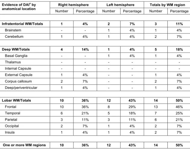
Table 4-3 Number/Percentage of Patients with T2* Evidence of Diffuse Axonal Injury ... 141
Table 4-4 TBSS Group Differences between the TBI and Control Groups ... 144
Table 4-5 TBSS Group Differences between the Microbleed and Non-microbleed TBI Groups ... 146
Table 4-6 TBSS Group Differences between the Non-microbleed TBI and Control Groups .... 148
Table 4-7 TBSS Group Differences between the Mild TBI and Control Groups ... 150
Table 4-8 Percentage/Total Skeletonised White Matter Showing Each Significant Group Difference ... 150
Table 5-1 Scores on Cognitive Measures of Theoretical Interest and Their Subcomponents . 179 Table 5-2 Correlations between the DTI Indices and Cognitive Measures ... 187
List of Figures
FIGURE Page
Figure 1-1‘Lesions’ in traumatic brain injury. ... 26
Figure 2-1 Anisotropic and isotropic diffusion ... 80
Figure 2-2 Fractional anisotropy (FA) modulated by V1 ... 81
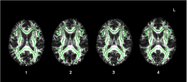
Figure 2-3 Alignment of the mean FA skeleton with individual patients’ white matter tracts. ... 85
Figure 3-1 Lesion distribution and probability ... 108
Figure 4-1 Axons before and after cytoskeletal disruption due to mild TBI ... 128
Figure 4-2 Distribution and frequency of focal lesions ... 140
Figure 4-3 Widespread white matter disruption following TBI ... 143
Figure 4-4 Patients with microbleed evidence of DAI (MB) show more extensive white matter damage than patients without microbleeds (Non-MB) ... 145
Figure 4-5 Patients without microbleeds show DTI evidence of white matter injury ... 147
Figure 4-6 Patients with TBI classified as ‘mild’ also show white matter abnormalities ... 149
Figure 5-1 White matter tracts putatively involved in verbal learning and memory and executive functions ... 169
Figure 5-2 Correlation between associative learning and memory and FA ... 181
Figure 5-3 Group-specific correlations between associative learning and memory and FA ... 182
Figure 5-4 Group-specific correlations between FA and Logical Memory I performance ... 183
Figure 5-5 Group-specific correlations between radial diffusivity (Drad) and set-shifting performance (TMT alternating-switch cost) ... 185
Figure 5-6 Group interaction: the relationship between radial diffusivity (Drad) and set-shifting performance (TMT alternating switch-cost) ... 186
List of abbreviations
Abbreviation Meaning First used on page
TBI traumatic brain injury 4
MRI magnetic resonance imaging 4
DTI diffusion tensor imaging 4
TBSS tract-based spatial statistics 4
CT computed tomography 17
DAI diffuse axonal injury 17
PTA post-traumatic amnesia 18
RTA road traffic accident 21
CSF cerebrospinal fluid 23
GCS Glasgow Coma Scale 27
PCS post-concussive syndrome 31
NICE National Institute for Health and Clinical Excellence 33
FLAIR fluid attenuated inversion recovery 35
SPECT single photon emission computed tomography 36
PET positron emission tomography 36
MRS MR spectroscopy 36
fMRI functional MRI 36
FA fractional anisotropy 38
MD mean diffusivity 38
Dax axial diffusivity 38
Drad radial diffusivity 38
PFC prefrontal cortex 42
DMN default mode network 43
TMT Trail Making Test 46
VBM voxel-based morphometry 47
WTAR Wechsler Test of Adult Reading 47
WAIS-III Wechsler Adult Intelligence Scale- Third Edition 48
WASI Wechsler Abbreviated Scale of Intelligence 48
LM Logical Memory 68
DS Digit Span 69
D-KEFS Delis-Kaplan Executive Function System 70
CRT choice-reaction task 72
T Tesla 73
SNR signal-to-noise ratio 74
T1 spin-lattice relaxation time 74
T2 spin-spin relaxation time 75
T2* T2 star relaxation 75
FT Fourier Transform 75
FMRIB Oxford Centre for the Functional Magnetic Resonance Imaging of the Brain 74
TR repetition time 74
TE echo time 74
FOV field of view 74
FSL FMRIB’s Software Library 78
FLIRT FMRBI’s Linear Image Registration Tool 79
BET Brain Extraction Tool 79
FDT FSL’s Diffusion Toolbox 79
SPSS Statistical Package for the Social Sciences 83
VLSM Voxel-based lesion-symptom mapping 84
FNIRT FMRBI’s Nonlinear Image Registration Tool 84
GLM General Linear Model/Modelling 87
EV explanatory variable 87
TFCE threshold-free cluster enhancement 88
MNI Montréal Neurological Institute 88
GOS Glasgow Outcome Scale 96
MARS Microbleed Anatomical Rating Scale 102
FDR False Discovery Rate 104
CHAPTER 1: Introduction
1.1 Motivation for the Research
Traumatic brain injury (TBI) is a major health and socioeconomic problem, accounting for the majority of deaths due to trauma and for a large proportion of lifelong disability globally (Azouvi et al., 2011; Dikmen, Machamer, Powell, & Temkin, 2003). Those who survive a TBI frequently experience marked cognitive impairment that significantly contributes to the disability (e.g. Berg, Tagliaferri, & Servadei, 2005; Green, Colella et al., 2008; van Velzen, van Bennekom, Edelaar, Sluiter, & Frings-Dresen, 2009; Whitnall, McMillan, Murray, & Teasdale, 2006). Traumatic brain injury thus places a substantial burden on public health and social care resources (Berg et al., 2005; Thurman, Alverson, Dunn, Guerrero, & Sniezek, 1999). In many cases, continued community care and rehabilitation is required subsequent to acute treatment (National Collaborating Centre for Acute Care, 2007). Furthermore, most survivors of TBI are young, meaning that some of the problems can be very long-term, and that important developmental processes may be interrupted, including obtaining qualifications, establishing a vocation, attaining financial independence and forming social networks (Fleminger & Ponsford, 2005; Thornhill et al., 2000). Thus, in addition to direct medical and non-medical costs, TBI is associated with significant indirect costs relating to lost productivity due to reduced/lost employability and personal costs including diminished quality of life (Berg et al., 2005). The burden of TBI on public health and social care is therefore substantial (Thurman et al., 1999).
Persistent functional limitations and failure to return to employment after TBI are particularly associated with cognitive impairments (Green, Colella et al., 2008; van Velzen et al., 2009; Whitnall et al., 2006). Amongst the most commonly affected domains of cognitive function are verbal learning and memory, executive function, and information processing speed (Draper & Ponsford, 2008; Levin et al., 1990; Ponsford & Kinsella, 1992; Salmond, Chatfield, Menon, Pickard, & Sahakian, 2005). Although many patients show improvement of cognitive function following their TBI, for a significant number of patients certain impairments persist in the long-term (Dikmen et al., 2009; Draper & Ponsford, 2008; Ruttan, Martin, Liu, Colella, & Green, 2008; Salmond, Menon, Chatfield, Pickard, & Sahakian, 2006). Given the scale of the problem and the costs associated with TBI, further research to investigate brain-behaviour relationships in TBI is highly motivated, and yet such research remains markedly under-resourced
(Lowenstein, 2009; Maas, Stocchetti, & Bullock, 2008). Understanding of the neural underpinnings of the cognitive impairments frequently observed after TBI therefore remains limited. Their identification is further complicated by the heterogeneity of TBI, and, together, these issues contribute to the difficulty in developing effective treatments. This is why further cross-disciplinary work in this field is needed to elucidate the pathophysiologal mechanisms associated with cognitive dysfunction after TBI. The present programme of research assesses a sample of TBI patients using a combination of neuroimaging and cognitive measures and explores their interrelationships.
Currently, the primary investigation of choice for the detection of clinically important brain injury is computed tomography (CT) imaging. Brain imaging is used in emergency departments to assess the acute neural consequences of head injury and to identify those patients who are likely to go on to develop serious clinical sequelae (National Collaborating Centre for Acute Care, 2007).
Standard brain imaging, however, cannot detect the diffuse axonal injury (DAI) that may cause subtle disruption of the structural integrity of white matter tissue (Symms, Jäger, Schmierer, & Yousry, 2004). Evidence is beginning to accumulate that individual differences in the structure of white matter may have behavioural relevance (see Johansen-Berg, 2010, for a review) and that loss of white matter integrity may contribute to cognitive impairment in neurological conditions (see Chapter 5, for further discussion). Diffusion tensor imaging (DTI; Basser, Matiello, & Le Bihan, 1994; see section 1.6, pp. 36-40) is an advanced magnetic resonance imaging (MRI) technique that holds the promise of substantially improving detection of white matter damage following TBI (Kou et al., 2010; see Chapter 4). The research programme discussed in the subsequent chapters investigates the relationships between the neural and cognitive sequelae of TBI using structural MRI, including DTI, and neuropsychological assessment. Specifically, the principal aim of the research is to identify, using DTI, the white matter correlates of the most prominent cognitive deficits in a group of TBI patients. Having the ability to accurately map after brain injury the functionally relevant changes in white matter structure could inform and considerably improve TBI assessment, clinical management, and outcome prediction.
The following sections provide brief overviews of the definition, epidemiology and aetiology, injury mechanisms and neuropathology, severity classification, and the radiological
investigation of TBI, in particular the potential of DTI in the investigation of white matter injury. The topic of cognitive impairment associated with TBI is then introduced, and the cognitive functions commonly found impaired briefly discussed. A particular emphasis will be on how DTI can be used to investigate the relationship between white matter injury and cognitive impairment following TBI. Finally, a summary of the overall aims, main research questions and hypotheses will conclude this Introduction.
1.2 What Is ‘Traumatic Brain Injury’?
1.2.1 Definition. Traumatic brain injury (TBI) is indicated by the combination of a head trauma and a subsequent constellation of neurological, cognitive and behavioural/emotional sequelae, resulting from a cascade of events triggered by the trauma (Kibby & Long, 1996). Traumatic brain injury is one type of an acquired brain injury; other types including, for example, stroke and anoxic/hypoxic injury. The occurrence of a head trauma does not always imply TBI, especially if there are no neurological signs (Maas et al., 2008). Furthermore, subjective symptoms or cognitive impairments can arise following a head trauma, some of which are related to other reasons (e.g. secondary to stress or mood disturbance), which may not in themselves be definitively diagnostic of TBI.
In general, however, a TBI can be diagnosed where there is objective neuroimaging or clinical evidence of neuropathological changes and/or post-injury signs and symptoms consistent with TBI. These typically include: 1) loss of consciousness, 2) confusion or disorientation, 3) post-traumatic amnesia (PTA), i.e. confused or absent memory for a period of time following the trauma, and 4) focal neurological signs such as decreased sensation or perceptual abilities, loss of balance, general weakness, difficulty walking, abnormal reflexes, persistent headache, seizures, and language problems (Carroll et al., 2004; National Collaborating Centre for Acute Care, 2007).
The Mayo Classification System for Traumatic Brain Injury Severity (Malec et al., 2007) was used in the current research to classify TBI as moderate/severe (definite), mild (probable) or symptomatic (possible), depending on the presence and degree of the above criteria. For further detail on the bases of these classifications and how the Mayo criteria were applied here,
the reader is referred to Chapter 2. Section 1.4.5 (pp. 27-29) of this chapter will discuss issues around TBI severity in general.
1.2.2 Characteristic sequelae. Typical physical, somatic, and sensory effects of TBI include seizures, loss of coordination, partial paralysis, sleep disturbance, fatigue, dizziness, headaches, nausea, visual disturbances, sensitivity to light and sound, and loss of hearing or sense of smell (e.g. Riggio & Wong, 2009). Common hormonal effects include single or multiple pituitary-target neuroendocrine disruption (Rothman, Arciniegas, Filley, & Wierman, 2007). The focus here, however, will be on the characteristic range of cognitive impairments following TBI, discussed below (section 1.7, pp. 40-48), as well as in empirical chapters 3 and 5.
Apart from the above, TBI can result in a variety of behavioural and emotional sequelae that amongst others can include personality changes, agitation and aggression, disinhibition, apathy, and motivational impairment (see e.g. Reeves & Panguluri, 2011). Mood disturbance is common after TBI, with clinically significant anxiety and depression frequently reported (Bowen Neumann, Conners, Tennant, & Chamberlain, 1998; Jorge & Starkstein, 2005). Some of these sequelae are not direct effects of the brain trauma, but rather psychogenic effects relating to factors such as psychological reaction to the consequences of TBI, psychosocial stressors, the individual’s coping strategies, or medicolegal issues. The degree of each of the sequelae may vary both according to TBI severity and as a function of time since the injury.
Associated with these diverse consequences (other than those which are of psychogenic origin) is damage to the physical integrity of nerve cells resulting from TBI, which is often widespread rather than locally restricted. A severe head injury can damage the brain in several ways, and this can lead to a variety of complications. Some types of brain damage are temporary, whilst others can result in permanent damage. Section 1.4 (pp. 22-32) discusses these injury mechanisms and TBI neuropathology.
1.3 Epidemiology and Aetiology of Traumatic Brain Injury
Epidemiological and aetiological data on TBI are currently rather patchy. With this in mind, Tagliaferri, Compagnone, Korsic, Servadei and Kraus (2006) aimed to compile European
data on brain injury epidemiology through a meta-analytic review of 23 national and regional studies in 13 countries. This task proved challenging, as prevalence of TBI (i.e. the total number of cases in a given population at a specific time), a key piece of epidemiological information, is rarely reported in the literature. Although it would be possible to derive an estimate of TBI prevalence based on its incidence (i.e. the total number of new cases during a specific period of time) multiplied by the length of lifespan post-TBI, it is difficult to derive an estimate of lifetime duration of TBI sequelae, partly as a result of the lack of clear and commonly agreed-upon definition for TBI (see Menon, Schwab, Wright, & Maas, 2010). Thus, no reliable data are available on TBI prevalence in Europe. However, using Tagliaferri and colleagues’ (2006) conservative estimate of an average of 10 years of TBI-related disability during a survivor’s lifetime, it is possible that in 2010 as many as 11,775,850 individuals were living within the European Union (population of 501.1 million; Eurostat, 2010) who were coping with the effects of TBI.
Incidence of TBI in developed countries, including the UK, has been estimated to be in the region of 200-300 new cases per 100,000 annually (Torner, Schootman, Rizzo, & Tranel, 1996). The incidence has a bimodal age distribution in that it peaks both in late adolescence/early adulthood and again after the age of 70 years (Marquez de la Plata et al., 2008). Advancing age has been found in several studies to affect outcome so that older TBI survivors in general have worse outcomes (e.g. Flanagan, Hibbard, & Gordon, 2005; Katz & Alexander, 1994). The reasons for this may include the brain’s limited plasticity and capacity for compensation as well as age-related progressive cognitive decline that would disproportionately affect the older survivors. Older individuals (≥65 years) are also more likely than the young to be hospitalised following their TBI (Rutland-Brown, Langlois, Thomas, & Xi, 2006). According to the National Collaborating Centre for Acute Care (2007), 70-88 per cent of all people in the UK who sustain a head injury are male, and overall, males are reported to experience a TBI about twice as often as females (Langlois, Rutland-Brown, & Thomas, 2004).
The TBI-related fatality rate has been reported to be two to three times lower in the UK than in several other developed countries, including France, Spain, Australia and the United States (Jennett, 1996). Tagliaferri et al. (2006) reported an aggregate European rate of 235 fatal and hospitalised TBIs in a population of 100,000 as well as an average mortality rate of 15 TBI-related deaths per a population of 100,000. The UK incidence of hospital admissions due to TBI
is estimated at 253 per 100,000 (Tagliaferri et al., 2006), with over 15,000 intensive care unit beds occupied by TBI survivors annually (National Collaborating Centre for Acute Care, 2007). Morbidity following TBI is disproportionately increased in the younger age groups, and long-term morbidity overall is increased in TBI survivors compared with the general population (Cameron, Purdie, Kliewer, & McClure, 2008).
Thirteen of the studies reviewed by Tagliaferri et al. (2006) provided data on aetiology. Across these studies, road traffic accidents (RTA) were found to be the most common cause of injury, falls the second, and violent assaults the third most common. However, the variation between studies carried out in different regions and countries was considerable. For instance, a comparison between three of the studies, one from the Glasgow region of the UK (Thornhill et al., 2000), another one from the Aquitaine region of Italy (Masson et al., 2001), and the third one from Finland (Alaranta, Koskinen, Leppänen, & Palomäki, 2000), reveals quite different proportions of the three most consistently reported causes, as shown in Table 1-1.
Whilst the top cause of TBI in the Thornhill et al. (2000) study as well as in the Alaranta et al. (2000) study was falls, it was RTAs in the Masson et al. (2001) study. Strikingly, violent assaults were reported to have caused a considerably larger proportion of TBIs in the Glasgow study than either in the Aquitaine or Finnish study. By contrast, the proportion of TBIs resulting from RTAs was much smaller in Glasgow than in Aquitaine, and also smaller than their proportion in Finland. Finally, TBIs caused by falls appear to be clearly more common in Finland than in either Glasgow or Aquitaine.
Table 1-1
Traumatic Brain Injuries Caused by Each Main Cause in Three European Regions
Region (Study) accidents (RTAs) Road traffic Falls Violent assaults Other causes Glasgow, Scotland (Thornhill et al., 2000) 11% 46% 28% 15% Aquitaine, Italy (Masson et al., 2001) 48% 42% 3% 7% Finland (Alaranta et al., 2000) 26% 61% 5% 8%
There is further variation in how these causes relate to TBI severity (see section 1.4.5, pp. 27-29). For example, in the UK overall, falls are estimated to cause 22-43 per cent, assaults
30-50 per cent, and RTAs approximately 25 per cent of mild head injuries. The proportion of RTA-related head injuries classified as moderate/severe is considerably greater (National Collaborating Centre for Acute Care, 2007).
These summary data highlight the difficulty of obtaining consistent estimates of TBI aetiology across studies even as it relates to the most commonly reported causes in developed European countries. Amalgamating data from several studies carried out in different regions is an extremely challenging task, plagued by problems of marked variation in aetiology as well as inconsistencies of TBI case definition, assessment, management and outcome evaluation (Menon et al., 2010). Furthermore, accurate epidemiological data cannot be derived from non-population-based studies. As noted by Cameron et al. (2008), conclusions drawn from the literature are limited by the design and methodological shortcomings of most TBI outcome studies. Apart from non-population-based sampling, these include small sample size, injury severity-specific sampling, lack of appropriate control groups, not controlling for potential confounders such as pre-TBI health status, inadequate length of follow-up and considerable loss of participants to follow-up that can bias findings. Thus, although the figures presented here do give some idea of the scale and nature of the problem, they should be considered as rough estimates only of the true extent of the burden from TBI.
1.4 Injury Mechanisms, Neuropathology and Severity of Injury
1.4.1 Physical mechanisms. The principal physical injury mechanism in TBI is impact loading (collision of the head with a solid object at speed) (Goldsmith, 1966; in Halliday, 1999), associated with a combination of contact and inertial forces. Contact force occurs at the time of impact if the head is prevented from moving, whereas inertial force occurs as the head is set in motion by the impact and rapidly accelerates. These forces can injure the brain if the tissue is compressed (i.e. compressive strain) or stretched (i.e. tensile strain) beyond its structural tolerance (Gennarelli & Meaney, 1996). A third type of tissue strain is shear strain, the least well tolerated type of tissue deformation that occurs as one tissue slides against another (Halliday, 1999). According to Goldsmith (1966), other types of physical injury mechanisms include impulsive loading that without significant physical impact results in sudden motion of the head
as well as static/quasistatic loading in which the speed of the occurrence is not critical (e.g. when the head is trapped between slowly moving rigid structures). These are much less common as causes of TBI than impact loading, however (Halliday, 1999).
The mechanical forces and the ensuing tissue strain can result in complex anatomic and physiologic abnormalities. Properties of the cranium and the intracranial components, including mechanically important features of the interior surface of the skull, subarachnoid space, cerebrospinal fluid (CSF), and intracranial soft tissue, predispose the brain to particular types of injury (Halliday, 1999).
1.4.2 Primary and secondary injury. Direct or indirect impact to the brain triggers a cascade of pathophysiological events. Some of these emerge within hours of TBI and resolve over time, whilst others can persist or gradually develop over long periods of time (Beretta, Gemma, Anzalone, 2008; Kou et al., 2010). Types of brain injury giving rise to the neuropathological changes in TBI are often classified as primary or secondary (delayed) injury, depending on at which point following the initial impact they occur. Where the impact causes the head to bend about the centre of angular acceleration, located in the lower or middle cervical spine, both contact and inertial forces are involved (Halliday, 1999). First, as the brain undergoes acceleration, the soft brain tissue is set in motion inside the skull, and contact and inertial forces cause focal injuries such as ‘coup’ contusions and intracerebral and subdural haematomas (see Figure 1-1A). The mechanical disruption of brain tissue due to the angular acceleration and its associated inertial forces can produce any type of injury apart from skull fractures and epidural haematomas, caused by laceration of veins and arteries by fractured bone edges (Halliday, 1999). Patients who sustain a skull fracture, caused by contact forces producing strain that exceeds the skull’s tolerance, are also at a high risk for intracranial injury (Borg et al., 2004; National Collaborating Centre for Acute Care, 2007).
Some hours following TBI, a process known as secondary injury that leads to cellular damage begins to develop due to factors such as hypoxia, oedema or raised intracranial pressure, and can still be ongoing several months, even years, later (Graham, Adams, Nicoll, Maxwell, & Gennarelli, 1995). For example, in a related study reported elsewhere (Ramlackhansingh et al., 2011), our group found evidence in chronic TBI patients of increased microglial activation in subcortical structures, most likely reflecting an ongoing inflammatory
response to TBI that was found up to 17 years post-injury. Brain microglial cells are sensitive to the neural insult in TBI and rapidly become activated, beginning to secrete cytokines (i.e. small protein molecules that act as cellular regulators), and act to prevent further damage to the neural tissue (see Gehrmann, 1996; Gehrmann, Matsumoto, & Kreutzberg, 1995, for reviews). These recent in vivo results corroborate previous post-mortem findings in humans, demonstrating the presence of a long-term inflammatory response following TBI (Gentleman et al., 2004), and suggest that instead of being a single event, TBI actually triggers a cascade of neuropathological events, some of which persist in the long-term.
In general, secondary injury refers to various neurochemical and molecular alterations that cause damage by disrupting cerebral blood flow, ion homeostasis and metabolism, or by having direct neurotoxic effects on brain cells (e.g. Raghupathi, Graham, & McIntosh, 2000). These processes may also contribute to overall brain tissue atrophy that can increase over time post-injury (see Bigler, 2001a, for a review), although there is also evidence to suggest that overall brain tissue density remains relatively stable after head injury (Salmond, Menon, Chatfield, Pickard et al., 2006).
1.4.3 ‘Focal’ injury. As Bigler (2001a) points out, use of the term ‘lesion’ when referring to brain damage resulting from a TBI can be misleading. What may seem like an isolated lesion on standard clinical neuroimaging of TBI (see section 1.5, pp. 33-36) can always involve more extensive damage to the brain. For example, the tissue and vascular disruption as part of primary injury often results in brain contusions that primarily cause damage to cerebral grey matter, but that can also extend into the white matter. So-called gliding contusions that affect subcortical structures, and are seen especially at grey-white matter junctions, can also result from the angular acceleration of the impact and cause displacement of brain structures (Halliday, 1999). ‘Coup’ contusions, often found under the site of the initial impact, are likely to arise from a situation in which a negative pressure is created by the deformation of the skull at the time of impact that is followed by a rapid return to its usual shape (Gennarelli & Meaney, 1996; Halliday, 1999). ‘Contrecoup’ contusions that resemble coup contusions pathologically affect a site remote from the original point of impact (often, but not necessarily, opposite to it), and are likely to arise from inertial loading that develops from the slight lag between the acceleration of the hard skull and dura and the soft brain (Halliday, 1999). The exact
mechanisms of cerebral contusion formation remain under debate, but their typical pattern following TBI is predominantly fronto-temporal, with regions most frequently affected including the anterior and inferior frontal and temporal regions and the medial temporal region/limbic system (Adams, Graham, Scott, Parker, & Doyle, 1980; Halliday, 1999; Povlishock & Katz, 2005; Ratnaike, Hastie, Gregson, & Mitchell, 2011).
Apart from contusions, the other main ‘focal-appearing’ neuropathological sequelae of closed-head TBI are subdural, epidural, and intracerebral haematomas. Figure 1-1A illustrates how these lesions can alter the original anatomical location of other brain structures, and may cause the midline of the brain to shift. In the case of haemorrhages that include subarachnoid, intraventricular, and tissue tear types, a simple division between focal and diffuse injury may not hold, and the same applies for cerebral swelling and ischemia. Axonal injury associated with structural disconnection, the neuropathological event of primary interest here, is characterised by its widespread distribution (Halliday, 1999; Saatman et al., 2008; see the next section). Chapter 3 will provide further detail regarding ‘focal’ injury following TBI, in particular the occurrence of brain contusions and microbleeds as observed in the current research sample.
1.4.4 Diffuse axonal injury (DAI). Microbleeds are petechial haemorrhages that are associated with DAI following TBI (De Coene et al., 1992; Scheid, Preul, Gruber, Wiggins, & von Cramon, 2003). However, there is no one-to-one correspondence between the number of microbleeds and the degree of severity of DAI (Scheid, Walther, Guthke, Preul, & von Cramon, 2006). Figure 1-1B shows a histopathology slide from a patient who has sustained a severe TBI (Meythaler, Peduzzi, Eleftheriou, & Novack, 2001), illustrating microbleeds and DAI as observed in the corpus callosum of the patient.
White matter structures located in different parts of the brain have different tissue densities and, thus, also distinct mechanical response characteristics. These characteristics determine the way that the injured structure responds to the application of mechanical energy (Meythaler et al., 2001). In a high-speed RTA inertial forces are generated due to the rotational acceleration of the brain, which can result in disarray of intracellular contents, also at sites distant from the original site of impact. However, although DAI can lead to widespread disruption of the structural integrity of white matter and is the predominant injury mechanism in a large proportion of TBIs, axonal fibres in certain regions are particularly susceptible to DAI
(Meythaler et al., 2001). White matter structures at a high risk include the corpus callosum, parasagittal and superior frontal white matter, frontal and temporal grey-white matter junctions, and the brain stem (Lux, 2007; Meythaler et al., 2001; Scheid et al., 2003).
Despite axons’ viscoelastic nature and normally substantial tolerance to stretching, an impact loading TBI can severely damage them (Tang-Schomer, Patel, Baas, & Smith, 2010). The diffuse pattern of injury that results from the shearing and tearing forces during rotational acceleration of the brain can destruct small cytoskeletal components of axons known as neurofilaments and microtubules (see Chapter 4). The localised mechanical damage causes primary axonal swelling, which is followed by disruption of the intracellular components, with potential to ultimately lead to axonal degeneration (Büki & Povlishock, 2006). This secondary degeneration is considered to be the critical mechanism of axonal disconnection for the majority of injured axons (see Li et al., 2010, for a review). The evolution of this process is illustrated in Figure 1-1C (Smith & Meaney, 2000).
Figure 1-1. ‘Lesions’ in traumatic brain injury. A) Types of haematoma. Retrieved March 17, 2011, from: https://doctor2008.wordpress.com/tag/epidural-hematoma. B) Diffuse axonal injury and microbleeds in the corpus callosum. Reprinted from Archives of Physical Medicine and Rehabilitation October 2001, 82/10, Meythaler, J. M., Peduzzi, J. D., Eleftheriou, E., & Novack, T. A., Current concepts: Diffuse axonal injury-associated traumatic brain injury, 1461-1471, Copyright (2011), with permission from Elsevier. C) The process of axonal disconnection. Reprinted from The Neuroscientist December 2000, 6/6, Smith, D. H., & Meaney, D. F., Axonal damage in traumatic brain injury, 483-495, Copyright (2011), with permission from Sage Publications.
A) B)
As the mechanical forces cause the axons to stretch and tear and the usually linear arrangement of the axonal cytoskeleton is lost, the normal flow of water molecules within the axons also becomes interrupted (Büki & Povlishock, 2006). These microscopic characteristics of axonal injury can now be probed using diffusion tensor imaging (DTI), an MRI technique applied to study the diffusion of water molecules in brain tissue (see section 1.6, pp. 36-40). This technique can detect abnormalities in damaged white matter that on standard clinical neuroimaging is deemed to appear normal (e.g. Rugg-Gunn, Symms, Barker, Greenwood, & Duncan, 2001).
Considering the potential of DAI to cause widespread damage to brain tissue, it may also critically contribute to cognitive impairment following TBI (Lux, 2007; Scheid et al., 2006; Sugiyama et al., 2007). A possible mechanism for this would be that DAI interrupts the structural integrity of white matter tracts interconnecting large-scale neural networks that support complex cognitive functions (Mesulam, 1998). For example, impairments of memory and executive function, which have been observed in TBI patients with DAI (Scheid et al., 2006), could relate to the widespread disruption caused to the axonal cytoskeleton by TBI and its associated mechanical forces (Meythaler et al., 2001). Using DTI, it has become possible to investigate the effects of brain injury on the structure of white matter tracts as well as to explore the behavioural relevance of these brain structural abnormalities. For a more detailed discussion of the use of DTI in the assessment of axonal injury following TBI and relationships with cognitive function the reader is referred to chapters 4 and 5.
1.4.5 Classification of injury severity. As mentioned above, inconsistency in the definition of TBI and discrepancy in research methods have made TBI epidemiology difficult to describe accurately. A further complication is the ongoing inconsistency in the classification of TBI severity.
Most typically, TBI severity is classified as mild, moderate or severe based on a patient’s score on the Glasgow Coma Scale (GCS; Teasdale & Jennett, 1974), recorded within 48 hours of injury. Based on the patient’s verbal responses, physical reflexes and ease of eye opening, GCS score ranging from 3 to 15 is assigned, with higher scores indicating less severe injuries. As highlighted by Zuercher et al. (2009), though, in a critical review of the use of the GCS in TBI assessment, inconsistency in the timing of the initial assessment and in the order in
which each item of the scale is administered, as well as the presence of various confounders, diminishes the reliability of the score in clinical and research settings. Moreover, while the GCS score is easy to record, and as such practical in the early clinical management of TBI, it does not provide information about the specific pathophysiologic mechanisms that underlie the observable neurological deficits (Saatman et al., 2008). Thus, the severity of injury in patients recruited for the present research was estimated using the Mayo Classification System for Traumatic Brain Injury Severity (Malec et al., 2007), a system that integrates information about the lowest recorded Glasgow Coma Scale score in the first 24 hours, length of PTA, duration of loss of consciousness, and initial neuroimaging results. Chapter 2 includes the details regarding the basis of determining the presence of TBI in the current patient sample and how the Mayo system was used for classifying injury severity.
Overall, mild TBIs are by far the most prevalent, with the percentage of moderate to severe injuries out of all hospitalised head injury cases being in the range of 10-30 per cent (Cassidy et al., 2004; Tagliaferri et al., 2006). However, because a significant proportion of patients with mild TBIs do not receive hospital treatment, TBI overall is likely to be a considerably larger problem than suggested by official records. Moreover, some of the patients who do receive medical attention initially are discharged from the emergency department without receiving further care and sometimes also without detailed documentation of their injury.
These issues make it difficult to collect accurate data on TBI severity, also acting to bias the epidemiological data. Fewer than half of the European studies reviewed by Tagliaferri et al. (2006) provided information regarding TBI severity. As expected, patients with TBIs classified as mild were the largest severity group in the majority of these studies. However, it is difficult to compare severity data between studies when the classification systems used for severity stratification vary as they did in the reviewed studies. This inconsistency and the potential inaccuracy in TBI diagnosis and severity classification are compounded by the inherent heterogeneity of TBI that further complicates outcome prediction.
Similar clinical characteristics and standard post-injury care can result in very different outcomes, and this significant individual variability includes long-term outcomes (Wagner, 2010). While most patients show at least some functional improvement, and some show dramatic improvement, during the first one to five years post-injury, others remain the same and a small minority seem to decline (Hammond et al., 2004). This presents a major challenge for
both treatment planning and outcome prediction and underlines the need for a valid and reliable multidimensional classification system that could be used to identify specific patterns of injury after TBI and match these with targeted treatments. This could then, potentially, lead to improved outcomes through more individualised interventions (Saatman et al., 2008). It is worth noting, though, that a range of other factors including diverse rehabilitative interventions, social circumstances, and psychiatric conditions also contribute to TBI outcome, which is why predicting it remains a challenge, even with better indices of injury severity.
However, a particular area where there is room for improvement in current TBI stratification systems is that they do not incorporate measures of the degree of white matter injury. The identification of relevant biomarkers that could be used to more fully characterise patterns of brain injury associated with the different degrees of TBI severity would be a step forward. Diffusion tensor imaging, in particular, could prove useful in providing such biomarkers, although its use in clinical diagnosis of TBI remains to be further validated (Huisman et al., 2004; Lee et al., 2008; see Chapter 4, for further discussion). Such research could answer important clinical questions about the mechanisms, prognosis, and treatment of TBI. For example, identifying a specific ‘signature’ of white matter damage after TBI associated with different types and degrees of cognitive impairment could have important implications for early outcome prediction and the planning of maximally effective TBI management and rehabilitation practices.
1.4.6 Relationships between injury severity, neuropathology and outcome. Brain injury is capable of altering the physical status of brain cells and vasculature, and can result from any degree of stretching, twisting or compression, but moderate to severe strain is required for more severe degrees of injury (Bigler, 2001b; Gennarelli & Meaney, 1996). Thus, the extent and severity of damage that ensues is related to the strength of the mechanical force applied during the impact (Kibby & Long, 1996). For example, different strain loads produced by by different injuries also mean varying degrees of damage to the cytoarchitecture of axons and, as a result of this, different degrees of severity of axonal damage (Bigler, 2001b; see Chapter 4). Furthermore, there is recent experimental evidence from an animal model of closed-head injury that the direction of head motion caused by rotational inertial forces plays a role in determining the severity of the neuropathological response, with motion along the sagittal plane
being particularly involved in more severe degrees of injury (Eucker, Smith, Ralston, Friess, & Margulies, 2011). This thesis primarily deals with the effects of TBI on the structure of axonal fibres, which is why the role of vascular disruption in the pathophysiology of TBI, associated with cerebral ischemia and infarction, is not discussed here in any more detail, but the interested reader is instead referred to DeWitt and Prough (2003) for a comprehensive overview of this topic.
Despite the problems relating to its classification, injury severity is widely regarded to be among the most significant predictors of TBI outcome (Mushkudiani et al., 2008; Schönberger, Ponsford, Reutens, Beare, & O'Sullivan, 2009). Schönberger et al. (2009) were the first to investigate whether injury severity together with age, another known predictor of TBI outcome, may also relate to the extent of residual brain injury observed in the chronic stages of TBI. Thus, they studied the relationships between age, injury severity (as indexed by GCS scores and PTA duration) and the extent of residual brain damage (degree of brain atrophy and lesion volumes) in 98 TBI patients 2.3 years post-injury on average. Controlling for gender, TBI aetiology, time since injury, and total brain tissue volume, their regression analyses revealed that older age at injury and longer PTA together were associated with larger grey and white matter lesion volumes in a range of brain regions. In addition, longer duration of PTA predicted smaller residual white matter volumes. They also found that older age predicted smaller ‘lesion-free’ grey matter volumes in several regions, particularly in all neocortical regions, and in the frontal lobe specifically. Surprisingly, though, in the light of a number of studies that have recently demonstrated a relationship between advancing age and breakdown of white matter integrity (see Madden, Bennett, & Song, 2009, for a review), Schönberger et al. (2009) did not find age to be associated with volumes of lesion-free white-matter. This may be partly explained by their use of conventional MRI, though: standard structural MRI is not capable of detecting subtle changes in white matter structure. These findings, as well as emphasising the associations in TBI between initial indirect (clinical) indices of injury severity and subsequent neural pathology, suggest that age may modulate these relationships.
The majority of patients with a mild TBI appear to show a good outcome, and are in general free of residual symptoms after the first year post-injury (Carroll et al., 2004). However, a proportion of such patients experience persistent ‘post-concussive symptoms’. A concussion is indicated by transient confusion following a head trauma, with full recovery normally expected
within two to three weeks (Anderson, Heitger, & Macleod, 2006). As there is no common agreement, the terms concussion and mild TBI are often not clearly distinguished in the literature, which complicates the interpretation of research findings (see Bigler, 2008, for a review). Here, concussion and mild TBI are viewed as follows: concussion refers to a symptom of head injury that has not resulted in loss of consciousness or PTA and in the Mayo system (Malec et al., 2007; see Chapter 2) corresponds to symptomatic (possible) TBI, whilst head injuries associated with at least momentary loss of consciousness (<30 min) or PTA (<24 h) are classified as mild (probable) TBIs.
‘Post-concussive syndrome’ (PCS) is characterised by a collection of markedly non-specific somatic, cognitive and behavioural/emotional symptoms including fatigue, headaches, dizziness, sleep problems, lowered tolerance for alcohol, problems with memory and concentration, irritability, and mood disturbance (Bigler, 2008; De Kruijk, Twijnstra, & Leffers, 2001). Whilst these complaints typically decline within the first few months post-injury, some patients experience subtle symptoms for longer (Williams, Potter, & Ryland, 2010). Post-concussive syndrome is aetiologically ambiguous, however, and many clinicians believe that persistent symptoms (even in cases involving momentary changes in consciousness) reflect psychological factors rather than organic damage. In other words, although mild head trauma is sometimes followed by ongoing problems in daily activities with memory and concentration, it remains controversial whether it can result in persisting sequelae other than those that can follow any traumatic experiences. In some patients there are also pre-injury factors (e.g. a psychiatric condition, alcohol or drug abuse or psychosocial problems) that may contribute to post-injury symptoms (e.g. Mathias & Coats, 1999). Mood disturbance is a common psychological reaction to traumatic events or to their practical consequences, and can also give rise to the kind of non-specific symptoms that fall under the umbrella of PCS, or that may be associated with brain injury.
Thus for instance Ponsford, Cameron, Fitzgerald, Grant and Mikocka-Walus (2011) assessed symptoms that characterise PCS and cognitive function in 123 patients with a suspected mild TBI, compared with 100 matched controls who had sustained traumatic injuries other than TBI. All participants were initially assessed in the emergency department and then at follow-ups first one week and then three months after the injury. Patients with suspected TBI showed significantly more severe post-concussive symptoms than non-TBI controls both initially
and one week post-injury. However, despite being more likely than controls to report ongoing cognitive problems in everyday life at the two follow-ups, the TBI group’s performance was impaired on only one test tapping visual memory. Considering the large number of outcome measures used in the study, it is possible that the visual memory result does not reflect a true deficit in the TBI group. These findings highlight the issue that although some patients report cognitive sequelae or other symptoms after mild head trauma, their correspondence with objectively or clinically identifiable brain injury can be poor. Although mild head injury can result in brain damage, in many such cases the results of standard brain imaging and neurological examination are normal. Such observations have contributed to the controversy regarding whether the lasting problems sometimes observed after mild TBI stem from organic sources (Kibby & Long, 1996; Bigler, 2008; Bigler & Bazarian, 2010). In some cases this issue is further complicated by personal injury litigation (see Belanger, Curtiss, Demery, Lebowitz, & Vanderploeg, 2005, for a review).
It is not clear to what extent the symptoms of PCS correspond to the cognitive impairments often observed following more severe TBI, including deficits of memory, executive function, and information processing speed (see section 1.7, pp. 40-48), and further research is needed to explore the factors underlying persistent cognitive dysfunction across the spectrum of TBI severity.
It is possible that current problems with identifying the relationships between the cognitive sequelae of TBI and the presence of brain injury are partly due to the lack of sufficiently accurate diagnostic methods (Bazarian et al., 2007). The degree of neuropathology in mild TBI especially remains a particular challenge to accurately assess using the standard structural neuroimaging techniques (Niogi & Mukherjee, 2010; see the next section). As stretch-induced DAI is a key injury mechanism in TBI, and may be involved in the development of cognitive impairments and other symptoms, a diagnostic technique able to detect such damage would be a substantial step forward (Bazarian et al., 2007). One promising neuroimaging technique for the assessment of more subtle brain structural abnormalities following TBI is DTI. Section 1.8.3 (pp. 51-54) gives a brief overview of how DTI has been so far used to investigate the structure of white matter after TBI.