BIROn - Birkbeck Institutional Research Online
Maghsood, A.H. and Sissons, J. and Rezaian, M. and Nolder, D. and
Warhurst, D. and Khan, Naveed Ahmed (2005) Acanthamoeba genotype T4
from the UK and Iran and isolation of the T2 genotype from clinical isolates.
Journal of Medical Microbiology 54 (8), pp. 755-759. ISSN 0022-2615.
Downloaded from:
Usage Guidelines:
Please refer to usage guidelines at
or alternatively
Correspondence
Naveed Ahmed Khan [email protected]
Received 30 November 2004
Accepted 15 March 2005
Acanthamoeba genotype T4 from the UK and Iran
and isolation of the T2 genotype from clinical isolates
Amir Hossein Maghsood,
1,2James Sissons,
2Mostafa Rezaian,
1Debbie Nolder,
3David Warhurst
3and Naveed Ahmed Khan
21School of Biological and Chemical Sciences, Birkbeck College, University of London,
London WC1E 7HX, UK
2Department of Medical Parasitology and Mycology, School of Public Health, Tehran University of
Medical Sciences, Tehran, 14155-6446, Iran
3Diagnostic Parasitology Laboratory, London School of Hygiene and Tropical Medicine, London, UK
The majority of the keratitis-causingAcanthamoebaisolates are genotype T4. In an attempt to determine whether predominance of T4 isolates inAcanthamoebakeratitis is due to greater virulence or greater prevalence of this genotype,Acanthamoebagenotypes were determined for 13 keratitis isolates and 12 environmental isolates from Iran. Among 13 clinical isolates, eight (61.5 %) belonged to T4, two (15.3 %) belonged to T3 and three (23 %) belonged to the T2 genotype. In contrast, the majority of 12 environmental isolates tested in the present study belonged to T2 (7/12, 58.3 %), followed by 4/12 T4 isolates (33.3 %). In addition, the genotypes of six newAcanthamoeba
isolates from UK keratitis cases were determined. Of these, five (83.3 %) belonged to T4 and one was T3 (16.6 %), supporting the expected high frequency of T4 inAcanthamoebakeratitis. In total, the genotypes of 24Acanthamoebakeratitis isolates from the UK and Iran were determined. Of these, 17 belonged to T4 (70.8 %), three belonged to T2 (12.5 %), three belonged to T3 (12.5 %) and one belonged to T11 (4.1 %), confirming that T4 is the predominant genotype (÷2¼4.167;
P¼0.0412) inAcanthamoebakeratitis.
INTRODUCTION
Acanthamoebaare ubiquitous organisms found in a variety of environments including soil, water, air, food items, humidifiers, dialysis units and healthy individuals (reviewed by Khan, 2003; Marciano-Cabral & Cabral, 2003; Schuster & Visvesvara, 2004). Based on their ecological distribution, there is circulation ofAcanthamoebastrains between humans and the environment. Acanthamoeba are opportunistic causative agents of nasopharyngeal and cutaneous infections, painful blinding keratitis and fatalAcanthamoeba granulo-matous encephalitis (Khan, 2003; Marciano-Cabral & Cab-ral, 2003; Schuster & Visvesvara, 2004). The difficulty in assigning an explicit role ofAcanthamoeba in infections is becauseAcanthamoebaare heterogeneous and only certain subgroups may be pathogenic. Recent advances in the taxonomy of Acanthamoeba have led to the identification of the pathogenic subgroups. The genusAcanthamoebahas been classified into 15 different genotypes (T1–T15) based on rRNA gene sequencing (Gast, 2001; Stothardet al., 1998; Schuster & Visvesvara, 2004). To date, only isolates belong-ing to the T3, T4, T6 and T11 genotypes have been associated
with Acanthamoeba keratitis (Booton et al., 2002; De Jonckheere, 2003; Khan et al., 2002; Ledee et al., 1996; Stothardet al., 1998; Walochniket al., 2000), and a large number of studies have identified T4 as the predominant keratitis-causing genotype. Indeed more than 90 % of
Acanthamoebakeratitis cases in literature reports have been caused by T4 isolates. As suggested before, the frequent involvement of T4 isolates inAcanthamoebakeratitis may be due to their greater abundance in the environment and thus they are most likely to be picked up by the susceptible hosts, or because T4 isolates possess properties that enable them to be more virulent, or both. In an attempt to clarify this, we determined the environmental and clinical distribution of
Acanthamoebain Iran. We tested 13Acanthamoebakeratitis cases presented during 1998–2003 at the Tehran University of Medical Sciences, Iran. In addition,Acanthamoebafrom environmental sources from widespread geographic loca-tions were isolated and identified at the genotype level using rRNA gene sequencing.
METHODS
of Parasitology and Mycology, School of Public Health, Tehran University of Medical Sciences, Tehran, Iran. A total of 52 keratitis cases was tested. To determine the presence ofAcanthamoeba, speci-mens (from contact lenses or corneal biopsies) were inoculated on to non-nutrient agar (Oxoid) plates seeded withEnterobacter aerogenes (formerly known asKlebsiella aerogenes). Plates were incubated at 308C and observed daily for the presence of amoebae as previously described (Khan & Paget, 2002).
Environmental isolates.Water samples from different cities in Iran (Table 1) were collected and tested for the presence ofAcanthamoeba. For each sample, approximately 100–500 ml water was filtered through a 0.45ìm pore-size filter. Filters were inoculated on to non-nutrient agar plates overlaid withE. aerogenesas described above.Acanthamoeba were identified at the genus level, based on morphological character-istics of trophozoites and cysts using phase-contrast microscopy. Acanthamoeba were axenically cultivated in PYG medium (0.75 %, w/v, proteose peptone; 0.75 %, w/v, yeast extract; and 1.5 %, w/v, glucose) at 308C as described previously (Khan & Paget, 2002).
Our previous studies have isolated Acanthamoeba from other geo-graphic locations including the UK (Khanet al., 2002) and these isolates were compared in the present study. In addition,Acanthamoebaspp. were isolated from six new keratitis patients in the UK (Table 1), identified at the genotype level using rRNA gene sequencing and used in comparative studies.
[image:3.612.72.574.282.691.2]PCR analysis. To confirm the identity of Acanthamoeba, PCR reactions were performed using genus-specific primers as previously described (Khanet al., 2001). Briefly, total DNA was extracted using the InstaGene matrix (Chelex; Bio-Rad) method as follows. Approximately 103cells (counted using a haemocytometer) were incubated with 30ìl Chelex. Tubes were incubated at 568C for 20 min, followed by a 10 min incubation at 99.98C and finally centrifuged at 10 000gfor 5 min. The supernatants containing DNA were used as the template for PCR. Primer sequences were 59-TTTGAATTCGCTCCAATAGCGTATATT AA-39and 59-TTTGAATTCAGAAAGAGCTATCAATCTGT-39(Kong & Chung, 1996). PCR was performed in a volume of 50ìl containing 1.25 UTaqpolymerase (Qiagen), 0.1–1.0 ng DNA, 200ìM dNTPs,
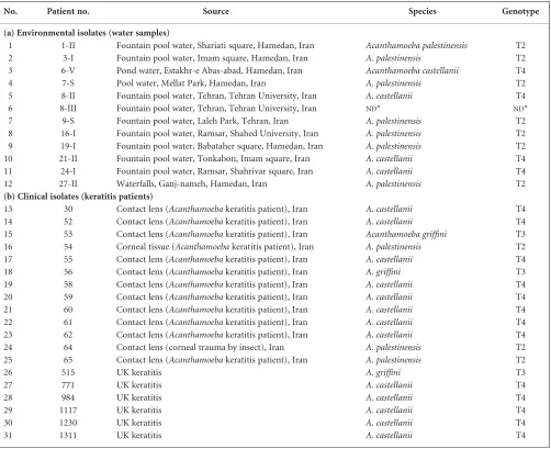
Table 1.Acanthamoebaisolates tested in the present study
No. Patient no. Source Species Genotype
(a) Environmental isolates (water samples)
1 1-II Fountain pool water, Shariati square, Hamedan, Iran Acanthamoeba palestinensis T2
2 3-I Fountain pool water, Imam square, Hamedan, Iran A. palestinensis T2
3 6-V Pond water, Estakhr-e Abas-abad, Hamedan, Iran Acanthamoeba castellanii T4
4 7-S Pool water, Mellat Park, Hamedan, Iran A. palestinensis T2
5 8-II Fountain pool water, Tehran, Tehran University, Iran A. castellanii T4
6 8-III Fountain pool water, Tehran, Tehran University, Iran ND* ND*
7 9-S Fountain pool water, Laleh Park, Tehran, Iran A. palestinensis T2
8 16-I Fountain pool water, Ramsar, Shahed University, Iran A. palestinensis T2
9 19-I Fountain pool water, Babataher square, Hamedan, Iran A. palestinensis T2
10 21-II Fountain pool water, Tonkabon, Imam square, Iran A. castellanii T4
11 24-I Fountain pool water, Ramsar, Shahrivar square, Iran A. castellanii T4
12 27-II Waterfalls, Ganj-nameh, Hamedan, Iran A. palestinensis T2
(b) Clinical isolates (keratitis patients)
13 30 Contact lens (Acanthamoebakeratitis patient), Iran A. castellanii T4
14 52 Contact lens (Acanthamoebakeratitis patient), Iran A. castellanii T4
15 53 Contact lens (Acanthamoebakeratitis patient), Iran Acanthamoeba griffini T3
16 54 Corneal tissue (Acanthamoebakeratitis patient), Iran A. palestinensis T2
17 55 Contact lens (Acanthamoebakeratitis patient), Iran A. castellanii T4
18 56 Contact lens (Acanthamoebakeratitis patient), Iran A. griffini T3
19 58 Contact lens (Acanthamoebakeratitis patient), Iran A. castellanii T4
20 59 Contact lens (Acanthamoebakeratitis patient), Iran A. castellanii T4
21 60 Contact lens (Acanthamoebakeratitis patient), Iran A. castellanii T4
22 61 Contact lens (Acanthamoebakeratitis patient), Iran A. castellanii T4
23 62 Contact lens (Acanthamoebakeratitis patient), Iran A. castellanii T4
24 64 Contact lens (corneal trauma by insect), Iran A. palestinensis T2
25 65 Contact lens (Acanthamoebakeratitis patient), Iran A. palestinensis T2
26 515 UK keratitis A. griffini T3
27 771 UK keratitis A. castellanii T4
28 984 UK keratitis A. castellanii T4
29 1117 UK keratitis A. castellanii T4
30 1230 UK keratitis A. castellanii T4
31 1311 UK keratitis A. castellanii T4
*Acanthamoeba, but species/genotype not determined. This sample was omitted from the statistical analysis of association with genotype, i.e. only 11 environmental samples were included.
A. H. Maghsood and others
4 mM MgCl2 and 0.5ìM primer. PCR reactions were performed at 948C for 1 min, 558C for 1 min and 728C for 2 min for 30 cycles, with a final elongation step of 10 min at 728C. Amplified DNA was electro-phoresed on a 2 % agarose gel, stained and visualized under UV illumination.
18S rRNA gene sequencing. For 18S rRNA gene sequencing, amplified DNA products were directly sequenced with an automated fluorescent sequencing system using a LI-COR 4200 DNA sequencer for diagnostic fragment 3 using primer 892C (59-GTCAGAGGTGAA ATTCTTGG-39) (Bootonet al., 2002) and identified at the genotype level by comparison with the availableAcanthamoebaDNA sequences in GenBank or by comparison with theAcanthamoeba DNA sequence database (Department of Molecular Genetics, The Ohio State Uni-versity, OH, USA). Diagnostic fragment 3 sequences for the new isolates are available upon request from N. A. Khan.
Cytotoxicity assay. For some Acanthamoeba isolates, cytotoxicity assays were performed as previously described (Khan & Tareen, 2003). Briefly, immortalized human corneal epithelial cells (HCECs) were grown to confluency in 24-well plates in supplementary hormonal epithelial medium (15 %, w/v, fetal bovine serum; 40ìg gentamicin ml1; 5ìg insulin ml1; 0.1ìg cholera toxin ml1; vitamins; 40 % Hanks F12; 40 % Dulbecco’s modified Eagle’s medium; 0.5 % DMSO; 10 ng epidermal growth factor ml1) (Invitrogen) at 378C in a 5 % CO
2 incubator as previously described (Araki-Sasakiet al., 2000). Acantha-moebaisolates (53105 amoebae per well) were incubated with cell monolayers in serum-free medium (RPMI 1640 containing 2 mM glutamine, 1 mM pyruvate and non-essential amino acids) at 378C in a 5 % CO2incubator for up to 24 h. At the end of this incubation period, supernatants were collected and cytotoxicity was determined by meas-uring lactate dehydrogenase release using a cytotoxicity detection kit (Roche Applied Science) as previously described (Khan & Tareen, 2003).
RESULTS AND DISCUSSION
Isolation ofAcanthamoebafrom the clinical specimens in Iran
Of 52 specimens taken from keratitis patients in Iran, 13 were found to contain Acanthamoeba, seven from women (53.9 %) and six from men (46.1 %); their ages ranged from 15 to 54 years (mean 22 years). Twelve patients (92.3 %) wore soft contact lenses and one patient (7.7 %) wore hard lenses. Patients were treated with topical application of propami-dine isethionate (Brolene) plus Neosporin (neomycin, poly-myxin B sulfates and bacitracin). Twelve patients responded to therapy with improved clinical symptoms and no report of recurrence, indicating successful treatment. One patient (#64) did not respond to anti-amoebic drugs and a corneal graft was performed. This patient wore hard lenses and while travelling on a motorcycle an insect had entered his right eye. He rubbed his eye while wearing a contact lens, which resulted in corneal trauma. The clinical signs of Acantha-moebakeratitis followed soon after and included severe pain, photophobia and stromal infiltrates. Within 20 days, he was diagnosed with Acanthamoeba keratitis and anti-amoebic therapy was initiated but showed no response. However, the symptoms cleared after a successful corneal graft, as indi-cated above.
Isolation ofAcanthamoebafrom the environmental samples in Iran
To determine the distribution of Acanthamoeba in the environmental samples in Iran, 12 Acanthamoeba were isolated from water samples obtained from different cities (Table 1).Acanthamoebawere isolated based on morpholo-gical characteristics of trophozoites and cysts. Overall, these results suggested a wide environmental distribution of
Acanthamoeba from various geographic locations in Iran. The identity of both clinical and environmental isolates was further confirmed using PCR analysis as described in Meth-ods (data not shown). Acanthamoeba were successfully cultured in PYG medium containing penicillin (100 U ml1)
and streptomycin (100ìg ml1) at 308C, except for one
environmental isolate (8-III).
Molecular typing of clinical isolates of Acanthamoebafrom Iran
In this study, 13Acanthamoebakeratitis cases were identified. Among these, eight (61.5 %) isolates belonged to the T4 genotype, two (15.3 %) belonged to T3 and three (23 %) belonged to the T2 genotype (Table 2). This is the first time thatAcanthamoeba isolates belonging to the T2 genotype have been linked withAcanthamoebakeratitis. Of interest, patient #64, who was non-responsive to anti-amoebic therapy, was infected with a T2 isolate. To determine the pathogenic potential of the three T2 isolates (#54, #64 and #65),in vitrocytotoxicity assays were performed. We ob-served that two Acanthamoeba isolates (#64 and #65) produced more than 90 % HCEC cytotoxicity and were considered as potential pathogens. However, #54 exhibited minimal HCEC cytotoxicity (,10 %) and was considered as a weak or non-pathogen. Overall, these data indicated that the majority of keratitis-causing isolates in Iran belong to the T4 genotype.
T2 is a widely distributed genotype in the environmental samples isolated from Iran
To determine the environmental distribution of Acantha-moebain Iran, amoebae were isolated from 12 water samples. Out of these, seven (58.3 %) belonged to the T2 genotype and four belonged to T4 (33.3 %), while theRnssequence of one (8-III) (8.3 %) was not determined (Table 2). These findings indicated that T2 is a widely distributed genotype (followed by T4) in the water samples tested from Iran.
Clinical and environmental distribution of Acanthamoebafrom other geographic locations
For comparison of clinical isolates, 11Acanthamoeba kera-titis isolates from the UK were used (Khanet al., 2002, plus the six newAcanthamoebakeratitis cases tested in the present study; Table 1). Of these, nine isolates (81.8 %) belonged to the T4 genotype, one belonged to T3 (9.1 %) and one belonged to T11 (9.1 %) (Table 2). In total, 24Acanthamoeba
three belonged to T3 (12.5 %) and one belonged to T11 (4.1 %), confirming that T4 is the predominant genotype (÷2¼4.167,P¼0.0412) inAcanthamoebakeratitis.
For comparison of the environmental isolates, nine Acantha-moeba isolates from the UK were tested as previously described (Khanet al., 2002; Khan & Paget, 2002). Among these, four isolates (44.4 %) belonged to T4, two belonged to T2 (22.2 %), one belonged to T3 (11.1 %) and two belonged to T7 (22.2 %) (Table 2). It is important to note that, except for the T4 isolates, the majority of amoebae were isolated from soil samples. Of interest, both T7 isolates were from soil samples.
Acanthamoebaare causative agents of painful eye infections, which can lead to blindness. They are one of the most
ubiquitous organisms and have been classified into 15 different genotypes. In an attempt to correlate pathogenicity with certain genotypes, several studies have identified T4 as the major keratitis-causing genotype. However, whether the increased ability of T4 isolates to produce keratitis is due to their greater virulence or their greater prevalence is some-what unclear. In this study, we tested clinical and environ-mental isolates of Acanthamoebafrom a variety of sources and various geographic locations in the UK and Iran. The majority of clinical isolates tested in our study belonged to the T4 genotype, consistent with previous studies. Among the environmental isolates, T4 was again the predominant genotype in the UK, but T2 was predominant in Iran within the limited number of samples tested.
One of the interesting findings in our study was the isolation of T2 isolates from three keratitis patients (#54, #64, #65). Of these, #65 exhibited the characteristics of Acanthamoeba
keratitis. In addition, #65 received anti-acanthamoebic chemotherapy with a successful outcome, indicating
Acanthamoeba as the causative agent. As quoted by De Jonckheere (2003) and others, ‘contact lens cases are the breeding grounds forAcanthamoeba’, and in the absence of a mixed infection, it is reasonable to presume that the T2 isolate was the causative agent. This is the first time that
Acanthamoeba isolates belonging to the T2 genotype have been linked withAcanthamoebakeratitis. This is in contrast to previous studies, which showed that a T2 isolate (CCAP 1501/3c) exhibited minimal binding and minimal cytotoxi-city to the host cells (Alsamet al., 2003; Khanet al., 2002) and was considered as a weak or non-pathogen. There are several possible explanations for these findings: the T2 genotype may consist of both pathogenic and weakly or non-pathogenic isolates (since rRNA gene sequence information is not a determinant for pathogenicity), or T2 isolates may have diverse virulence properties and individual strains (current or new isolates) should be tested for their virulence before designating them pathogens or non-pathogens. Alterna-tively, a more feasible scheme will be to subdivide the T2 genotype into two separate sequence types based on theirRns
sequences. In support of this, Stothard et al. (1998) have shown that, within the T2 genotype, a sequence dissimilarity of 4.9 % exists between CCAP 1501/3c and ATCC 30871. Comparison of CCAP 1501/3c with another T2 isolate (ATCC 50252) also revealed similar differences (4.5 %) (Stothard et al., 1998). These differences are very close to the current cut-off limit of 5 % between different genotypes. Thus we propose that T2 isolates should be subdivided into two separate groups, T2A and T2B, with a sequence dissim-ilarity of 4 % or more. This may help to differentiate pathogenic and non-pathogenic isolates within this geno-type. Further studies involvingAcanthamoebabelonging to the T2 genotype with CCAP 1501/3c-likeRnssequences and more detailed analysis of their properties will clarify the need for two separate groups in this genotype.
[image:5.612.70.310.82.454.2]Another intriguing finding was the inability of one Acantha-moebaisolate (#54 from the contact lens of a keratitis patient) to produce host-cell cytotoxicity, suggesting that it has weak
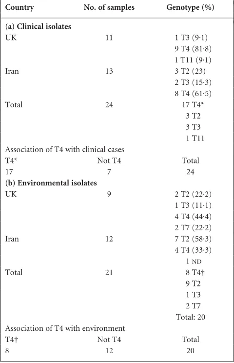
Table 2.Comparison of the clinical and environmental
distribu-tion ofAcanthamoebain the UK and Iran
Country No. of samples Genotype (%)
(a) Clinical isolates
UK 11 1 T3 (9.1)
9 T4 (81.8) 1 T11 (9.1)
Iran 13 3 T2 (23)
2 T3 (15.3) 8 T4 (61.5)
Total 24 17 T4*
3 T2 3 T3 1 T11 Association of T4 with clinical cases
T4* Not T4 Total
17 7 24
(b) Environmental isolates
UK 9 2 T2 (22.2)
1 T3 (11.1) 4 T4 (44.4) 2 T7 (22.2)
Iran 12 7 T2 (58.3)
4 T4 (33.3) 1ND
Total 21 8 T4†
9 T2 1 T3 2 T7 Total: 20 Association of T4 with environment
T4† Not T4 Total
8 12 20
*÷2¼4.167 with 1 degree of freedom. The two-tailedPvalue equals 0.0412, indicating significant association of T4 with clinical cases.
†÷2¼0.800 with 1 degree of freedom. The two-tailedPvalue equals 0.3711, indicating no significant association of T4 in the environmental isolates.
A. H. Maghsood and others
and/or non-pathogenic properties (,10 % cell death, as compared with more than 70 % cell death with other clinical isolates). One explanation for this is that there may have been a mixed infection and T2 was isolated from the contact lens even though it was not the causative agent. Future studies are in progress to address these issues.
Despite the occasional cases ofAcanthamoebakeratitis due to T2 (the present study), T3 (Ledeeet al., 1996), T6 (Waloch-niket al., 2000) and T11 (Khanet al., 2002), the majority of cases are due to the T4 genotype. This was further confirmed in our study, as more than 80 % of Acanthamoebaisolates (UK keratitis) belonged to the T4 genotype. In addition, we also determined that T4 was widely distributed in the environmental samples isolated from the UK. These results, together with previous findings, suggest thatAcanthamoeba
isolates belonging to the T4 genotype naturally occur in the environment and present potential reservoirs and therefore sources of infection to susceptible hosts. As indicated above, the repeated appearance of T4 isolates in keratitis may be due to their greater pathogenicity (virulence properties), greater transmissibility (i.e. they may be widely distributed in the habitat where the probability of being picked up by humans is very high), or a combination of both. To this end, we determined T2 as the most abundant genotype, followed by T4, in the environmental samples from Iran, even though the number of T4 Iranian keratitis isolates was higher than that of T2 isolates (61.5 vs 23 %). These findings demonstrated that the abundance of T4 isolates in Acanthamoeba keratitis patients is most likely due to their greater virulence and/or properties that enhance their transmissibility, as well as their ability to bind to contact lenses and their decreased suscept-ibility to disinfectants. This raises additional questions about the properties of T2 and T4 genotypes that enable them to be the most widespread genotype in a given environment among 15 different genotypes and whether these properties play any role(s) in infection. Future studies that identify virulence traits and genetic markers limited to certain genotypes may help to clarify these issues.
Overall, there is a clear need for more detailed knowledge about the distribution of each genotype in different environ-ments and for detailed analyses of direct and indirect virulence factors of environmental and clinical isolates of each genotype. These findings will help us understand the basis of differential distribution of genotypes in natural habitats, and to some extent in clinical cases, and may identify their differential properties contributing to disease, which should allow the development of pre-emptive or therapeutic measures against these serious infections.
ACKNOWLEDGEMENTS
We are grateful to Dr Sim Webb, Norwich Eye Research Group, School of Biological Sciences, University of East Anglia, UK, for providing immortalized human corneal epithelial cells and Dr Gregory Booton,
Department of Molecular Genetics, The Ohio State University, USA, for critical reviewing of the manuscript. This work was supported by grants from The Nuffield Foundation.
REFERENCES
Alsam, S., Kim, K. S., Stins, M., Rivas, A. O., Sissons, J. & Khan, N. A. (2003).Acanthamoebainteractions with human brain microvascular endothelial cells.Microb Pathog35, 235–241.
Araki-Sasaki, K., Aizawa, S., Hiramoto, M. & 7 other authors (2000).
Substance P-induced cadherin expression and its signal transduction in a cloned human corneal epithelial cell line.J Cell Physiol182, 189–195.
Booton, G. C., Kelly, D. J., Chu, Y.-W., Seal, D. V., Houang, E., Lam, D. S. C., Byers, T. J. & Fuerst, P. A. (2002).18S ribosomal DNA typing and tracking ofAcanthamoebaspecies isolates from corneal scrape speci-mens, contact lenses, lens cases, and home water supplies of Acantha-moebakeratitis patients in Hong Kong.J Clin Microbiol40, 1621–1625.
De Jonckheere, J. F. (2003).Epidemiological typing ofAcanthamoeba strains isolated from keratitis cases in Belgium.Bull Soc Belge Ophtalmol 287, 27–33.
Gast, R. J. (2001).Development of anAcanthamoeba-specific reverse dot-blot and the discovery of a new ribotype.J Eukaryot Microbiol48, 609–615.
Khan, N. A. (2003).Pathogenesis ofAcanthamoebainfections.Microb Pathog34, 277–285.
Khan, N. A. & Paget, T. A. (2002).Molecular tools for speciation and epidemiological studies ofAcanthamoeba.Curr Microbiol44, 444–449.
Khan, N. A. & Tareen, N. K. (2003).Genotypic, phenotypic, biochemical, physiological and pathogenicity-based categorisation ofAcanthamoeba strains.Folia Parasitol50, 97–104.
Khan, N. A., Jarroll, E. L. & Paget, T. A. (2001).Acanthamoebacan be differentiated clinically by the polymerase chain reaction and simple plating assays.Curr Microbiol43, 204–208.
Khan, N. A., Jarroll, E. L. & Paget, T. A. (2002). Molecular and physiological differentiation between pathogenic and nonpathogenic Acanthamoeba.Curr Microbiol45, 197–202.
Kong, H. H. & Chung, D. I. (1996).PCR and RFLP variation of conserved region of small subunit ribosomal DNA amongAcanthamoebaisolates assigned to eitherA. castellaniiorA. polyphaga.Korean J Parasitol34, 127–134.
Ledee, D. R., Hay, J., Byers, T. J., Seal, D. V. & Kirkness, C. M. (1996).
Acanthamoeba griffini. Molecular characterization of a new corneal pathogen.Invest Ophthalmol Vis Sci37, 544–550.
Marciano-Cabral, F. & Cabral, G. (2003).Acanthamoebaspp. as agents of disease in humans.Clin Microbiol Rev16, 273–307.
Schuster, F. L. & Visvesvara, G. S. (2004).Free-living amoebae as opportunistic and non-opportunistic pathogens of humans and ani-mals.Int J Parasitol34, 1001–1027.
Stothard, D. R., Schroeder-Diedrich, J. M., Awwad, M. H., Gast, R. J., Ledee, D. R., Rodriguez-Zaragoza, S., Dean, C. L., Fuerst, P. A. & Byers, T. J. (1998).The evolutionary history of the genusAcanthamoebaand the identification of eight new 18S rRNA gene sequence types.J Eukaryot Microbiol45, 45–54.