ISSN Online: 2160-5874 ISSN Print: 2160-5866
A Motor Programming Task Activates the
Prefrontal Cortex More than a
Sensitivity-to-Interference Task or an Inhibitory
Control Task in Older Adults
Masahiro Toyoda1*, Yuko Yokota1, Susan Rodiek2
1Graduate School of Landscape Design and Management, University of Hyogo, Awaji, Japan 2Center for Health Systems & Design, Texas A&M University, College Station, TX, USA
Abstract
The objectives of this study were to detect age-related differences in activation of the prefrontal cortex (PFC) during the tasks of hand motions and to determine an activ-ity-related task type activating the PFC. PFC activation during three tasks, three sub-tests of the Frontal Assessment Battery (FAB), was investigated in 77 healthy adults by using near-infrared spectroscopy (NIRS). The tasks were a motor programming task (FAB 3), a sensitivity-to-interference task (FAB 4) and an inhibitory control task (FAB 5). We divided participants into three age groups of Younger (20 - 39 years), Middle-aged (40 - 59 years), and Older (60 - 81 years), and compared relative changes in oxygenated hemoglobin concentration in the PFC during the tasks. The activation in the frontal pole (FP) and the dorsolateral prefrontal cortex (DLPFC) during a motor programming task and a sensitivity-to-interference task showed no main effects by age. The results indicated that they were not likely to be affected by age-related cognitive decline compared to an inhibitory control task. In addition, in the Older group, a motor programming task induced significantly greater activation than a sensitivity-to-interference task at eleven channels out of twelve on which we focused (p < 0.05). It was suggested that some characteristic factors included in the motor programming task such as repetition of a series of hand motions and attention to action have the potential to contribute to PFC activation in older adults. These findings provide a clue to understanding daily activities available to suppress cogni-tive decline of older adults by activating the PFC.
Keywords
Frontal Assessment Battery (FAB), Working Memory (WM), Attention to Action, Cognitive Decline, Near-Infrared Spectroscopy (NIRS)
How to cite this paper: Toyoda, M., Yoko-ta, Y. and Rodiek, S. (2016) A Motor Pro-gramming Task Activates the Prefrontal Cortex More than a Sensitivity-to-Interfe- rence Task or an Inhibitory Control Task in Older Adults. Journal of Behavioral and Brain Science, 6, 433-447.
http://dx.doi.org/10.4236/jbbs.2016.611040 Received: August 30, 2016
Accepted: October 10, 2016 Published: October 13, 2016
Copyright © 2016 by authors and Scientific Research Publishing Inc. This work is licensed under the Creative Commons Attribution International License (CC BY 4.0).
http://creativecommons.org/licenses/by/4.0/
1. Introduction
In 2015, Alzheimer’s Disease International reported that 46.8 million people worldwide were living with dementia. This number is expected to almost double every 20 years, to 74.7 million in 2030 and 131.5 million in 2050 [1]. Dementia prevalence in Japan (in-cluding community-dwelling as well as care facilities) was estimated to be 15% of the total population in 2012, when the estimated number of people with dementia in Japan was 4.62 million [2]. Some studies have reported on impaired working memory (WM) of people with mild cognitive impairment (MCI) and Alzheimer’s Disease (AD), and declined blood flow response in the dorsolateral prefrontal cortex (DLPFC) activation in cognitively normal aging individuals [3] [4]. It is known that age-related changes in cognitive function occur even in individuals without dementia as well, and executive function typically declines with aging. Decrease in information processing speed [5], decline of WM function [6] and decline of inhibitory control function [7] are consi-dered to be the main factors in the age-related decline of executive function.
Other studies have reported the effectiveness of training to enhance frontal lobe function: overall frontal lobe function improved in healthy adults in their 20’s to 30’s after specific WM training by using tasks such as the Stroop interference task associated with selective attention, inhibition, and cognitive flexibility, and the span-board task in which the participants were required to recall the location and order of the presented cues [8]. In older adults diagnosed with MCI, memory training was found to improve overall brain plasticity [9]. Van Halteren-van Tilborg, Scherder & Hulstijn reviewed previous studies targeting AD patients with the use of implicit learning tasks that can be mastered without awareness as a procedure in the absence of verbalization [10]. These studies suggest that activities stimulating frontal lobe function can be effective for maintaining and improving cognitive function, and that appropriate training has the potential to maintain or improve cognitive function in a wide range of the popula-tion: healthy younger adults in their 20’s and 30’s, healthy older adults, people with MCI, and AD patients. The question remains, how can we apply such research findings to the daily living of older adults as activities that may help reduce the risk of cognitive decline?
A recent comprehensive review of the existing literature on behavioral factors sug-gests that physical activities, cognitive engagement, and leisure activities can decrease the risk of AD or cognitive decline in older adults [11]. Currently, games, physical ex-ercises, household activities, music, handicrafts, coloring books, gardening etc. are pro-vided as activities for dementia prevention in healthcare facilities for older adults in Ja-pan. However, it is difficult to find similarities or common components between such activities and the previously-mentioned tasks for training cognitive function [8]-[10].
However, there have been few earlier studies that investigated the impact of such activi-ties on brain function by using the tasks relevant to executive functions of the brain. In addition, although there have been several near-infrared spectroscopy (NIRS) studies using cognitive tests that demand hand motions [12]-[14], no research has been found that specifically investigated blood flow during performance of the Frontal Assessment Battery (FAB) [15] tasks. To clarify the impact of such tasks on PFC activation poten-tially helps to suppress age-related cognitive decline by daily activities stimulating the frontal lobe effectively.
In this study, we selected three subtests of the FAB—FAB 3, FAB 4 and FAB 5—as the tasks to be performed by the participants. People often engage in movements simi-lar to these three tasks in their daily lives. If some of these tasks prove to be effective in promoting PFC activation, the findings will provide important clues to suppression of age-related cognitive decline.
The objectives of this study were to examine whether aging makes any differences in PFC activation during three FAB tasks with hand motions, and to determine what types of task activate the PFC of older adults effectively, as measured by NIRS.
2. Materials and Methods
2.1. Participants
We recruited participants through the University of Hyogo website, and also from the attendees of lectures for the general public held at the university between August and September 2007. Seventy-seven participants were given the Mini-Mental State Exami-nation (MMSE) to check their cognitive function prior to the experiment. MMSE is a screening tool for cognitive impairment, with a maximum possible total score of 30 points and a cut-score of 23 points [16]. Scoring 23 points or below indicates the like-lihood of cognitive impairment [17]. There were no participants who scored 23 points or be-low, and all of them were accepted for the study. All participants were right- handed. All received an explanation of the study in advance, and provided written in-formed consent.
and protection of privacy. Written informed consent was obtained from all partici-pants.
2.2. Tasks
This study was conducted in a quiet room at the University of Hyogo in October 2007 (room size: approximately 60 square meters, average temperature: 23.6˚C, average hu-midity: 63.0%). The participants were instructed to enter the room individually, un-dergo MMSE, and perform three FAB tasks in a sitting posture with NIRS optodes po-sitioned on the head. A NIRS optode is an optical sensor device to measure the local changes in oxygenated hemoglobin (Oxy-Hb), deoxygenated hemoglobin (Deoxy-Hb) and total hemoglobin (Total-Hb).
The FAB is a standardized measure to assess frontal lobe functions, and consists of six subtests examining conceptualization, mental flexibility, motor programming, sen-sitivity to interference, inhibitory control, and environmental autonomy. The partici-pants engaged in three tasks selected out of these subtests: a motor programming task (FAB 3), a sensitivity-to-interference task (FAB 4), and an inhibitory control task (FAB 5). These tasks have common characteristics of using WM function and accompanying hand motions to be carried out. A participant performs FAB 3 by imitating and memo-rizing an instructed series of three hand motions with his/her right hand (fist- edge-palm) on his/her left palm. In FAB 4, a participant taps his/her finger on the desk while recalling given instructions to tap twice when the examiner taps once, and tap once when the examiner taps twice. In FAB 5, a participant is required to tap his/her finger on the desk, or to inhibit tapping while recalling given instructions to tap once when the examiner taps once, and to refrain from tapping when the examiner taps twice. While FAB 4 and FAB 5 are explicit learning tasks, FAB 3 is an implicit learning task that requires continuous attention to a series of hand motions.
2.3. Procedure
Three trials were run for each task, and the order of the three tasks was the same as the standard FAB test. The protocol was as follows: positioning of optodes, instructions for FAB 3 (60 seconds), the baseline period (10 seconds), (FAB 3 [15 seconds] and rest [20 seconds]) × 3 sets, instructions for FAB 4 (60 seconds), the baseline period (10 seconds), (FAB 4 [15 seconds] and rest [20 seconds]) × 3 sets, instructions for FAB 5, the baseline period (10 seconds) and (FAB 5 [15 seconds] and rest [20 seconds]) × 3 sets. During the baseline period and the rest period, we asked participants to gaze blankly at an x-mark on a piece of white paper on the ivory-colored wall in front of them, so they could recover enough to be stable.
2.4. NIRS Measurements
BOLD (blood oxygenation level dependent) signals are only sensitive at the small ven-ous vessel level. They suggested that NIRS measurements are more directly correlated to neuronal activities compared with fMRI [19].
NIRS measures relative changes in the concentration of Oxy-Hb, Deoxy-Hb and To-tal-Hb. Hoshi, Kobayashi & Tamura noted that HbO2 (Oxy-Hb) is the most sensitive indicator of changes in rCBF (regional cerebral blood flow) in NIRS measurements, and the direction of the change in Deoxy-Hb is determined by changes in both venous oxygenation and blood volume [20]. We used the variations in the Oxy-Hb concentra-tion as indicators of changes in the regional cerebral blood flow, because Oxy-Hb was considered to be more sensitive than Deoxy-Hb as a parameter for measuring the blood flow relevant to PFC activation.
The NIRS measurements were performed by means of Hitachi ETG-4000, a 24- channel NIRS system (Hitachi Medical Corporation, Tokyo, Japan) using two wave-lengths of near-infrared light (695 nm and 830 nm). Absorption of near-infrared light was measured with a time resolution of 0.1 seconds. Each channel consisted of a pair of optodes—1 emitter (or light source optode) and 1 detector (or detection optode)—and the distance between them was 3.0 cm from each other.
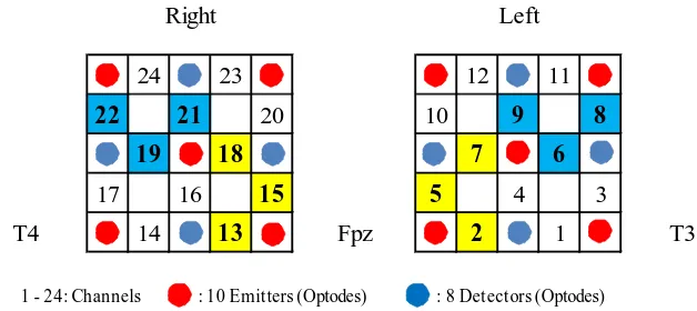
[image:5.595.214.528.488.628.2]Optodes were positioned on the participant’s forehead according to the International 10 - 20 system, a standard for Electroencephalography (EEG) electrode positioning [21]. The front edge of the optode line was positioned along the T3-Fpz-T4 line of the system, which means the center of the lowest row of the two holders with 9 optodes set in a 3 × 3 lattice pattern was placed on Fpz (the middle of the forehead) as shown in Figure 1.
We used virtual registration to ensure that probe channels corresponded to specific brain regions [22] based on the studies of virtual spatial registration [23] [24], and au-tomated Talairach Atlas Labels for functional brain mapping [25]. The regions meas-ured by NIRS in this study included the fronto-polar area (or the frontal pole [FP]), the
Figure 1. Location of 18 optodes and position of 24 channels in NIRS. (One channel consists of 1 emitter and 1 detector at a distance of 3.0 cm from each other. Two holders with 9 optodes were attached to the forehead of the participant according to the International 10 - 20 system. We fo-cused on the channels in yellow squares: 2, 5, 7, 13, 15, and 18 reflecting activation of the FP 100% and those in blue squares: 6, 8, 9, 19, 21, and 22 reflecting activation of the DLPFC 100% or more than 90%).
24 23 12 11
22 21 20 10 9 8
19 18 7 6
17 16 15 5 4 3
T4 14 13 Fpz 2 1 T3
Right Left
DLPFC, the inferior frontal gyrus (including the orbitofrontal cortex), the pars triangu-laris Broca’s area, and the frontal eye field. We focused on the channels (CHs) reflecting activation of the FP 100% (CHs 2, 5, and 7 in the left hemisphere, and CHs 13, 15, and 18 in the right hemisphere) and the channels which reflect activation of the DLPFC 100% (CH 6 in the left hemisphere, and CHs 19 and 21 in the right hemisphere) or more than 90% (CH 8 [90.5%] and CH 9 [99.1%] in the left hemisphere, and CH 22 [91.9%] in the right hemisphere).
2.5. Data Analysis
Task performance of FAB 3, FAB 4 and FAB 5 was scored according to the specified criteria of the FAB [15]. We used the mean score of three trials of each task for data analysis. The ETG-4000 analyzing software was used in the integral mode for analyzing the mean values of changes in Oxy-Hb concentration at each channel while a partici-pant was performing each task three times. Statistical analyses were performed using Excel 2010 (Microsoft, USA) with the add-in software SSRI, Version 1.02, 2012, (Social Survey Research Information Co., Ltd.), two sided test with p < 0.05 considered statis-tically significant.
The data were evaluated by the nonparametric Kruskal-Wallis one-way ANOVA with the post-hoc Steel-Dwass multiple comparison tests. Differences in mean Oxy-Hb values among tasks in each group were tested by the nonparametric Friedman re-peated-measure ANOVA with the post-hoc Scheffe multiple comparison tests. Age-re- lated differences in mean changes in Oxy-Hb values at each channel among groups were evaluated by the nonparametric Kruskal-Wallis one-way ANOVA with the post- hoc Steel-Dwass multiple comparison tests.
3. Results
3.1. Scores in FAB 3, FAB 4 and FAB 5
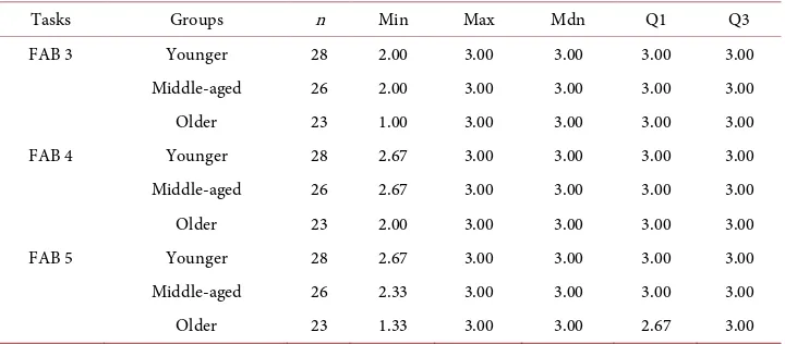
[image:6.595.195.555.526.684.2]Table 1 shows scores of FAB 3, FAB 4, and FAB 5 in each age group. The median, the
Table 1. Age-related differences of FAB scores among three age groups.
Tasks Groups n Min Max Mdn Q1 Q3
FAB 3 Younger 28 2.00 3.00 3.00 3.00 3.00
Middle-aged 26 2.00 3.00 3.00 3.00 3.00
Older 23 1.00 3.00 3.00 3.00 3.00
FAB 4 Younger 28 2.67 3.00 3.00 3.00 3.00
Middle-aged 26 2.67 3.00 3.00 3.00 3.00
Older 23 2.00 3.00 3.00 3.00 3.00
FAB 5 Younger 28 2.67 3.00 3.00 3.00 3.00
Middle-aged 26 2.33 3.00 3.00 3.00 3.00
Older 23 1.33 3.00 3.00 2.67 3.00
first quartile and the third quartile were 3.0 points with the only exception of the first quartile of FAB 5 in the Older group (2.67). As a result of the Kruskal-Wallis ANOVA, no main effects of age were detected in FAB 3 (χ2 = 0.153, df = 2, p = 0.927) and FAB 4 (χ2 = 0.531, df = 2, p = 0.767). Whereas, the main effect of age was verified in FAB 5 (χ2 = 9.981, df = 2, p = 0.007). The results of post hoc analysis by Steel-Dwass multiple com-parison tests showed that the score of FAB 5 in the Older group was significantly lower than that in the Younger group (t = 2.911, p = 0.010).
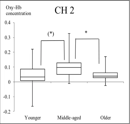
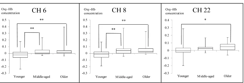
3.2. Comparison of Changes in Oxy-Hb Values among Three Age Groups The nonparametric Kruskal-Wallis one-way ANOVA showed no main effects of age in FAB 3 and FAB 4 at every channel. However, main effects of age were observed in FAB 5 at CH 2 (χ2 = 8.59, df = 2, p = 0.014), CH 6 (χ2 = 13.23, df = 2, p = 0.001), CH 8 (χ2 = 13.76, df = 2, p = 0.001), and CH 22 (χ2 = 9.74, df = 2, p = 0.008).
At CH 2, significantly greater increase in Oxy-Hb values was observed in the Mid-dle-aged group than the Older group (p = 0.017). The Younger group showed smaller Oxy-Hb values than the Middle-aged group with a p-value of 0.051, which was margi-nally significant. There was a tendency that activation in the Younger group was small compared to that in the Middle-aged group. The Middle-aged group showed signifi-cantly greater increase in Oxy-Hb than the Younger group at CHs 6 and 8 (p < 0.05). The Older group showed significantly greater increase than the Younger group at CHs 6, 8 and 22 (p < 0.05) (Figure 2 and Figure 3).
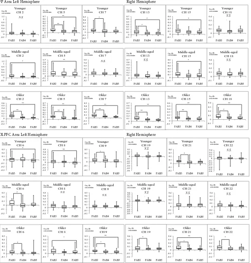
[image:7.595.267.479.460.662.2]3.3. Comparison of Changes in Oxy-Hb Values among Three FAB Tasks in Each Group
Figure 4 shows the results of the repeated measure ANOVA and the Scheffe multiple
-0.2 -0.1 0 0.1 0.2 0.3 0.4
Younger Middle-aged Older
CH 2
Oxy-Hb concentration
(*) *
Figure 3. Comparison of Oxy-Hb values during FAB 5 among three age groups at CH 6, CH 8 and CH 22. (CH 6 and CH 8 reflect activation of the DLPFC in the left hemisphere 100% and 90.5% respectively. CH 22 reflects activation of the DLPFC in the right hemisphere 91.9%; Numbers on the longitudinal axis represent Oxy-Hb concentration in mmol∙mm/0.1 s: *p < 0.05, **p < 0.01).
comparison tests of Oxy-Hb mean values among tasks in each group. The upper half of Figure 4 shows the boxplots of Oxy-Hb values at the channels reflecting activation of the FP 100% (CHs 2, 5, 7, 13, 15, and 18), and the lower half shows those at the channels reflecting activation of the DLPFC 100% or more than 90% (CHs 6, 8, 9, 19, 21, and 22).
As the boxplots on the upper half of Figure 4 show, in the Younger group, the Oxy-Hb values in the FP during FAB 3 were significantly greater than those of FAB 4 and/or FAB 5 at CHs 5, 7, 13, and 15 (p < 0.05). In the Middle-aged group, the Oxy-Hb values during FAB 3 were significantly greater than those of FAB 4 and/or FAB 5 at CHs 2, 5, and 15 (p < 0.05). In the Older group, the Oxy-Hb values during FAB 3 were significantly greater than those of FAB 4 and FAB 5 at all channels reflecting activation of the FP.
Meanwhile, the boxplots on the lower half of Figure 4 show that the Oxy-Hb values in the DLPFC during FAB 3 were significantly greater than those of FAB 4 and/or FAB 5 at CHs 6, 8, 9, and 21 in the Younger group (p < 0.05). In the Middle-aged group, the Oxy-Hb value during FAB 3 was significantly greater than that of FAB 4 (p < 0.05) only at CH 6. There was no significant difference detected between FAB 3 and FAB 5. In the Older group, the Oxy-Hb values during FAB 3 were significantly greater than those of FAB 4 and/or FAB 5 at all channels reflecting DLPFC activation more than 90%.
4. Discussion
In all groups, FP and DLPFC activation during FAB 3 was significantly greater than or similar to that during FAB 4 and/or FAB 5 (p < 0.05). In other words, FAB 3 activated the FP and the DLPFC of the participants at any age compared to FAB 4 and FAB 5. We speculated that repetition of a sequence of hand motions and attention to action, which are distinctive elements included only in FAB 3, caused such activation.
FP Area Left Hemisphere Right Hemisphere -0.2 0 0.2 0.4 0.6 0.8 1 1.2
FAB3 FAB4 FAB5
Younger CH 2 -0.2 0 0.2 0.4 0.6 0.8 1 1.2
FAB3 FAB4 FAB5
Middle-aged CH 2 * Oxy-Hb concentration -0.2 -0.1 0 0.1 0.2 0.3 0.4 0.5 0.6 0.7
FAB3 FAB4 FAB5
Middle-aged CH 5 ** Oxy-Hb concentration -0.2 -0.1 0 0.1 0.2 0.3 0.4 0.5 0.6 0.7
FAB3 FAB4 FAB5
Older CH 5 ** ** Oxy-Hb concentration -0.2 -0.1 0 0.1 0.2 0.3 0.4 0.5
FAB3 FAB4 FAB5
Younger CH 7 * Oxy-Hb concentration -0.2 -0.1 0 0.1 0.2 0.3 0.4 0.5
FAB3 FAB4 FAB5
Middle-aged CH 7 N.S. Oxy-Hb concentration -0.2 -0.1 0 0.1 0.2 0.3 0.4 0.5
FAB3 FAB4 FAB5
Older CH 7 ** ** Oxy-Hb concentration N.S. -0.2 -0.1 0 0.1 0.2 0.3 0.4 0.5 0.6 0.7
FAB3 FAB4 FAB5
Younger CH 5 ** ** -0.2 -0.1 0 0.1 0.2 0.3 0.4 0.5 0.6
FAB3 FAB4 FAB5
Older CH 2 ** ** Oxy-Hb concentration Oxy-Hb concentration Oxy-Hb concentration -0.1 0 0.1 0.2 0.3 0.4 0.5 0.6 0.7
FAB3 FAB4 FAB5
Younger CH 13 * * Oxy-Hb concentration -0.1 0 0.1 0.2 0.3 0.4 0.5 0.6 0.7
FAB3 FAB4 FAB5
Middle-aged CH 13 N.S. Oxy-Hb concentration -0.1 0 0.1 0.2 0.3 0.4 0.5 0.6
FAB3 FAB4 FAB5
Younger CH 15 * Oxy-Hb concentration -0.1 0 0.1 0.2 0.3 0.4 0.5 0.6
FAB3 FAB4 FAB5
Middle-aged CH 15 * Oxy-Hb concentration -0.1 0 0.1 0.2 0.3 0.4
FAB3 FAB4 FAB5
Younger CH 18 N.S . Oxy-Hb concentration -0.1 0 0.1 0.2 0.3 0.4
FAB3 FAB4 FAB5
Middle-aged CH 18 N.S. Oxy-Hb concentration -0.1 0 0.1 0.2 0.3 0.4 0.5 0.6 0.7
FAB3 FAB4 FAB5
Older CH 13 ** ** Oxy-Hb concentration -0.1 0 0.1 0.2 0.3 0.4 0.5 0.6
FAB3 FAB4 FAB5
Older CH 15 ** ** Oxy-Hb concentration -0.1 0 0.1 0.2 0.3 0.4
FAB3 FAB4 FAB5
Older CH 18 * ** Oxy-Hb concentration
DLPFC Area Left Hemisphere Right Hemisphere
-0.3 -0.2 -0.1 0 0.1 0.2 0.3 0.4 0.5
FAB3 FAB4 FAB5
Younger CH 8 ** Oxy-Hb concentration -0.2 -0.1 0 0.1 0.2 0.3 0.4 0.5
FAB3 FAB4 FAB5
Middle-aged CH 8 N.S. Oxy-Hb concentration -0.2 -0.1 0 0.1 0.2 0.3
FAB3 FAB4 FAB5
Younger CH 9 * Oxy-Hb concentration -0.2 -0.1 0 0.1 0.2 0.3 0.4 0.5
FAB3 FAB4 FAB5
Middle-aged CH 9 N.S. Oxy-Hb concentration -0.1 0 0.1 0.2 0.3 0.4 0.5
FAB3 FAB4 FAB5
Older CH 8 * Oxy-Hb concentration -0.1 0 0.1 0.2 0.3 0.4 0.5
FAB3 FAB4 FAB5
Older CH 9 ** * Oxy-Hb concentration -0.3 -0.2 -0.1 0 0.1 0.2 0.3
FAB3 FAB4 FAB5
Younger CH 6 * Oxy-Hb concentration -0.2 -0.1 0 0.1 0.2 0.3
FAB3 FAB4 FAB5
Middle-aged CH 6 * Oxy-Hb concentration -0.1 0 0.1 0.2 0.3 0.4 0.5
FAB3 FAB4 FAB5
Older CH 6 * Oxy-Hb concentration -0.6 -0.5 -0.4 -0.3 -0.2 -0.10 0.1 0.2 0.3 0.4
FAB3 FAB4 FAB5
Middle-aged CH 19 N.S . Oxy-Hb concentration -0.2 -0.1 0 0.1 0.2 0.3 0.4
FAB3 FAB4 FAB5
Younger CH 21 * Oxy-Hb concentration -0.2 -0.1 0 0.1 0.2 0.3 0.4
FAB3 FAB4 FAB5
Middle-aged CH 21 N.S. Oxy-Hb concentration -0.6 -0.5 -0.4 -0.3 -0.2 -0.1 0 0.1 0.2 0.3 0.4
FAB3 FAB4 FAB5
Younger CH 22 N.S. Oxy-Hb concentration -0.2 -0.1 0 0.1 0.2 0.3 0.4
FAB3 FAB4 FAB5
Middle-aged CH 22 N.S. Oxy-Hb concentration -0.2 -0.1 0 0.1 0.2 0.3 0.4
FAB3 FAB4 FAB5
Older CH 19 * Oxy-Hb concentration -0.2 -0.1 0 0.1 0.2 0.3 0.4
FAB3 FAB4 FAB5
Older CH 21 * ** Oxy-Hb concentration -0.2 -0.1 0 0.1 0.2 0.3 0.4
FAB3 FAB4 FAB5
Older CH 22 * Oxy-Hb concentration -0.6 -0.5 -0.4 -0.3 -0.2 -0.1 0 0.1 0.2 0.3
FAB3 FAB4 FAB5
Younger CH 19
N.S.
[image:9.595.45.555.73.607.2]Oxy-Hb concentration
Figure 4. Comparison of Oxy-Hb values among three tasks in three age groups at channels reflecting FP activation 100% and DLPFC activation 100% or more than 90%. (CHs 2, 5, 7, 13, 15 and 18 reflect activation of the FP 100% and CHs 6, 8, 9, 19, 21 and 22 reflect acti-vation of the DLPFC 100% or more than 90%. Numbers on the longitudinal axis represent Oxy-Hb concentration in mmol∙mm/0.1 s; *p < 0.05, **p < 0.01).
WM tasks have found results that differ from those of simple retention studies in two important ways [26]. First, they pointed out that prefrontal activations often occur bi-laterally, independent of the type of material used in the task as one important point. Second, the activations often occur in regions dorsal to those found in studies of simple retention (e.g., BAs [Brodmann Area] 9 and 46) [26].
Bilateral activation observed during FAB 3, FAB 4 and FAB 5 in the this study was considered to indicate that these tasks were such types of “more difficult WM tasks” as Rypma et al. mentioned.
To summarize, three FAB tasks used in this study required bilateral PFC activation to some extent. On the other hand, Dubois et al. reported that the group of healthy adults (mean age 58.0 ± 14.4) scored approximately 18 points, the perfect score of the FAB [15]. This means that any adult can perform the three tasks with no or few errors.
4.2. The Age-Related Difference in FAB Scores and Oxy-Hb Values The three age groups in this study showed no significant difference in the scores of FAB 3 and FAB 4, and the main effects of age were not detected at any channels between the Oxy-Hb values during FAB 3 and those during FAB 4.
The results indicate that the motor programing task and the sensitivity-to-interfe- rence task in this study have the potential to activate the FP and the DLPFC regardless of age, although both tasks were simple enough for most adults to perform with few or no errors. On the other hand, the score of FAB 5 in the Older group was significantly lower than that in the Younger group. This indicates that FAB 5 related to inhibitory control is more likely for older adults to make errors compared to younger adults, which is consistent with age decline of the inhibitory control function reported by Hasher et al. [7].
Age-related differences were also observed in Oxy-Hb values at one channel in the FP area during FAB 5: The Oxy-Hb value at CH 2 of the Middle-aged group was higher than that of the Younger group, while that of the Older group were lower than that of the Middle-aged group (Figure 2). Although the Oxy-Hb value of the Older group was expected to increase as did that of the Middle-aged group, the result was different. Age-related decline of PFC function may have affected this insufficient activation in the Older group. The differences in Oxy-Hb values among age groups were also observed at some channels in the DLPFC area: At CH 6 and CH 8, Oxy-Hb values of both the Mid-dle-aged group and the Older group were higher than those of the Younger group. Moreover, at CH 22, the Oxy-Hb value of the Older group was higher than that of Younger group. Specifically, the Middle-aged and the Older group needed more activa-tion at in the DLPFC in the left hemisphere than the Younger group, and the Older group needed additional activation in the DLPFC in the right hemisphere compared to the Younger group.
On the other hand, activation in the FP in the left hemisphere was lower in older adults over 60 than middle-aged adults.
Insufficient activation in the FP in the left hemisphere appears to have some relation with the degraded performance of FAB 5 in the Older group.
4.3. Differences of Activation during Three FAB Tasks in Each Group The FAB 3motor programing task has characteristics that activate the FP and the DLPFC more than the other FAB tasks, regardless of participant age. To perform the implicit learning tasks of FAB 3 correctly, a participant must pay continuous attention to a series of hand motions demonstrated by the examiner. In the explicit learning tasks of FAB 4 and FAB 5, a participant must pay attention to the sound of the examiner’s tap. It was reported that the FP and the DLPFC were activated when the participant learned a motor sequence accompanied by finger movements by using a keypad with four keys, and that the left DLPFC was activated when the participant performed the same motor sequence paying attention to the pre-learned motions [27]. Rowe, Friston, Frackowiak & Passing ham showed that attention to action specifically enhances the ef-fective connectivity between the dorsal prefrontal cortex and premotor cortex [28]. Ac-tivation in the FP and the DLPFC during FAB 3 is considered to have resulted from the same motor sequence and attention to hand action.
This study indicates that a motor programming task has the potential to activate the FP and the DLPFC compared to a sensitivity-to-interference task and an inhibitory control task in older adults aged 60 years or over.
4.4. Application to Clinical Care and Daily Life for Older People
From the results of FAB 5 in the Older group, the activities including inhibitory control were found to be rather difficult for older adults aged over 60 years to do without er-rors. Doing such activities would be useful for maintaining the inhibitory control ability of older adults. However, generally speaking, they tend to avoid engaging in activities they cannot do well in their daily lives, because the experience of failure can lead to loss of confidence or low self-esteem. Especially, older adults with declined cognitive func-tion can easily make mistakes. Once they lose confidence, they may give up doing even the activities they are still able to do. Therefore, if the activities are at a level of difficulty that most people can do without failure, older people can easily engage in the activities continuously.
and so on.
It was previously reported that the PFC was only activated during new motor se-quence learning for young adults with a mean age of 32.5 years [29]. Strange, Henson, Friston & Dolan found that the FPPC [fronto-polar prefrontal cortex] is engaged dur-ing intentional or explicit rule induction, but once a rule is learnt, more posterior pre-frontal areas mediate rule application, in their study using young adults with a mean age of 27.4 years [30]. If their findings can be applied to cognitive training for older adults, it would be meaningful and important for older adults to engage in a large va-riety of simple motor programming activities, or to do one or two activities that include many different elements of simple motor programming process.
This study suggests that doing activities such as housekeeping or gardening fre-quently and consistently in their daily lives is meaningful in activation of the FP and the DLPFC, and can potentially to help suppress decline of the PFC function in older adults.
5. Limitations and Further Studies
In this study, we used a 24-channel NIRS system, which can reflect activation in the DLPFC region, but cannot detect activation in the whole FP. We used the subtests of the standard FAB test as the tasks requiring WM function that appeared to be included in activities in daily living. Further studies will be necessary on PFC activation during practical activities.
6. Conclusion
PFC activation during three FAB tasks was compared by means of a 24-channel NIRS system among three groups: the Younger group aged 20 - 39 years, the Middle-aged group aged 40 - 59 years, and the Older group aged 60 – 81 years. It is generally known that PFC activation tends to decline in older adults; however, this study showed that ac-tivation in the FP and the DLPFC during a motor programming task and a sensitivity- to-interference task were not likely to be affected by age-related decline compared to an inhibitory control task. In addition, a motor programming task induced greater activa-tion than a sensitivity-to-interference task in the Older group, at eleven channels out of twelve on which we focused. These findings suggest that activities including repetition of a series of simple hand motions accompanied by attention to action have the poten-tial to activate the PFC, and practicing such activities in daily living may be helpful to older adults in suppressing cognitive decline.
Acknowledgements
Graduate School of Medicine, Kyoto University, for providing thought-provoking input.
References
[1] Alzheimer’s Disease International (2015) World Alzheimer Report 2015: The Global Impact of Dementia. An Analysis of Prevalence, Incidence, Costand Trends.
https://www.alz.co.uk/research/WorldAlzheimerReport2015.pdf
[2] Asada, T. (2013) 都市部における認知症有病率と認知症の生活機能障害への対応
[Dementia Prevalence in Urban Areas and Countermeasures against Functioning Disabili-ties of Dementia] (MHLW No. 201218011B).
http://www.tsukuba-psychiatry.com/wp-content/uploads/2013/06/H24Report_Part1.pdf [3] Gagnon, L.G. and Belleville, S. (2011) Working Memory in Mild Cognitive Impairment and
Alzheimer’s Disease: Contribution of Forgetting and Predictive Value of Complex Span Tasks. Neuropsychology, 25, 226-236. http://dx.doi.org/10.1037/a0020919
[4] Kwee, I.L. and Nakada, T. (2003) Dorsolateral Prefrontal Lobe Activation Declines Signifi-cantly with Age—Functional NIRS Study. Journal of Neurology, 250, 525-529.
http://dx.doi.org/10.1007/s00415-003-1028-x
[5] Salthouse, T.A. (1996) The Processing-Speed Theory of Adult Age Differences in Cogni-tion. Psychological Review, 103, 403-428. http://dx.doi.org/10.1037/0033-295X.103.3.403 [6] Cherry, K.E., Park, D.C., Frieske, D.A. and Smith, A.D. (1996) Verbal and Picotorial
Elabo-rations Enhance Memory in Young and Older Adults. Aging, Neuropsychology, and Cog-nition, 3, 15-29. http://dx.doi.org/10.1080/13825589608256609
[7] Hasher, L., Stoltzfus, E.R., Zacks, R.T. and Rypma, B. (1991) Age and Inhibition. Journal of Experimental Psychology: Learning, Memory, and Cognition, 17, 163-169.
http://dx.doi.org/10.1037/0278-7393.17.1.163
[8] Olesen, P.J., Westerberg, H. and Klingberg, T. (2004) Increased Prefrontal and Parietal Ac-tivity after Training of Working Memory. Nature Neuroscience, 7, 75-79.
http://dx.doi.org/10.1038/nn1165
[9] Belleville, S., Clément, F., Mellah, S., Gilbert, B., Fontaine, F. and Gauthier, S. (2011) Training-Related Brain Plasticity in Participants at Risk of Developing Alzheimer’s Disease. Brain, 134, 1623-1634. http://dx.doi.org/10.1093/brain/awr037
[10] vanHalteren-van Tilborg, I.A., Scherder, E.J. and Hulstijn, W. (2007) Motor-Skill Learning in Alzheimer’s Disease: A Review with an Eye to the Clinical Practice. Neuropsychology Review, 17, 203-212. http://dx.doi.org/10.1007/s11065-007-9030-1
[11] Williams, J.W., Plassman, B.L., Burke, J., Holsinger, T. and Benjamin, S. (2010) Preventing Alzheimer’s Disease and Cognitive Decline (Evidence Report/Technology Assessment No. 193, AHRQ Publication No. 10-E005). Agency for Healthcare Research and Quality, Rock-ville.
http://www.ahrq.gov/sites/default/files/wysiwyg/research/findings/evidence-based-reports/ alzcog-evidence-report.pdf
[12] Nakahachi, T., Ishii, R., Iwase, M., Canuet L., Takahashi, H., Kurimoto, R., et al. (2008) Frontal Activity during the Digit Symbol Substitution Test Determined by Multichannel Near-Infrared Spectroscopy. Neuropsychobiology, 57, 151-158.
http://dx.doi.org/10.1159/000147467
[14] Sumitani, S., Tanaka, T., Tayoshi, S., Ota, K., Kameoka, N., Ueno, S. and Ohmori, T. (2006) Activation of the Prefrontal Cortex during the Wisconsin Card Sorting Test as Measured by Multichannel Near-Infrared Spectroscopy. Neuropsychobiology, 53, 70-76.
http://dx.doi.org/10.1159/000091722
[15] Dubois, B., Slachevsky, A., Litvan, I. and Pillon, B. (2000) The FAB: A Frontal Assessment Battery at Bedside. Neurology, 55, 1621-1626. http://dx.doi.org/10.1212/WNL.55.11.1621 [16] Folstein, M.F., Folstein, S.E. and McHugh, P.R. (1975) “Mini-Mental State”. A Practical
Method for Grading the Cognitive State of Patients for the Clinician. Journal of Psychiatric Research, 12, 189-198. http://dx.doi.org/10.1016/0022-3956(75)90026-6
[17] Mungas, D. (1991) In-Office Mental Status Testing: A Practical Guide. Geriatrics, 46, 54-67. https://www.ncbi.nlm.nih.gov/pubmed/2060803
[18] Treitz, F.H., Heyder, K. and Daum, I. (2007) Differential Course of Executive Control Change during Normal Aging. Aging Neuropsychology, and Cognition, 14, 370-393. http://dx.doi.org/10.1080/13825580600678442
[19] Shoyama, M., Nishioka, T., Okumura, M., Kose, A., Tsuji, T., Ukai, S. and Shinosaki, K. (2011) Brain Activity during the Clock-Drawing Test: Multichannel Near-Infrared Spec-troscopy Study. Applied Neuropsychology, 18, 243-251.
http://dx.doi.org/10.1080/09084282.2011.595450
[20] Hoshi, Y., Kobayashi, N. and Tamura, M. (2001) Interpretation of Near-Infrared Spectros-copy Signals: A Study with A Newly Developed Perfused Rat Brain Model. Journal of Ap-plied Physiology, 90, 1657-1662. http://jap.physiology.org/content/90/5/1657.short [21] Pivik, R.T., Broughton, R.J., Coppola, R., Davidson, R.J., Fox, N. and Nuwer, M.R. (1993)
Guidelines for Recording and Quantitative Analysis of Electroencephalographic Activity in Research Contexts. Psychophysiology, 30, 547-558.
http://onlinelibrary.wiley.com/doi/10.1111/j.1469-8986.1993.tb02081.x/full http://dx.doi.org/10.1111/j.1469-8986.1993.tb02081.x
[22] Center for Development of Advanced Medical Technology (N.D.) Results for Virtual Re-gis-Tration: Holder Type 3 x 5 by Hitachi Medical Corporation. Functional Brain Science lab: Virtual Registration. Jichi Medical University,Tochigi.
http://www.jichi.ac.jp/brainlab/virtual_registration/Result3x5_E.html
[23] Singh, A.K., Okamoto, M., Dan, H., Jurcak, V. and Dan, I. (2005) Spatial Registration of Multichannel Multi-Subject fNIRS Data to MNI Space without MRI. NeuroImage, 27, 842- 851. http://dx.doi.org/10.1016/j.neuroimage.2005.05.019
[24] Tsuzuki, D., Jurcak, V., Singh, A.K., Okamoto, M., Watanabe, E. and Dan, I. (2007) Virtual Spatial Registration of Stand-Alone fNIRS Data to MNI Space. NeuroImage, 34, 1506-1518. http://dx.doi.org/10.1016/j.neuroimage.2006.10.043
[25] Lancaster, J.L., Woldorff, M.G., Parsons, L.M., Liotti, M., Freitas, C.S., Rainey, L., et al. (2000) Automated Talairach Atlas Labels for Functional Brain Mapping. Human Brain Mapping, 10, 120-131. http://www.talairach.org/Lancaster_HBM_00.pdf
http://dx.doi.org/10.1002/1097-0193(200007)10:3<120::AID-HBM30>3.0.CO;2-8
[26] Rypma, B., Prabhakaran, V., Desmond, J.E., Glover, G.H. and Gabrieli, J.D.E. (1999) Load-Dependent Roles of Frontal Brain Regions in the Maintenance of Working Memory. NeuroImage, 9, 216-226. http://dx.doi.org/10.1006/nimg.1998.0404
[27] Jueptner, M., Stephan, K.M., Frith, C.D., Brooks, D.J., Frackowiak, R.S. and Passingham, R.E. (1997) Anatomy of Motor Learning. I. Frontal Cortex and Attention to Action. Journal of Neurophysiology, 77, 1313-1324.
[28] Rowe, J., Friston, K., Frackowiak, R. and Passingham, R. (2002) Attention to Action: Spe-cific Modulation of Corticocortical Interactions in Humans. NeuroImage, 17, 988-998. http://dx.doi.org/10.1006/nimg.2002.1156
[29] Jenkins, I.H., Brooks, D.J., Nixon, P.D., Frackowiak, R.S. and Passingham, R.E. (1994) Mo-tor Sequence Learning: A Study with Positron Emission Tomography. The Journal of Neu-roscience, 14, 3775-3790.http://www.jneurosci.org/content/14/6/3775.short
[30] Strange, B.A., Henson, R.N.A., Friston, K.J. and Dolan, R.J. (2001) Anterior Prefrontal Cortex Mediates Rule Learning in Humans. Cerebral Cortex, 11, 1040-1046.
http://dx.doi.org/10.1093/cercor/11.11.1040
Submit or recommend next manuscript to SCIRP and we will provide best service for you:
Accepting pre-submission inquiries through Email, Facebook, LinkedIn, Twitter, etc. A wide selection of journals (inclusive of 9 subjects, more than 200 journals)
Providing 24-hour high-quality service User-friendly online submission system Fair and swift peer-review system
Efficient typesetting and proofreading procedure
Display of the result of downloads and visits, as well as the number of cited articles Maximum dissemination of your research work