preserves function in a mouse model of
neovascularization-associated oxidative stress
Michael I. Dorrell, … , Gary Siuzdak, Martin Friedlander
J Clin Invest.
2009;119(3):611-623. https://doi.org/10.1172/JCI35977.
In several disease states, abnormal growth of blood vessels is associated with local
neuronal degeneration. This is particularly true in ocular diseases such as retinal
angiomatous proliferation (RAP) and macular telangiectasia (MacTel), in which, despite the
absence of large-scale leakage or hemorrhage, abnormal neovascularization (NV) is
associated with local neuronal dysfunction. We describe here a retinal phenotype in mice
with dysfunctional receptors for VLDL (
Vldlr
–/–mice) that closely resembles human retinal
diseases in which abnormal intra- and subretinal NV is associated with photoreceptor cell
death. Such cell death was evidenced by decreased cone and, to a lesser extent, rod opsin
expression and abnormal electroretinograms. Cell death in the region of intraretinal
vascular abnormalities was associated with an increased presence of markers associated
with oxidative stress. Oral antioxidant supplementation protected against photoreceptor
degeneration and preserved retinal function, despite the continued presence of abnormal
intra- and subretinal vessels. What we believe to be novel, Müller cell–based, virally
mediated delivery of neurotrophic compounds specifically to sites of NV was also
neuroprotective. These observations demonstrate that neuronal loss secondary to NV can
be prevented by the use of simple antioxidant dietary measures or cell-based delivery of
neurotrophic factors, even when the underlying vascular phenotype is not altered.
Research Article
Ophthalmology
Find the latest version:
Research article
Antioxidant or neurotrophic factor treatment
preserves function in a mouse model
of neovascularization-associated
oxidative stress
Michael I. Dorrell,1 Edith Aguilar,1 Ruth Jacobson,1 Oscar Yanes,2 Ray Gariano,1
John Heckenlively,3 Eyal Banin,4 G. Anthony Ramirez,5 Mehdi Gasmi,5 Alan Bird,6
Gary Siuzdak,2 and Martin Friedlander1
1Department of Cell Biology and 2Department of Molecular Biology, Scripps Center for Mass Spectrometry, The Scripps Research Institute, La Jolla, California,
USA. 3Department of Ophthalmology, Kellogg Eye Institute, University of Michigan, Ann Arbor, Michigan, USA. 4Department of Ophthalmology,
Hadassah-Hebrew University Medical Center, Jerusalem, Israel. 5Ceregene, San Diego, California, USA. 6Institute of Ophthalmology,
Moorfields Eye Hospital, University College London, London, United Kingdom.
In several disease states, abnormal growth of blood vessels is associated with local neuronal degeneration.
This is particularly true in ocular diseases such as retinal angiomatous proliferation (RAP) and macular
tel-angiectasia (MacTel), in which, despite the absence of large-scale leakage or hemorrhage, abnormal
neovas-cularization (NV) is associated with local neuronal dysfunction. We describe here a retinal phenotype in mice
with dysfunctional receptors for VLDL (
Vldlr
–/–mice) that closely resembles human retinal diseases in which
abnormal intra- and subretinal NV is associated with photoreceptor cell death. Such cell death was evidenced
by decreased cone and, to a lesser extent, rod opsin expression and abnormal electroretinograms. Cell death in
the region of intraretinal vascular abnormalities was associated with an increased presence of markers
associ-ated with oxidative stress. Oral antioxidant supplementation protected against photoreceptor degeneration
and preserved retinal function, despite the continued presence of abnormal intra- and subretinal vessels. What
we believe to be novel, Müller cell–based, virally mediated delivery of neurotrophic compounds specifically to
sites of NV was also neuroprotective. These observations demonstrate that neuronal loss secondary to NV can
be prevented by the use of simple antioxidant dietary measures or cell-based delivery of neurotrophic factors,
even when the underlying vascular phenotype is not altered.
Introduction
Oxidative damage is associated with the pathogenesis of multiple degenerative disorders of the CNS, including Parkinson disease (1), Huntington disease (2), and Alzheimer disease (3, 4). The pathogenesis of these and other degenerative diseases is associ- ated with oxidative stress due to multiple factors, including exces-sive neovascularization (NV) (5). Although the direct relationship between NV and neurodegeneration remains unclear, the presence of excess vessels may exacerbate the underlying neurodegeneration and contribute to progression of the disease (1). A growing body of evidence suggests that abnormalities in small blood vessels can have a deleterious effect on neighboring neurons. This effect may be the result of disruption of vascular/parenchymal trophic inter-actions (6), deleterious activity of inflammatory cells associated with NV (7), or accumulation of reactive oxygen species. Cone pho-toreceptors can be especially sensitive to oxidative damage, at least in part because of the stressful and naturally oxidative environ-ment of the retina (8–10). In the presence of abnormal NV, normal
oxidative stress defense mechanismsmay no longer be adequate, leading to cell death.
The growth of blood vessels in the mammalian eye is highly regulated, but abnormal retinal angiogenesis is frequently associ-ated with the most common causes of vision loss in industrialized nations (11). In a relatively uncommon, but probably underdiag-nosed, form of retinal NV called macular telangiectasia (MacTel; also referred to as idiopathic parafoveal telangiectasia), intrareti-nal telangiectatic vessels proliferate in the central portion of the inner retina and grow into the normally avascular outer retina (12–14). In the NV form of age-related macular degeneration (AMD), abnormal vessels typically arise from the choroid and invade the subretinal space (15). However, in a subset of patients with AMD, intraretinal and subretinal NV arises from the inner retinal vessels, a condition known as retinal angiomatous prolif- eration (RAP) (16). Both MacTel and RAP exhibit glial cell abnor- malities and varying degrees of photoreceptor degeneration asso-ciated with abnormal intra- and subretinal NV (12, 14).
Subretinal NV originating from the intraretinal vessels can be generated by blocking R-cadherin–mediated endothelial cell migratory cues (17, 18) or by overexpressing VEGF in photore-ceptors (19). Similarly misdirected vascular growths occur in the retinas of Vldlr–/– mice, which have a defective gene for the VLDL
receptor (VLDLR; refs. 20–22). Aberrant retinal vessels in these mice extend from the inner retina through the photoreceptors to the subretinal space. Here we describe the retinal phenotype in eyes
Conflict of interest: G. Anthony Ramirez and Mehdi Gasmi are employees of Ceregene.
Nonstandard abbreviations used: AAV-2, serotype 2 adeno-associated virus; AMD, age-related macular degeneration; ERG, electroretinography, electroretinographies; GFAP, glial fibrillary acidic protein; INL, inner nuclear layer; MacTel, macular telangi-ectasia; NT4, neurotrophin-4; NV, neovascularization; ONL, outer nuclear layer; OP, oscillatory potential; RAP, retinal angiomatous proliferation; RPE, retinal pigment epithelium; VLDLR, VLDL receptor.
from Vldlr–/– mice and its striking similarity to that observed in
human patients with RAP or MacTel, including focal disruption of photoreceptors coincident with the abnormal penetrating vessels. Using the Vldlr–/– mouse model, we also demonstrated a correlation
between intraretinal vascular abnormalities, oxidative stress, and cone-dominated neuronal degeneration, an association that may also be observed in other neuronal degenerative disorders. Target-ed delivery of neurotrophic factors to sites of abnormal NV or oral antioxidant supplementation significantly reduced photoreceptor degeneration and protected against visual dysfunction in Vldlr–/–
mice. We hypothesize that diet-responsive, NV-induced oxidative stress may similarly link retinal angiogenesis and neuronal loss in human diseases. In addition, we demonstrated that intravitreally injected glial fibrillary acidic protein (GFAP) promoter–driven serotype 2 adeno-associated viruses (AAV-2s) targeted to activated Müller glia delivered neurotrophic factors to photoreceptors locat-ed in the outer retina via the retinal spanning Müller cell processes. Our findings reveal what we believe to be a novel treatment modal- ity by which ocular gene therapy may be used to treat similar ocu-lar vascular diseases with associated neuronal degeneration.
Results
Characterization of abnormal retinal NV in Vldlr–/– mice. Vldlr–/– mice
appear outwardly normal, and are viable and fertile (23), but exhibit abnormalities in the retinal vasculature (20, 21). Systematic evalua-tion of neonatal Vldlr–/– mice revealed normal development of retinal
vessels through the first postnatal week. During the second postna-tal week, Vldlr–/– retinas exhibited transient hyperproliferation of the
inner vascular plexus and associated astrocytes, particularly in newly vascularized regions near the retinal periphery (Figure 1, A–D). This associated astrocytosis is consistent with previous studies dem-onstrating a direct correlation between vessels and astrocytes in the inner retina (17, 24, 25). By the third postnatal week, vessel and astrocyte density in the superficial plexus receded to normal levels.
Dramatic abnormalities were observed during formation of the deep vascular plexuses in Vldlr–/–
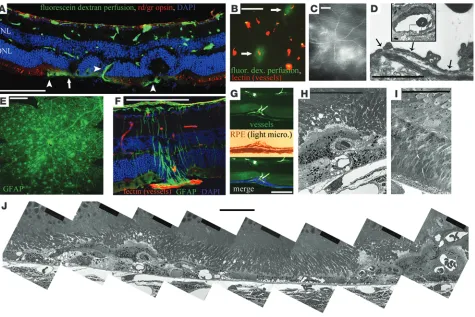
mice. During the second postna-tal week, abnormal intraretinal vessels sprouted from the deep and intermediate plexuses and penetrated through the outer nuclear layer (ONL) to the subretinal space (Figure 1, E–J). By 4 weeks of age, numerous intraretinal vessels extended to form a subretinal vascular network (Figure 1F). Choroidal vessels were normal at this time, and breaks in Bruch’s membrane and anastomoses of the intraretinal vessels with the choriocapillaris were not observed in mice less than 6 months of age, confirming the intraretinal ori-gin of subretinal NV (Figure 1, I and J). Although the numbers of subretinal sprouts were reduced in older mice, most intra- and sub-retinal vessels persisted throughout the life of the Vldlr–/– mice.
[image:3.585.62.528.80.323.2]The subretinal vessels were patent and perfused (Figure 1I and Figure 2, A and B). Modest extravasation of 43-kDa fluorescein dextran was observed from a subset of subretinal vascular sprouts (Figure 2, A–C), indicating a mild dysfunction of the blood-retinal barrier normally formed by tight junctions between nonfenestrated vascular endothelial cells and close association with perivascular glia (26). Electron microscopic analysis of intra- and subretinal NV
Figure 1
research article
in Vldlr–/– mouse retinas revealed fenestrated endothelia (Figure
2D), providing an anatomic basis for the abnormal permeability observed in these vessels. Müller glial cell activation, as demonstrated by GFAP expression, was selectively associated with, but secondary to, the formation of intraretinal NV (Figure 2, E and F).
By the fifth postnatal week, the normal retinal pigment epithe- lium (RPE) monolayer was disrupted in areas surrounding subreti-nal vascular lesions, and multiple layers of RPE cells enveloped the neovascular complexes in Vldlr–/– retinas (Figure 2, G and H). These
abnormalities lead to focal distortion and scarring of the retina, and were found to be associated with focal loss of photoreceptor inner and outer segments, as demonstrated by abnormal retinal morphology and an absence of red/green opsin staining in regions with subretinal NV (Figure 2, A and J). Neuronal abnormalities never preceded the appearance of intra- and subretinal NV. At P12, just prior to the onset of abnormal NV, all retinal layers were intact and identical to those of age-matched WT C57BL/6J con-trols when evaluated by electron and confocal microscopy (Figure
2I). Thus, neuronal abnormalities in Vldlr–/– mouse retinas appear
to be the result, rather than a cause, of NV.
Subretinal NV in Vldlr–/– retinas results from increased VEGF. In order
to gain insight into a possible relationship between VLDLR defi-ciency and the development of abnormal intraretinal vessels, we analyzed VLDLR expression in WT mouse retinas. The ganglion cell layer, retinal vessels, inner nuclear layer (INL), ONL, photoreceptor inner and outer segments, and RPE were isolated using laser cap-ture microdissection techniques. Each of these fractions is highly enriched for the indicated retinal layer, although on occasion, neighboring cells may have been inadvertently captured as a result of technical limitations with the technique. RNA was purified and analyzed by quantitative RT-PCR to assess the relative levels of VLDLR expression in each retinal layer. At P14, when intraretinal NV began in Vldlr–/– retinas, VLDLR expression in WT mice was
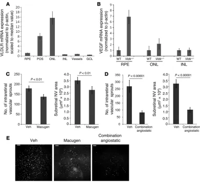
[image:4.585.59.534.81.398.2]greatest in the photoreceptors, with the RPE also expressing high levels of VLDLR mRNA. Expression in other retinal tissues, includ-ing retinal vessels, was observed at relatively low levels (Figure 3A).
Figure 2
Recently it has been reported that VEGF expression is increased in Vldlr–/– mouse retinas (27). We confirmed this finding and tested
for a direct correlation between VLDLR and VEGF expression in individual retinal layers at P14, when subretinal NV begins in Vldlr–/–
mice. In WT mice, VEGF expression was highest in the ONL, and smaller amounts of VEGF mRNA were found in the RPE and INL (Figure 3B). In Vldlr–/– retinas, VEGF expression was upregulated
compared with WT retinas more than 5-fold in the RPE and at least 2-fold in the ONL. No difference in expression levels was found in other retinal layers (Figure 3B).
It is likely that increased VEGF expression is responsible for new vessel growth. We tested this by using anti-VEGF agents to suppress subretinal NV in Vldlr–/– retinas. Intravitreal injection of Macugen,
an RNA aptamer that blocks VEGF165
, reduced intra- and subreti-nal NV confirming that VEGF upregulation in the deep retina is critical for abnormal vessel formation in the Vldlr–/– mouse (Figure
3C). In order to further evaluate effects of antiangiogenic therapy on subretinal vessels, we used a combination of angiostatic agents
previously shown to synergistically inhibit retinal angiogenesis (28): Macugen, an integrin αvβ3/αvβ5 antagonist, and T2-TrpRS, a
fragment of tryptophan transfer RNA synthetase with angiostatic activity (29–31). Using combination therapy, we observed substan-tial reductions in intraretinal vascular sprouts and total area of subretinal NV (70% and 60%, respectively; Figure 3, D and E).
Photoreceptor degeneration in Vldlr–/– mouse eyes
. Our initial morpho-logical studies suggested that neuronal abnormalities are directly associated with NV in the Vldlr–/– mouse retinas (Figure 2A). These
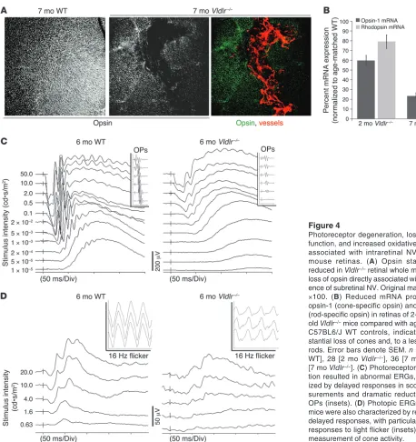
abnormalities consisted of rosettes within the ONL, thinning of the ONL and, to a lesser extent, the INL, and reduced red/green opsin in the areas directly associated with subretinal NV (Figure 4A). To assess photoreceptor degeneration in Vldlr–/– mice further, real-time
[image:5.585.97.487.81.436.2]RT-PCR was performed to analyze opsin-1 (cone-specific) and rho-dopsin (rod-specific) mRNA expression in whole retinas. Although an indirect correlation, we believe this measure to be correlated with photoreceptor viability and health, and it allowed analysis of degeneration in whole retinas. In 2-month-old mice, a 40% decrease
Figure 3
research article
in opsin-1 expression was observed in the Vldlr–/– retinas compared
with retinas of age-matched WT controls (Figure 4B). Rhodopsin expression was decreased in Vldlr–/– mouse retinas by only 20%. In
7-month-old mice, cone opsin-1 expression decreased approximately 70% in Vldlr–/– retinas compared with those of age-matched WT
controls, while rhodopsin expression was reduced by 30% (Figure 4B). Thus, both rod and cone photoreceptor abnormalities were associated with the vascular phenotype in Vldlr–/– mice, with cones
affected earlier and to a greater extent than rods.
Electroretinography (ERG) was performed to compare visual function in Vldlr–/– mice and age-matched WT controls. The ERGs
of 6-month-old Vldlr–/– mice exhibited delayed signaling, reduced
oscillatory potentials (OPs), and reduced and delayed cone responses. Amplitudes of scotopic rod-dominated responses were
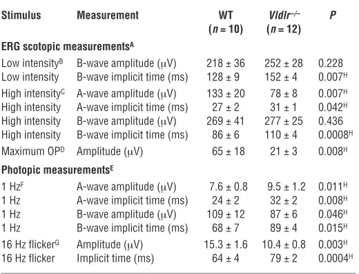
largely preserved. However, maximal dark-adapted B-wave implicit times in Vldlr–/– mice were consistently prolonged (Figure 4C and
Table 1), implying generalized rod dysfunction. Photopic cone responses were also delayed in Vldlr–/–
mice, but unlike the scoto-pic signals, amplitudes were significantly reduced as well (Figure 4D and Table 1). Abnormalities in the cone-specific responses to light flicker at 16 Hz were even more pronounced in the Vldlr–/–
mice, with delayed implicit times as well as reduced signal ampli-tudes (Figure 4D, insets, and Table 1). OPs were also significantly reduced in all 6-month-old Vldlr–/– ERGs (Figure 4C, insets, and
Table 1), indicating broad retinal signaling defects and suggestive of specific defects in the INL neurons.
[image:6.585.45.503.77.563.2]Targeted delivery of neurotrophin-4 protects retinas from neuronal degen-eration. Although we observed a highly significant reduction in the
Figure 4
Photoreceptor degeneration, loss of retinal function, and increased oxidative stress are associated with intraretinal NV in Vldlr–/– mouse retinas. (A) Opsin staining was reduced in Vldlr–/– retinal whole mounts, with loss of opsin directly associated with the pres-ence of subretinal NV. Original magnification,
formation of subretinal NV using combination angiostatic therapy, this effect was only transient, and subretinal NV returned to normal
Vldlr–/–
levels within 2–3 weeks of treatment. This is similar to clini- cal observations that report temporary beneficial effects of angio-static therapy. Thus, additional strategies for protecting the retinal neurons in ocular vascular diseases are still required (32). We next evaluated the potential effects of neurotrophic factors specifically delivered to sites of retinal NV and associated retinal degeneration. Because we observed GFAP activation in Müller cells located adja-cent to subretinal NV in the Vldlr–/– mouse retina (Figure 2, E and F),
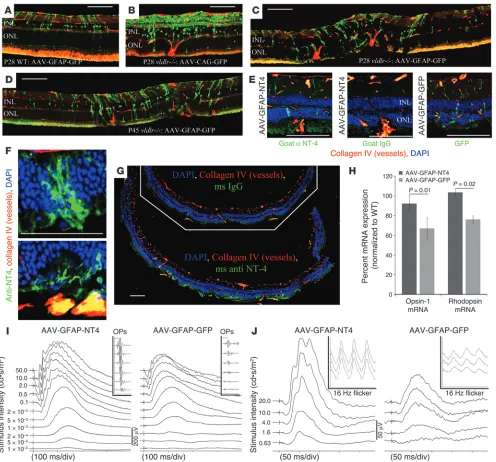
we obtained an AAV-2 vector containing transgenes driven by a GFAP promoter. Intravitreal injection of the GFP-encoding vector AAV-GFAP-GFP demonstrated that expression was strictly limited to cells of the inner retina in WT mice (Figure 5A); GFP expression in Müller cell processes was observed, but was limited to the gan-glion cell layer and the Müller cell bodies. However, in Vldlr–/– mice,
GFP expression was observed in a greater number of Müller cells, specifically within the processes of the activated Müller cells adja- cent to areas of subretinal NV. Importantly, the GFP signal extend-ed throughout both inner and outer retinas of Vldlr–/– mice in these
activated Müller cells (Figure 5, C and D). In contrast, a control vector with a ubiquitously expressed CAG promoter–driven trans-gene, AAV-CAG-GFP, demonstrated nonspecific expression of GFP in the inner retina mainly localized to ganglion cells, with mini-mal expression in the outer retina (Figure 5B). Similar expression patterns were observed when the AAV-2 vectors were injected into 5-month-old adult mice (data not shown). By targeting activated Müller glia using the AAV-2 vector with a GFAP promoter, we were able to specifically deliver transgene products to the outer retina in areas directly adjacent to subretinal NV.
Neurotrophin-4 (NT4) has been shown to protect neurons in sev- eral models of neuronal degeneration, including retinal degenera-
tion (33, 34). Similar to the GFP vectors, AAV-2 vectors with GFAP-driven expression of NT4 (AAV-GFAP-NT4) resulted in NT4 production in activated Müller cell bodies and, more importantly, outer retinal processes specifically adjacent to intraretinal NV (Figure 5, E and F). This caused accu- mulation of NT4 in the outer retina near areas of subreti-nal NV throughout the central two-thirds of the retina (Figure 5, F and G). Use of AAV-GFAP-NT4 protected the retina from neuronal degeneration, as demonstrated by opsin and rhodopsin mRNA expression (Figure 5H), and protected the Vldlr–/– retinas from the characteristic loss
of retinal function, resulting in more normalized ERGs (Figure 5, I and J, and Table 2). This protection, provided by the selective delivery of a neurotrophic factor to reti- nal areas with subretinal NV, supports a direct correla-tion between abnormal NV and neuronal degeneration in the retina. Furthermore, because activation of Müller glia is associated with numerous retinal diseases, par-ticularly those associated with abnormal NV and retinal degeneration, our findings provide data supporting the use of activated Müller glia for viral-mediated (GFAP vec-tors) delivery of therapeutic gene products to the outer retina using intravitreal injections.
Vldlr–/– retinas demonstrate increased oxidative stress. We
hypothesized that photoreceptor degeneration might be caused by oxidative stress generated by the presence of abnormal intra- and subretinal vessels. Metabolomic anal-ysis comparing retinas from 4-month-old Vldlr–/– and WT
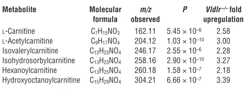
mice demonstrated a significant increase in metabolites commonly associated with oxidative stress (Table 3 and Supplemental Figure 1).
l-Carnitine, l-acetylcarnitine, and their derivatives are naturally
occurring antioxidants (35, 36) that are upregulated in response to increased oxidative stress (35, 37, 38). The observed increase in these metabolites in the Vldlr–/– retinas indicates an endogenous
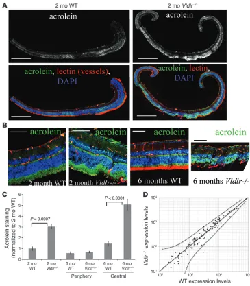
cellular response to oxidative stress. Acrolein is a commonly used marker of lipid peroxidation resulting from oxidative stress (9, 10, 39, 40). Acrolein staining was clearly greater in Vldlr–/–
retinas com-pared with age-matched WT controls (Figure 6, A and B). In the peripheral retina, acrolein levels were similar between Vldlr–/– and
WT retinas, while acrolein levels were greatly increased in the cen-tral two-thirds of Vldlr–/– retinas compared with age-matched WT
controls (Figure 6C). In Vldlr–/–
eyes, the vast majority of intrareti-nal NV occurs in the central two-thirds of the retina, whereas the peripheral retinal vasculature remains normal. Thus, increased oxidative stress measured by acrolein staining in Vldlr–/– retinas
appears to be limited to regions with subretinal NV, supporting a direct relationship between subretinal NV and oxidative stress.
The transient effect of antiangiogenic treatments in the Vldlr–/–
[image:7.585.53.313.111.310.2]retinas rendered us unable to prevent recurrence of subretinal NV using a single angiostatic treatment, and multiple, weekly injections led to phthisis. Thus, it is not possible with this experimental model to determine whether subretinal NV directly causes the observed oxidative stress and associated neuronal degeneration. An alterna-tive hypothesis that might explain the loss of photoreceptors in the presence of increased markers of oxidative stress would be a defect in natural oxidative stress defense mechanisms associated with VLDLR deficiency. To test this hypothesis, we used large-scale genomic anal-ysis to examine the expression of genes encoding factors involved in protection against oxidative stress. Of 210 gene products exam-ined, 109 were found to be expressed in the retina, and 108 of these were equally expressed in 3-week-old Vldlr–/– and WT retinas within
Table 1
Responses to light stimuli in 6-month-old WT and Vldlr–/– mice
Stimulus Measurement WT Vldlr–/– P
(n = 10) (n = 12)
ERG scotopic measurementsA
Low intensityB B-wave amplitude (μV) 218 ± 36 252 ± 28 0.228
Low intensity B-wave implicit time (ms) 128 ± 9 152 ± 4 0.007H
High intensityC A-wave amplitude (μV) 133 ± 20 78 ± 8 0.007H
High intensity A-wave implicit time (ms) 27 ± 2 31 ± 1 0.042H
High intensity B-wave amplitude (μV) 269 ± 41 277 ± 25 0.436 High intensity B-wave implicit time (ms) 86 ± 6 110 ± 4 0.0008H
Maximum OPD Amplitude (μV) 65 ± 18 21 ± 3 0.008H
Photopic measurementsE
1 HzF A-wave amplitude (μV) 7.6 ± 0.8 9.5 ± 1.2 0.011H
1 Hz A-wave implicit time (ms) 24 ± 2 32 ± 2 0.008H
1 Hz B-wave amplitude (μV) 109 ± 12 87 ± 6 0.046H
1 Hz B-wave implicit time (ms) 68 ± 7 89 ± 4 0.015H
16 Hz flickerG Amplitude (μV) 15.3 ± 1.6 10.4 ± 0.8 0.003H
16 Hz flicker Implicit time (ms) 64 ± 4 79 ± 2 0.0004H
Values are mean ± SEM. ADark-adapted measurements performed to determine rod and mixed cone-rod function. B0.005 cd•s/m2. C2 cd•s/m2. D10 cd•s/m2. ELight-adapted measurements performed to determine cone function. F20 cd•s/m2. G10 cd•s/m2. HStatistically significant difference between WT and Vldlr–/–
research article
95% confidence intervals, the single exception being 9-cis-retinol dehydrogenase (Figure 6D and Supplemental Table 1). While not absolute, these data suggest that neuronal defects in Vldlr–/– mice
are not caused by a generalized failure of oxidative stress defense and support the concept of a direct relationship between excessive NV, oxidative stress, and neuronal degeneration.
Antioxidant treatment protects against retinal damage in Vldlr–/– mice. If
oxidative stress is associated with neuronal damage in the Vldlr–/–
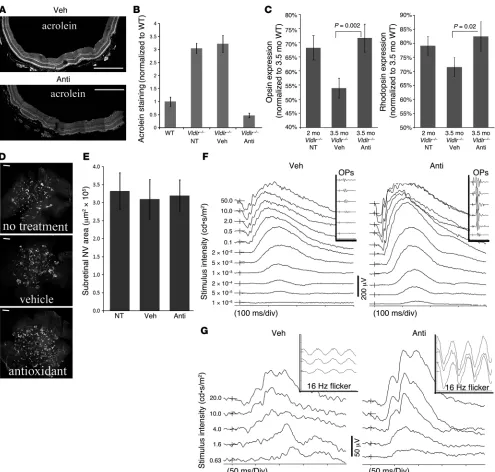
retina, we reasoned that treatment with commonly used antioxi-dants may attenuate this degeneration. Daily oral administration of an antioxidant cocktail consisting of ascorbic acid, α-lipoic acid, and reduced l
-glutathione for 6 weeks dramatically reduced the lev-Figure 5
[image:8.585.47.543.81.543.2]els of oxidative stress in Vldlr–/– mouse retinas (Figure 7, A and B).
This correlated with protection of the photoreceptors from degen-eration. Furthermore, 6 weeks of antioxidant treatment initiated at 2 months of age, well after abnormal subretinal NV is established, significantly improved the expression levels of opsin and rho-dopsin in the retinas of 3.5-month-old Vldlr–/– mice (Figure 7C).
Opsin and rhodopsin expression levels remained similar to those of untreated 2-month-old Vldlr–/–
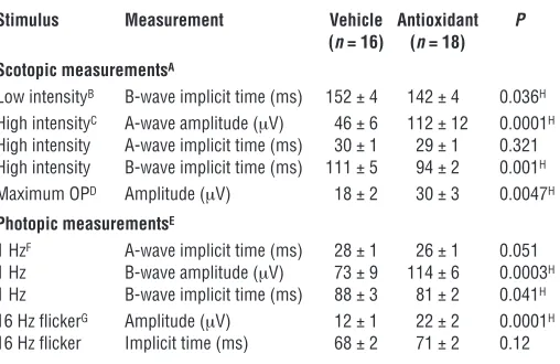
mice, which suggests that anti-oxidant treatment prevented further loss of cones and rods from the time treatment was initiated. Antioxidant treatment did not have any effect on the formation and persistence of subretinal NV (Figure 7, D and E). Thus, the observed activity was not caused by prevention or elimination of NV, but rather by protection of the neurons from secondary neurodegeneration associated with NV. Normalized ERGs were also observed in Vldlr–/–
mice on an anti-oxidant diet compared with vehicle-treated animals. Importantly, significant preservation of retinal function was observed even when treatments were initiated at 2 months of age, an age at which intra- and subretinal NV was well established and oxidative stress was already apparent (Figure 7, F and G, and Table 4), consistent with the results obtained from the RT-PCR experiments. ERGs of animals in which treatment was initi-ated at an earlier age were similar to those observed in WT animals (data not shown), which indicates that early dietary measures may provide nearly full protection from neuronal degeneration in the retina. The profound protective effect of antioxidant dietary supplementation on retinal degenera-tion further supports a direct link between oxidative stress and neuronal degeneration in the Vldlr–/– mouse retina.
Discussion
Paucity and dysfunction of blood vessels in tissues leading to ischemia is a well-recognized pathological process that often leads to irreversible injury, particularly in neuronal
tissue. At the other extreme, excessive vessels (NV) can also compromise tissue function, usually as a result of leakage, hemorrhage, or scarring caused by the abnormal new vessels. In this study, we provide evidence for yet another mechanism of injury associated with NV, even in the absence of clinically sig-nificant leakage or hemorrhage: neuronal cell death as a result of increased oxidative stress caused by proximity to the abnormal vessels.
In several diseases that can cause irreversible vision loss, such as RAP (16) and MacTel (14, 41), abnormal vessels extend from the highly vascularized inner retina into the normally avascular outer retina. The reti-na of the Vldlr–/– mouse exhibits
vascular changes very similar to those observed in patients with MacTel and RAP and thus is a good model for studying the general relationship between NV and neurodegeneration. The new vessels observed in the human disease and the Vldlr–/– mouse exhibit relatively mild permeability defects
and are accompanied by glial activation and disruption of the RPE. Another feature common to RAP, MacTel, and the Vldlr–/– retina is
that the nonuniformly distributed focal vascular lesions are directly associated with neuronal degeneration separated by relatively nor-mal regions. While other retinal degenerative models, such as rd1 or rds mice, have associated vascular abnormalities and neuronal loss, many of these result in uniform degeneration and relate more closely to genetic diseases, such as retinitis pigmentosa, rather than to ocular vascular disease. Taken together, these similarities support the use of the Vldlr–/– mouse as a compelling model to investigate
selected human retinal diseases (e.g., MacTel) as well as other, non-ocular neurodegenerative disorders with associated NV.
VEGF plays a critical role in normal retinal vascularization and retinal angiogenic diseases (42–44). In the Vldlr–/– retina, VEGF
upregulation is observed in photoreceptors and RPE and partici-Table 2
ERG measurements in 4-month-old WT and untreated, AAV-GFAP-GFP control–treated, and AAV-GFAP-NT4–treated Vldlr–/– mice 3.5 months after injection
Stimulus Measurement WT Vldlr–/– Vldlr–/– Vldlr–/– P
(n = 12) untreated GFP NT4 (NT4 vs. GFP)
(n = 10) (n = 15) (n = 15)
Scotopic measurementsA
Low intensityB B-wave implicit time (ms) 121 ± 5 130 ± 4 126 ± 4 118 ± 2 0.003H
High intensityC A-wave amplitude (μV) 136 ± 25 80 ± 17 68 ± 13 114 ± 18 0.011H
High intensity A-wave implicit time (ms) 24 ± 1 24 ± 1 24 ± 1 22 ± 1 0.067
High intensity B-wave implicit time (ms) 80 ± 4 89 ± 2 93 ± 4 75 ± 2 0.0002H
Maximum OPD Amplitude (μV) 71 ± 10 39 ± 7 36 ± 7 62 ± 12 0.013H
Photopic measurementsE
1 HzF A-wave implicit time (ms) 19 ± 2 23 ± 1 22 ± 1 18 ± 1 0.005H
1 Hz B-wave amplitude (μV) 105 ± 9 83 ± 6 80 ± 11 96 ± 9 0.022H
1 Hz B-wave implicit time (ms) 62 ± 4 71 ± 3 78 ± 3 58 ± 2 0.0001H
16 Hz flickerG Amplitude (μV) 19 ± 3 16 ± 2 15 ± 2 19 ± 2 0.041H
16 Hz flicker Implicit time (ms) 55 ± 3 58 ± 1 58 ± 1 56 ± 1 0.175
[image:9.585.56.403.122.302.2]Values are mean ± SEM. ADark-adapted measurements performed to determine rod and mixed cone-rod func-tion. B0.005 cd•s/m2. C2 cd•s/m2. D10 cd•s/m2. ELight-adapted measurements performed to determine cone func-tion. F20 cd•s/m2. G10 cd•s/m2. HStatistically significant difference between contralaterally AAV-GFAP-NT4– and AAV-GFAP-GFP–treated eyes.
Table 3
Metabolite analysis of 6-month-old WT and Vldlr–/– mouse retinas
Metabolite Molecular m/z P Vldlr–/– fold
formula observed upregulation
l-Carnitine C7H15NO3 162.11 5.45 × 10⁻⁶ 2.58
l-Acetylcarnitine C9H17NO4 204.12 1.03 × 10⁻¹⁰ 3.00
Isovalerylcarnitine C12H23NO4 246.17 2.55 × 10⁻⁶ 2.28
Isohydrosorbylcarnitine C13H23NO4 258.16 2.90 × 10⁻¹⁰ 3.27
Hexanoylcarnitine C13H25NO4 260.18 1.58 × 10⁻⁷ 2.18
Hydroxyoctanoylcarnitine C15H29NO4 304.21 6.66 × 10⁻⁷ 3.39
Shown are metabolites with an increased presence in Vldlr–/– retinas,
[image:9.585.281.531.621.712.2]research article
pates in the stimulation of subretinal NV. Normal VLDLR expres-sion within the avascular outer retina, and upregulation of VEGF coincident with formation of abnormal subretinal vessels in these areas of VLDLR-deficient mice, suggests a mechanism linking VLDLR with proangiogenic factors. No obvious abnormalities were observed in the choroid, suggesting potential RPE polarity of the VEGF overexpression toward the inner retina. VLDLR is a negative regulator of wnt signaling during retina development;
upregulation of the wnt coreceptor LRP5/6 and increased down-stream wnt signaling occurs in
Vldlr–/– mouse retinas (27). VEGF
is a well-known product of wnt signaling, and VLDLR may nor-mally participate in turning off wnt signaling once retinal vas-cular development is complete. In the absence of VLDLR, wnt signaling persists, leading to increased levels of VEGF expres-sion in photoreceptor and RPE layers and the angiogenic exten- sion of abnormal intra- and sub-retinal vessels. Whether such a mechanism operates in diseases such as MacTel and RAP is uncer- tain, although anti-VEGF thera-py has previously been tested in both conditions (45, 46). Initial efforts to treat RAP lesions using combination angiostatics are also promising (47). However, in our experience with the Vldlr–/–
mouse, antiangiogenic therapy is only transiently effective, subreti-nal NV returns within 2 weeks, and therapy must be initiated prior to NV formation. Thus, the usefulness of antiangiogenic therapy in human ocular diseases with attributes similar to those of
Vldlr–/– mice may be limited.
A major finding in this study is that neuronal abnormalities and impaired retinal function devel-oped in association with NV. We were able to prevent focal degen-eration of photoreceptors in the regions of vascular abnormalities through the selective delivery of neurotrophic molecules by tak-ing advantage of cellular changes observed in this condition. Müller glia, with cytoplasmic processes spanning the inner and outer retina, were activated in response to vascular changes such as those observed in the Vldlr–/– retina.
[image:10.585.40.405.81.498.2]Upon activation, these cells upreg-ulate GFAP, and their appearance and location precisely correlate, both temporally and spatially, with subretinal NV and associated neuronal degeneration in the outer retina. By targeting the acti-vated Müller cells and taking advantage of their retina-spanning cytoplasmic processes, NT4 was specifically delivered to sites of vas-cular abnormalities after intravitreal injection of AAV-GFAP-NT4. This strategy using endogenous cells (e.g., activated Müller cells) to deliver gene therapy products to the outer retina could be useful clinically to avoid the need for subretinal injection of the viral vec-tor, a procedure that can have deleterious effects on already diseased
Figure 6
retinas. In addition, we were able to dramatically attenuate retinal degeneration in the Vldlr–/– mouse retina, supporting a link between
the presence of subretinal vessels and neuronal degeneration.
Vldlr–/– retinas exhibit increased levels of oxidative stress (35, 37,
38). Excessive oxygen levels can generate reactive oxygen species that cause molecular damage to lipids, proteins, and nucleic acids, which
[image:11.585.47.542.79.551.2]can subsequently lead to cell death unless neutralized by the antioxi-dant defense system (48). The retina, with its high fat content and its exposure to light, is particularly susceptible to such injury. Cone loss is associated with oxidative damage in other models of primary reti-nal degeneration, such as the rd1 mouse (9, 10), and oxidative damage has been linked with AMD (40). Recent studies using adaptive optics
Figure 7
research article
also demonstrate focal photoreceptor loss associated with vascular abnormalities in diseases such as MacTel, traditionally considered a retinal vascular disease (A. Roorda, unpublished observations). We attempted to directly assess the role of abnormal NV in causing sub-sequent neuronal degeneration by blocking NV in the Vldlr–/– mice.
However, even the most potent angiostatic combinations were only transiently effective, while the neurodegeneration was progres-sive over the course of several weeks to months after initiation of subretinal NV. Thus, prolonged elimination of subretinal NV was impossible without repeated injections, a process which at best leads to injury and variability in the small mouse eye, and often leads to phthisis. However, based on the strong association between subreti-nal NV and neurodegeneration, the correlation between subretinal NV and oxidative stress, and the neuroprotective results obtained from treatment with antioxidants, we hypothesize that increased vascularization and abnormal localization of the subretinal NV leads to increased oxidative stress and subsequent neuronal damage. No defects were found in the cellular oxidative stress defense pathways of the Vldlr–/– mice, further supporting our hypothesis.
These findings demonstrating protective effects of antioxidant treatment on neuronal degeneration associated with abnormal NV support prior epidemiological studies in humans showing that an antioxidant-rich diet protects against AMD (40) and diminishes its progression (49). However, it should be noted that only moderate levels of protection were observed in these epidemiological studies, particularly when dietary supplementation is initiated after NV is already present. Such dietary measures may be a simple and effec-tive method of preventing neuronal cell loss associated with certain retinal vascular diseases such as MacTel and RAP. In instances of associated neuronal degeneration with substantial ERG abnor-malities, gene therapy using intravitreally injected AAV-2 vectors encoding neurotrophic transgenes may also serve to preserve visual function. These findings using the Vldlr–/– mouse retina model also
support other studies that demonstrate benefits of antioxidant-rich diets in relation to other neurodegenerative disorders of the CNS (50). Thus, our present findings may also have application to other
neurodegenerative disorders, such as Huntington disease, Parkinson disease, and Alzheimer disease. Further stud-ies will be required to better understand the link between vascular abnormalities such as those observed in retinal NV, oxidative stress, and neuronal degeneration.
Methods
Retina dissection and staining. All animal studies were approved by the animal protocols review committee at The Scripps Research Institute. Vldlr–/– mice (23, 51) were obtained and backcrossed in
the C57BL/6J background for at least 10 generations. WT mice were normal, nonknockout C57BL/6J mice. Retinas were dis-sected and prepared for whole mounts or sectioning as previously described (17). For preparation of retinal cross-sections, dissected retinas were laid flat with 4 radial relaxing incisions, placed in 4% PFA, and incubated at 4°C overnight. Retinas were then placed in 20% sucrose at 4°C for 4 hours and embedded in Tissue-Tek OCT compound (Sakura FineTechnical) for cryosectioning.
Fluorescently conjugated isolectin GS from Griffonia simplici-folia (Invitrogen) or antibodies against collagen IV (Chemicon), GFAP (DAKO), and red/green opsin (Chemicon) were used for immunohistochemistry. For analysis of vascular leak, FITC-dextran (43,200 MW; Sigma-Aldrich) was perfused through the left ventricle of deeply anesthetized mice using 150 μl of 50 mg/ml solution in PBS.
Electron microscopy analysis of Vldlr–/– retinas . Eye cups containing the reti-na and choroid from each mouse were fixed in 4% paraformaldehyde plus 1.5% glutaraldehyde in 0.1 M cacodylate buffer overnight at 4°C followed by rinsing in 0.1 M Na cacodylate buffer for 1 hour. The eye cups were then postfixed in 1% OsO4 in 0.1 M cacodylate buffer for 2 hours, followed
by another 1-hour wash and dehydration with graded ethanol solutions. Samples were incubated overnight in a 1:2 mixture of propylene oxide and Epon/Araldite (Sigma-Aldrich) and then placed in 100% resin followed by embedding. The blocks were sectioned and used for high-magnification electron microscopy analysis.
Laser capture . All steps were carefully performed using RNAse-free condi-tions. Whole eyes were isolated without fixation and embedded in OCT. After serial sectioning, laser capture microdissection was performed using an Arcturus PixCell II Laser Capture Microdissection microscope. The ganglion cell layer, INL, ONL, photoreceptor inner/outer segments, and retinal pigment epithelium were isolated separately from histologically stained retinas. Segments highly enriched for vessel fragments were also isolated after a brief stain with isolectin GS (Invitrogen). RNA was isolated from the microdissected retina using microRNA kits (Qiagen) and, after ensuring the quality of each RNA preparation, was reverse transcribed into cDNA for analysis by quantitative RT-PCR.
Quantitative RT-PCR analysis. Retinas were dissected under RNAse free conditions, and each retina was immediately lysed in separate tubes con-taining RLTplus buffer (Qiagen) with β-mercaptoethanol (Sigma-Aldrich). RNA was isolated using the RNeasy plus mini kit (Qiagen) protocol, which incorporates a genomic DNA elimination step. Total RNA (0.5 μg from each retina) was reverse transcribed into cDNA using the Quantitect reverse transcription kit (Qiagen). SYBR green–based real-time quantita- tive RT-PCR was performed using the iCycler machine (BioRad) to deter-mine mRNA expression levels of various gene products. Expression levels from each retina were normalized to β-actin.
[image:12.585.56.310.113.278.2]Primers. The following primers were used: cone opsin forward, 5′ -CAAGCCCTTTGGCAATGTGA-3′; cone opsin reverse, 5′ -GCTCCAAC-CAAAGATTGGTGG-3′; rhodopsin forward, 5′ -TCATGGTCTTCGGAG-GATTCAC-3′; rhodopsin reverse, 5′-TCACCTCCAAGTGTGGCAAAG-3′;
Table 4
ERG measurements of antioxidant- and vehicle-treated Vldlr–/– mice
Stimulus Measurement Vehicle Antioxidant P
(n = 16) (n = 18)
Scotopic measurementsA
Low intensityB B-wave implicit time (ms) 152 ± 4 142 ± 4 0.036H
High intensityC A-wave amplitude (μV) 46 ± 6 112 ± 12 0.0001H
High intensity A-wave implicit time (ms) 30 ± 1 29 ± 1 0.321 High intensity B-wave implicit time (ms) 111 ± 5 94 ± 2 0.001H
Maximum OPD Amplitude (μV) 18 ± 2 30 ± 3 0.0047H
Photopic measurementsE
1 HzF A-wave implicit time (ms) 28 ± 1 26 ± 1 0.051
1 Hz B-wave amplitude (μV) 73 ± 9 114 ± 6 0.0003H
1 Hz B-wave implicit time (ms) 88 ± 3 81 ± 2 0.041H
16 Hz flickerG Amplitude (μV) 12 ± 1 22 ± 2 0.0001H
16 Hz flicker Implicit time (ms) 68 ± 2 71 ± 2 0.12
VLDLR forward, 5′-TTCCTAGCTCATCCTCTTGCAC-3′; VLDLR reverse, 5′-CTGACCCAGTGAATTTATTGGC-3′; VEGF forward, 5′ -GGAGA-CTCTTCGAGGAGCACTT-3′; VEGF reverse, 5′ -GGCGATTTAGCAGCA-GATATAAGAA-3′. For β -actin normalization, the Mm_Actb_2_SG Quan-tiTect Primer Assay was used (Qiagen).
Treatment of murine eyes with antiangiogenics . Intravitreal injections of Macu-gen, combination angiostatics, or vehicle controls were performed at P12, just prior to formation of the subretinal vascular sprouts in the Vldlr–/– retinas. All intravitreal injections were performed using a Hamilton syringe fitted with a 33-gauge needle (Hamilton), injecting 0.5 μl of solutions containing 1.5 μg Macugen (3.0 mg/ml; Pfizer), a triple angiostatic combination, or PBS vehicle. The triple combination solution consisted of Macugen (3.0 mg/ml), integrin αvβ3 and αvβ5
antagonist EMD472523 (20 mg/ml; Merck), and T2-TrpRS (0.5 mg/ml; Angiosyn). T2-TrpRS is a fragment of human TrpRS that has previously been shown to have angiostatic activity (30). The retinas were analyzed 1 week later at P20 for the formation of subretinal vessels. Whole-mount preparations were stained for the vasculature, the subretinal space was imaged using confocal microscopy, and the number of subretinal vascular sprouts as well as the area of subretinal NV staining was quantified.
ERG. After overnight dark adaptation, mice were anesthetized under dim red light by intraperitoneal injection of 15 mg/kg ketamine and 7 mg/kg xylazine. Full-field ERGs were recorded from the corneal surface of each eye after pupil dilation (1% tropicamide and 2.5% phenylephrine) using a gold loop electrode together with a mouth reference and tail ground electrode, similar to previously published methods (52). A computerized system with an electronically controlled Ganzfeld dome was used (Espion E2 with Colordome; Diagnosys). In the dark-adapted state, we recorded rod and mixed cone/rod responses to a series of white flashes of increasing intensities (1 × 10⁻⁵ to 50 cd•s/m2). In the light-adapted state, with a 30
cd/m2 background, cone responses to 1-Hz (0.63 to 20 cd•s/m2) and 30-Hz
(3.98, 10, and 20 cd•s/m2) flicker stimuli were recorded. All ERG responses
were filtered at 0.3–500 Hz, and signal averaging was applied.
AAV-2 vector constructs.The AAV-2 vector genome backbone consisting of AAV-2 inverted terminal repeats flanking a neurotrophic factor transgene expression cassette with the ubiquitous CAG promoter and the human growth hormone gene polyadenylation signal (Stratagene) was described pre- viously (53). The construction of the AAV-GFAP-NT4 vector genome was per-formed in 2 steps. First, human NT4 cDNA was isolated from a human cDNA library (Clontech) by PCR and inserted into the AAV-CAG-Neurturin (NTN) in place of the NTN cDNA (53) to generate AAV-CAG-NT4. Second, a mini-mal 0.35-kb glial cell–specific GFAP promoter consisting of the A/B (–1757 to –1488) and D (–132 to –56) sequences of the human GFAP promoter (54) was isolated by PCR from human genomic DNA (Novagen), spliced together, and inserted into AAV-CAG-NT4 in place of the CMV enhancer and chicken β-actin promoter elements of the CAG promoter. Plasmid clone identities were confirmed by restriction digestions and nucleotide sequence determination.
Cell culture and AAV-2 vector production. AAV-2 vectors were produced by standard triple plasmid transfection technique in human embryonic kid-ney 293 cells and purified by multiple chromatography and filtration steps as described previously (53). After purification, vectors were resuspended in formulation buffer (PBS plus 2 mM MgCl2
), and vector titer was deter-mined by quantitative PCR and expressed as vector genomes/ml.
Treatment of murine eyes with AAV-2 vectors. Solutions (0.5 μ l) of AAV-2 par-ticles containing AAV-CAG-GFP (CAG promoter–driven GFP expression), AAV-GFAP-GFP (GFAP promoter–driven GFP expression), or AAV-GFAP-NT4 (GFAP promoter–driven NT4 expression), with titers of approximately 1 × 10¹³ vector genomes/ml, were injected intravitreally into the eyes of
2-week-old Vldlr–/– mice. Viral transfection was assessed by analyzing GFP expression at 1 and 2 months after injection. Effects on the neurodegenera-tive phenotype of Vldlr–/– mice were assessed 3 months after injection.
Acrolein staining for oxidative stress. Retinas were stained as described above, either with antibodies against acrolein (1:200 dilution, catalog no. ab37110; Abcam) or with appropriate control IgGs. After staining was complete, all ret- inas were imaged using confocal microscopy using identical settings, includ-ing laser power, iris opening, and detector sensitivity. Thus, differences in intensity correlated directly to differences in the amount of acrolein present. A baseline staining threshold level was established, and corresponding images from retinal sections within the central two-thirds or retinal periphery were quantified by comparing areas of acrolein staining above this threshold.
Metabolite extraction and mass spectrometry analysis. Proteins were precipitated and metabolites extracted from 12 Vldlr–/– and 12 WT retinas. After removal of proteins and isolation of metabolites, specimens were subjected to reversed phase and hydrophilic interaction chromatography. Metabolites with diffe-rential concentrations in Vldlr–/– compared with age-matched WT retinas were identified by exact mass (m/z) using METLIN (http://metlin.scripps. edu/), Human Metabolome Project (http://metabolomics.ca/), Lipid Maps (http://www.lipidmaps.org/), Biological Magnetic Resonance Data Bank (http://www.bmrb.wisc.edu/), and KEGG ligand (http://www.genome.jp/ kegg/ligand.html) databases. For a complete description of the materials and methods used for metabolomics analysis, see Supplemental Methods.
Microarray analysis . RNA was isolated from 3 sets of pooled retinas (6 reti-nas per set) from 3-week-old WT or Vldlr–/–mice, yielding triplicate samples accounting for both biological and technical variance. Samples were pre-pared and hybridized to Mu74Av2 gene chips (Affymetrix) according to the manufacturer’s instructions, and data were analyzed using GeneSpring software (Silicon Genetics). Genes related to oxidative stress pathways were analyzed for retinal expression, and only those above background in WT or
Vldlr–/– retinas with statistically consistent results amongst the replicates were analyzed further. The expression levels were compared between the WT and Vldlr–/– groups for each individual gene. Raw data values obtained from the Affymetrix analysis are included in Supplemental Table 1.
Antioxidant treatment. Littermates of Vldlr–/– mice were randomly separated into control and treated groups. Treated groups were fed a mixture of anti-oxidants that included 200 mg/kg ascorbic acid, 20 mg/kg α-lipoic acid, and 10 mg/kg l-glutathione (Sigma-Aldrich) in 1% aqueous sucrose. Controls were fed vehicle consisting of 1% aqueous sucrose. All mice were fed by orally pipetting 10–30 μl of solution and monitoring swallowing. Daily feedings were initiated at 2 months of age and were continued for 6 weeks.
Statistics. All data shown are mean ± SEM. All P values reported were derived using a standard Student’s t test with a 1-tailed distribution, and 2 sample equal variance for treatment groups using unpaired eyes or paired values for studies that used contralateral eyes to compare various treat-ments. A P value less than 0.05 was considered significant.
Acknowledgments
We thank Malcolm Wood for technical assistance with electron microscopy and Christopher Aderman, Kip Conner, and Lois Smith (Harvard Medical School, Boston, Massachusetts, USA) for assistance with laser capture microdissection. This work was funded by National Eye Institute, NIH, grant R01 EY011254 (to M. Friedlander), by the MacTel Foundation, and by the V. Kann Rasmussen Foundation. M.I. Dorrell is supported by a fellowship from the California Institute for Regenerative Medicine.
Received for publication June 30, 2008, and accepted in revised form December 10, 2008.