0095-1137/06/$08.00
⫹
0
doi:10.1128/JCM.00320-06
Copyright © 2006, American Society for Microbiology. All Rights Reserved.
Multilocus Sequence Typing for Analyses of Clonality of
Candida albicans
Strains in Taiwan
Kuo-Wei Chen,
1Yee-Chun Chen,
2Hsiu-Jung Lo,
3Frank C. Odds,
4Tzu-Hui Wang,
1Chi-Yang Lin,
1and Shu-Ying Li
1*
Laboratory for Mycology, Division of Research and Diagnostics, Center for Disease Control, Taipei, Taiwan
1; Department of
Internal Medicine, National Taiwan University Hospital, Taipei, Taiwan
2; Division of Clinical Research,
National Health Research Institutes, Taipei, Taiwan
3; and Aberdeen Fungal Group, Institute of
Medical Sciences, University of Aberdeen, Aberdeen AB25 2ZD, United Kingdom
4Received 14 February 2006/Returned for modification 22 March 2006/Accepted 1 April 2006
Multilocus sequence typing (MLST) was used to characterize the genetic profiles of 51
Candida albicans
isolates collected from 12 hospitals in Taiwan. Among the 51 isolates, 16 were epidemiologically unrelated, 28
were isolates from 11 critically ill, human immunodeficiency virus (HIV)-negative patients, and 7 were long-term
serial isolates from 3 HIV-positive patients. Internal regions of seven housekeeping genes were sequenced. A
total of 83 polymorphic nucleotide sites were identified. Ten to 20 different genotypes were observed at the
different loci, resulting, when combined, in 45 unique genotype combinations or diploid sequence types (DSTs).
Thirty (36.1%) of the 83 individual changes were synonymous and 53 (63.9%) were nonsynonymous. Due to the
diploid nature of
C. albicans
, MLST was more discriminatory than the pulsed-field gel
electrophoresis–BssHII-restricted fragment method in discriminating epidemiologically related strains. MLST is able to trace the
microevolution over time of
C. albicans
isolates in the same patient. All but one of the DSTs of our Taiwanese
strain collections were novel to the internet
C. albicans
DST database (http://test1.mlst.net/). The DSTs of
C.
albicans
in Taiwan were analyzed together with those of the reference strains and of the strains from the United
Kingdom and United States by unweighted-pair group method using average linkages and minimum spanning
tree. Our result showed that the DNA type of each isolate was patient specific and associated with ABC type
and decade of isolation but not associated with mating type, anatomical source of isolation, hospital origin, or
fluconazole resistance patterns.
Invasive
Candida
infections continue to cause major
prob-lems of morbidity and mortality in a diverse range of
debili-tated and immunocompromised hosts and constitute an
impor-tant public health problem (12, 13, 20, 41, 42).
Candida
species
were the leading pathogens of nosocomial bloodstream
infec-tion in a large teaching hospital in Taiwan, with
Candida
albi-cans
being the leading cause of
Candida
infections (5, 6). The
infections caused by
C. albicans
result in increased lengths of
hospital stays and medical costs (31, 34). Furthermore, an
outbreak of
C. albicans
fungemia in a neonatal intensive care
unit (16) and yeast carriage on hands of healthcare workers in
that intensive care unit were identified (15). The increasing
frequency of invasive candidal infections in Taiwan and their
severe outcome has underscored the importance of the
under-standing of the molecular epidemiology of fungal infections.
Molecular typing methods used to assess clonality of
C.
albicans
include pulsed-field gel electrophoresis (PFGE)-based
typing methods (4), restriction fragment length polymorphism
(10, 29), restriction fragment length polymorphism followed by
Ca3 probe hybridization (19, 40), and randomly amplified
poly-morphic DNA analysis (33). More recently developed typing
methods include amplified fragment length polymorphism (1),
multilocus microsatellite gene analysis (11, 25), and analysis of
variable numbers of tandem repeats (24). The choice of
ap-propriate typing methods depends on the purpose of the study.
Molecular typing methods should be reproducible,
discrimina-tory, digitally portable, and amenable to standardization and
have high throughput (35).
Recently, multilocus sequence typing (MLST) has been
de-veloped to meet the increasing need for global surveillance and
comparison of genotypes in a central database via the internet.
MLST is based on the sequencing of 6 to 7 selected
house-keeping genes and identification of polymorphic nucleotide
sites. Combination of the alleles at the different loci results in
unique diploid sequence types (DSTs) that can be used to
discriminate
C. albicans
strains (2, 3, 37). MLST provides a
robust and unambiguous characterization system to evaluate
the worldwide diversity and epidemiology of pathogens and is
truly portable between laboratories. MLST for typing of
patho-genic fungal species has been developed, such as
Candida
glabrata
(9),
Candida tropicalis
(38),
Histoplasma capsulatum
,
Aspergillus flavus
,
Coccidioides immitis
(21), and the
Fusarium
oxysporum
complex (28). A consensus set of 7 genes encoding
housekeeping functions comprising the fragments
AAT1a
,
ACC1
,
ADP1
,
MPIb
,
SYA1
,
VPS13
, and
ZWF1b
is
recom-mended for MLST with
C. albicans
(http://test1.mlst.net) (3).
In this study, we use MLST to assess the clonality of
C.
albicans
and to ascertain whether different characteristics (e.g.,
fluconazole resistance, patient or hospital origin, source, or
decade of isolation) can be attributed to certain specific MLST
DSTs in Taiwan. The purpose of this study was to evaluate the
usefulness of MLST relative to PFGE-BssHII fingerprinting
tools for discriminating among strains of
C. albicans
. The data
* Corresponding author. Mailing address: Laboratory for Mycology,
Division of Research and Diagnostics, Center for Disease Control,
No. 161, Kun-Yang Street, Taipei, Taiwan. Phone: 886 2 26531388.
Fax: 886 2 26513572. E-mail: [email protected].
2172
on May 16, 2020 by guest
http://jcm.asm.org/
obtained in this study will also contribute to the global
data-base and serve as a platform for comparison of domestic as
well as international fungal genotypes.
MATERIALS AND METHODS
Fungal isolates.A total of 51Candida albicansclinical isolates and 2 reference strains were used in this study. Information on each isolate was collected, in-cluding MICs of fluconazole, hospital origin, and body site origin. The 51C. albicansclinical isolates can be divided into 3 groups. Group 1 comprised 16 isolates (P1 to P16) as part of the collections of the Taiwan Surveillance of Antimicrobial Resistance of Yeasts Project, which collected clinical isolates from 22 hospitals located in different geographic areas in Taiwan from 15 April to 15 June 1999 (44). In group 1, only one isolate was accepted during each episode of infection, therefore they were epidemiologically unrelated. Group 2 consisted of 28 isolates (P17 to P27) from 11 human immunodeficiency virus (HIV)-negative patients during a 6-month surveillance study in adult intensive care units of a large teaching hospital (7). Group 3 contained seven oral isolates collected from three HIV-positive patients (P28 to P30) between 1999 and 2002 (17, 22). The identification of all isolates was done by the germ tube test followed by VITEK Yeast Biochemical Card and API-32C systems (43). The MICs of fluconazole for theC. albicansisolates were determined by the microdilution broth method, according to the guidelines of the Clinical and Laboratory Standards Institute (CLSI, formerly NCCLS) document M27-A as described previously (43).
PFGE of BssHII-restricted fragments.Preparation of plugs and restriction enzyme digestion were conducted as described previously (4). PFGE was per-formed with a Biometra Rotaphor at a pulse time of 5 to 60 s, an angle of 120°, and 180 V in 0.8% agarose gel with 0.5⫻TBE (50 mM Tris, 45 mM boric acid, 0.5 mM EDTA) for 36 h. After electrophoresis, the gel was stained in ethidium bromide solution for 15 min and destained in distilled water. DNA fragments were imaged with the IS-1000 digital imaging system (Alpha Innotech Corpora-tion, San Leandro, Calif.). Dendrograms were analyzed with BioNumerics soft-ware, version 4.0 (Applied Maths, Kortrijk, Belgium) as described previously. Isolates were assigned different PFGE genotypes when the band similarity value was less than 95% (4).
DNA extraction.The total genomic DNA of the strain was extracted by means of the PUREGENE DNA purification kit (Gentra, Minneapolis, Minn.) and was described previously (14). The concentration of DNA extracted fromC. albicans
isolates was measured with a spectrophotometer (A260). DNA was stored at
⫺80°C until used.
MLST.MLST was based on seven housekeeping genes, including lociAAT1a,
ACC1,ADP1,MPIb,SYA1,VPS13, andZWF1b(3). PCRs were carried out with mixtures containing 2l of extracted DNA (10 ng/l), 4l of each primer (5 M), 10l of distilled water, and the TEMPLY PCR kit (LTK BioLaboratories, Taipei, Taiwan). PCRs were performed with an initial 2-min denaturation step at 94°C, followed by 25 cycles of 94°C for 1 min, 52°C for 1 min, and 72°C for 1 min, with a final extension step of 10 min at 72°C; PCRs were performed in a PTC-200 96-well thermal cycler (MJ Research). DNA sequencing was performed by using the same primers used in PCR and on both strands.
Sequences and computations.Sequences of both strands were aligned with BioNumerics. Sequences were compared with data in the central database, and the sequence and DST identifiers were obtained there from http://test1.mlst.net/. To compare the relationship of Taiwanese isolates with those from other coun-tries, MLST data of isolates from the United Kingdom and United States were obtained from the publication by Tavanti et al. (39); one isolate was randomly chosen from each cluster and added to the strain panel for computation. Phy-logenetic relationships among isolates were then assessed by cluster analysis, using the unweighted-pair group method using average linkages and minimal spanning tree algorithm of the BioNumerics software applied to modified se-quence data. The sese-quence data of the seven housekeeping genes were trans-formed as described by Tavanti et al. (37). Briefly, the results for the variable sites from the seven gene fragments sequenced were concatenated into a single sequence. To cope with heterozygous code data, each base in the concatenated sequences of the polymorphic sites was transformed into two bases: the same if the base is homozygous code, so, e.g., the sequence ACGT would emerge as AACCGGTT, and as the component bases for heterozygous codes, so, e.g., AWST would come out as AAATCGTT.
Mating type-like locus status and ABC typing.PCR for determination of mating type-like locus status, heterozygous (a/␣) or homozygous (a/a or␣/␣), was conducted as previously described (39). PCRs were carried out with 50-l PCR volumes containing 100 ng of genomic DNA, 2.5 U of DyNAzyme II DNA polymerase (Finnzymes), 5l of 10⫻reaction buffer (supplied with the enzyme),
200M deoxynucleoside triphosphate mix, and 5M concentrations of each of the forward and reverse primers. The reactions were performed with an auto-mated thermal cycler (Biometra T3000) with a first cycle of denaturation for 3 min at 94°C, followed by 30 cycles of denaturation at 94°C for 1 min, annealing at 52°C for 1 min, elongation at 72°C for 1 min, and a final extension step of 10 min at 72°C. For ABC typing, PCRs for the 25S rRNA gene transcribed spacer region were done as previously described with modification (27). The volume and composition of the PCR mixture and PCR machine were the same as described above, only 1M concentrations of each of the forward and reverse primers were added. DNA samples were denatured by incubation for 3 min at 94°C, followed by 30 cycles of 94°C for 1 min, 65°C for 1 min, and 72°C for 2.5 min, and a final extension step of 10 min at 72°C. PCR with the pair of primers, CA-INT-L/CA-INT-R, resulted in a single product forC. albicansgenotypes A (⬃450 bp) and B (⬃840 bp), butC. albicansgenotype C isolates had two PCR products (⬃450 and⬃840 bp) that were identical in size to the respective products fromC. albicansgenotypes A and B.
Stability of the MLST method.To evaluate the stability of the MLST method, 6 isolates from patients and the two reference strains were subcultured three times at 7- to 10-day intervals. One subclone was chosen from the last subculture and subjected to MLST.
MST.The minimum spanning tree (MST) was constructed with BioNumerics software. The categorical coefficient was used to calculate the MST. When solutions with identical calculated distances were obtained, BioNumerics soft-ware applies a priority rule based on criteria other than distance. The highest number of single-locus variants (when two types have an equal distance to a linkage position in the tree, the type that has the highest number of single-locus variants is linked first) is applied.
RESULTS
Sequence variability.
A total of 2,883 bp from the 7 MLST
loci (
AAT1a
,
ACC1
,
ADP1
,
MPIb
,
SYA1
,
VPS13
, and
ZWF1b
)
were sequenced in each of the 51 isolates and the two
refer-ence strains. Eighty-three (2.9%) nucleotide sites were found
to be polymorphic; all were found to be heterozygous in at
least one isolate. The number of polymorphisms per locus was
6 in the
ACC1
locus, followed by 10 in the
AAT1a
and
ADP1
loci, 13 in the
SYA1
and
ZWF1b
loci, 15 in the
MPIb
locus, and
16 in the
VPS13
locus. Polymorphic amino acids per locus were
1 in the
AAT1a
locus, followed by 2 in the
ACC1
locus, 3 in the
ZWF1b
locus, 4 in the
ADP1
and
SYA1
loci, and 8 in the
MPIb
and
VPS13
loci. The percentage of polymorphic sites per gene
was as follows, in increasing order: 1.5% (
ACC1
), 2.3%
(
ADP1
), 2.7% (
AAT1a
and
ZWF1b
), 3.3% (
SYA1
), and 4%
(
MPIb
and
VPS13
). The polymorphisms defined were 10
(
ADP1
), 11 (
ACC1
), 14 (
MPIb
), 17 (
ZWF1b
), 18 (
AAT1a
), and
20 (
SYA1
and
VPS13
) genotypes per locus. Among the seven
fragments sequenced,
ACC1
gave the highest discriminatory
ratio, yielding 11 different genotypes from just 6 polymorphic
sites, followed by
ATT1a
(18 genotypes from 10 variable sites),
SYA1
(20 genotypes from 13 variable sites),
ZWF1b
(17
geno-types from 13 variable sites),
VPS13
(20 genotypes from 16
variable sites),
ADP1
(10 genotypes from 10 variable sites), and
MPIb
(14 genotypes from 15 variable sites). The
ACC1
(2),
ADP1
(1),
MPIb
(2),
SYA1
(8),
VPS13
(6), and
ZWF1b
(7)
fragments generated more new genotypes, which have now
been added to the database (37), than genotypes on the
inter-net. Thirty (36.1%) of the 83 individual changes were
synony-mous and 53 (63.9%) were nonsynonysynony-mous. Of the 30 amino
acid changes, 20 were substantive changes.
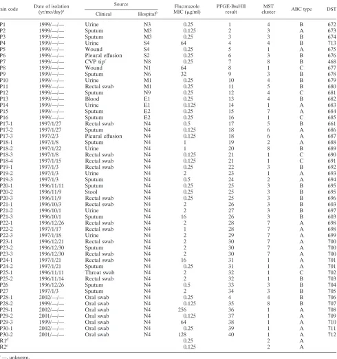
In Table 1, details are given of the ABC type and DST for
each isolate, together with the hospital origin and anatomical
source. For the 51 clinical isolates, 45 DSTs were defined by
the genotypes identified from the seven loci. With the sole
exception of type 468, all DSTs of our Taiwanese strain
on May 16, 2020 by guest
http://jcm.asm.org/
lections were novel to the internet DST database. A
dendro-gram generated to include the data from Table 1 and data
available from the internet MLST database showed that only 9
of the isolates in the present study (DSTs 676, 680, 661, 688,
694 to 696, and 704) coclustered with isolates presently
as-signed to clade 1, 3, or 4 among the four major
C. albicans
clades (37). No isolates coclustered with strains in clade 2.
[image:3.585.52.542.82.603.2]In this study, genetic profiles of 51
C. albicans
clinical
iso-lates and 2 reference strains were obtained by PFGE of
BssHII-restricted fragments and MLST analysis (Table 1). All
isolates were typeable by these two methods. For the 51 clinical
isolates, MLST generated 45 DSTs and PFGE-BssHII
gener-ated 40 DNA patterns in distinguishing isolates. For 16
unre-lated isolates, both typing methods generated 16 genotypes.
TABLE 1. Isolate descriptions and MLST results
Strain code Date of isolation (yr/mo/day)a
Source Fluconazole
MIC (g/ml)
PFGE-BssHII result
MST
cluster ABC type DST
Clinical Hospitalb
P1
1999/—/—
Urine
N3
0.25
1
4
B
672
P2
1999/—/—
Sputum
M3
0.125
2
3
A
673
P3
1999/—/—
Sputum
M3
0.25
3
3
B
674
P4
1999/—/—
Urine
S4
64
4
4
B
713
P5
1999/—/—
Wound
S4
0.25
5
1
A
675
P6
1999/—/—
Pleural effusion
S2
0.25
6
3
B
676
P7
1999/—/—
CVP tip
cN8
0.25
7
8
B
468
P8
1999/—/—
Wound
N1
64
8
1
C
677
P9
1999/—/—
Sputum
N6
32
9
3
B
678
P10
1999/—/—
Urine
M1
0.25
10
4
B
679
P11
1999/—/—
Rectal swab
M1
0.25
11
5
B
680
P12
1999/—/—
Sputum
N9
0.25
12
4
C
681
P13
1999/—/—
Blood
E1
0.25
13
4
B
682
P14
1999/—/—
Urine
E1
0.125
14
1
A
683
P15
1999/—/—
Sputum
E2
0.25
15
7
A
684
P16
1999/—/—
Sputum
E2
0.25
16
1
C
685
P17-1
1997/1/27
Rectal swab
N4
0.5
17
5
B
661
P17-2
1997/1/27
Sputum
N4
0.125
18
6
A
686
P17-3
1997/2/3
Pleural effusion
N4
0.125
18
6
A
687
P18-1
1997/1/8
Sputum
N4
1
19
2
A
688
P18-2
1997/1/22
Urine
N4
1
20
8
B
689
P18-3
1997/1/8
Rectal swab
N4
0.125
21
1
C
690
P18-4
1997/1/15
Rectal swab
N4
0.125
21
1
C
691
P19-1
1997/1/3
Rectal swab
N4
0.25
22
3
B
692
P19-2
1997/1/3
Urine
N4
2
23
1
A
693
P19-3
1997/1/3
Sputum
N4
0.5
24
2
A
694
P20-1
1996/11/11
Sputum
N4
0.25
25
3
B
695
P20-2
1996/11/9
Stool
N4
0.25
25
3
B
695
P20-3
1996/11/9
Rectal swab
N4
0.25
25
3
B
696
P21-1
1996/10/3
Rectal swab
N4
2
26
3
B
603
P21-2
1996/10/1
Urine
N4
2
27
3
B
697
P21-3
1996/10/1
Sputum
N4
16
26
3
B
603
P22-1
1996/12/26
Rectal swab
N4
2
28
7
A
698
P22-2
1997/1/17
Rectal swab
N4
1
28
7
A
698
P22-3
1997/1/18
Urine
N4
2
29
7
A
699
P23-1
1996/12/21
Rectal swab
N4
2
30
7
A
700
P23-2
1996/12/30
Sputum
N4
2
30
7
A
700
P23-3
1996/12/30
Rectal swab
N4
2
30
7
A
700
P24-1
1997/1/21
Rectal swab
N4
16
31
1
A
701
P24-2
1997/1/21
Sputum
N4
0.25
31
1
A
701
P25-1
1996/11/11
Throat swab
N4
2
32
1
C
702
P25-2
1996/11/14
Rectal swab
N4
2
32
1
B
703
P26
1996/12/26
Sputum
N4
0.5
33
3
B
704
P27
1997/1/3
Sputum
N4
2
34
3
B
705
P28-1
2002/—/—
Oral swab
N4
0.25
4
4
B
706
P28-2
1999/—/—
Oral swab
N4
0.125
35
8
B
707
P29-1
2002/—/—
Oral swab
N4
256
36
1
A
708
P29-2
2001/—/—
Oral swab
N4
0.125
37
1
A
709
P29-3
1999/—/—
Oral swab
N4
64
38
1
A
710
P30-1
2002/—/—
Oral swab
N4
0.25
39
1
A
711
P30-2
2001/—/—
Oral swab
N4
128
40
1
A
712
R1
d0.25
2
A
R2
e0.125
2
A
a—, unknown.
bN, north; S, south; M, middle; E, east. cCVP, central venous pressure line. dATCC 14053.
eATCC 90028.
on May 16, 2020 by guest
http://jcm.asm.org/
For 35 epidemiologically related isolates, MLST generated 29
genotypes and PFGE-BssHII generated 25 genotypes.
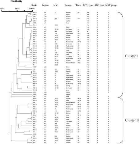
Relationships between the isolates.
The dendrogram in Fig. 1
indicates the similarities of 51
C. albicans
isolates determined
by MLST with 7 gene fragments. The dendrogram shows all
isolates can be divided into 2 clusters (I and II). All isolates
from HIV patients, except P28-1, belonged to cluster I. Isolates
from the same patient clustered together relatively closely. For
long-term isolates from the same HIV patients, isolates from
different years belonged to different genotypes. Neither PFGE
type nor MLST DST correlated with fluconazole resistance or
hospital origin.
Epidemiologically unrelated group 1 isolates (P1 to P16)
[image:4.585.67.519.73.543.2]displayed different PFGE-BssHII genotypes and MLST DSTs.
No genotypes were correlated with specimen types, hospital
origins, and fluconazole resistance. Group 2 strains were
col-lected from intensive care unit patients within a short period of
time. In this group, most strains from the same patient
dem-onstrated the same PFGE-BssHII genotypes. However, few
strains with the same PFGE-BssHII genotypes also exhibited
the same DSTs: they differed in from 0 to 2 alleles. This may be
due to the microevolution of the persistent strain within the
same patient. Isolates P28 to P30 were collected from the oral
cavity of HIV-positive patients over a period spanning many
years. Serial isolates of the same patient exhibited different
PFGE-BssHII genotypes and MLST DSTs. For MLST, within
FIG. 1. Dendrogram indicating the similarities of 63
C. albicans
isolates determined by MLST with 7 gene fragments.
on May 16, 2020 by guest
http://jcm.asm.org/
the same patient, the numbers of different alleles accumulated
with time. This was especially apparent in strain P29-1 to -3,
P29-3 differs from P29-2 by 1 allele in 2 years and differed from
P29-3 by 2 alleles 3 years later. The long time period and
perhaps the antifungal regimens have resulted in the
accumu-lation of microevolutionary events. Three isolates, 18-1, 18-2,
and 18-3 from the sputum, urine, and rectal swab, respectively,
from one intensive care unit patient, P18, possessed different
PFGE and MLST types. This is further confirmed by the
find-ing that these three isolates belonged to different 25S rRNA
types (A, B, and C types, respectively). These data
demon-strate that different clones can be isolated from different body
sites, especially nonsterile sites, in one patient.
ABC typing.
Isolates from the United Kingdom and the
United States belonged to cluster I, except UK2 and US2. All
United Kingdom and U.S. isolates were type A, except for
UK2 and US2, which were type B. Compared with isolates
from other countries, such as the United Kingdom and United
States, where isolates are predominantly type A, with type C
being very rare (37), the Taiwanese isolates had similar
pro-portion of types A (22/51, 43.1%) and B (23/51, 45.1%). There
were only 6 type C strains (11.8%). All isolates from HIV
patients were type A, except for 28-1 and 28-2, which were type
B. All isolates were heterozygous at the mating type locus,
except for P29-3, which has an MLT type
a
/
a
.
The MST (Fig. 2) showed the phylogenetic relationships
between isolates. Based on MST, all isolates can be grouped
into 8 groups (groups 1 to 8). The MST grouping correlates
with ABC typing. Groups 2, 6, and 7 are type A. Groups 5 and
8 are type B. Groups 3 and 4 are predominantly type B. Group
1 is predominantly a composite of types A and C. Strains from
the United Kingdom and United States fall predominantly into
cluster 2. The MST grouping correlates very well with the
unweighted-pair group method using average linkage
cluster-ing, with MST groups 1, 2, 6, 7, and 8 in cluster I and MST
groups 3, 4, and 5 in cluster II.
Stability of the MLST method.
The in vitro stability of the
MLST method was demonstrated by the finding that all of the
consecutive subcultured clones of each 6 isolates from patients
and the two reference strains showed same MLST DST.
DISCUSSION
[image:5.585.67.516.62.414.2]The increased incidence of transmission of pathogens
through international travel, global food chain supply, or even
deliberate terror attack have highlighted the importance of
FIG. 2. MST of 51 Taiwanese clinical isolates and strains from the United Kingdom and United States as well as reference strains. Each isolate
is represented by a circle. Relationships between the strains were depicted as connections between isolates and the lengths of the branches linking
them. Angles of line connection are of no relevance.
on May 16, 2020 by guest
http://jcm.asm.org/
global collaborative surveillance networking in control of
in-fectious diseases. Choosing appropriate molecular typing
methods is indispensable for fulfilling such needs. MLST
al-lows the exchange of molecular typing information via the
internet for global epidemiology. We evaluated MLST
meth-odology to ascertain its potential for outbreak investigation
and to know whether different characteristics (patient origin,
drug resistance, geographic origin, and source of isolation) can
be attributed to certain specific molecular types in Taiwan. The
data obtained in this study will contribute to our attempt to
establish a central genetic database of fungal pathogens in
Taiwan.
PFGE of restricted fragments represents a whole-genome
scanning method to reflect mutation events such as
polymor-phism in the recognition site, translocation (18),
reorganiza-tion of rRNA gene-containing chromosomes, or the
non-reciprocal reorganization of rRNA gene cistrons in the rRNA
gene-containing chromosomes (30). MLST is based on
vari-ability within particular housekeeping genes due to mutation
or recombination events; thus, it provides many genetic types
per locus and these can be utilized to define the allelic profile
or sequence type and determine the relatedness of strains (26,
36). In bacteria, PFGE-restricted methods are more
discrimi-natory than MLST methods. However, our data suggested that
the discriminatory ability of MLST for the typing of
C. albicans
offers advantages over PFGE typing, as the differential power
has been greatly enhanced by MLST. This may due to the large
genome size of yeast pathogens (16 Mb for diploid
C. albicans
),
which is on average about 4 to 8 times the average size of
bacterial genomes (gram-positive bacteria, 4.5 Mb;
gram-neg-ative bacteria, 2.2 Mb). The best resolution for PFGE is
lim-ited for a certain size range and number of fragments (e.g., 30
to 35 bands). This may limit the resolutive power of PFGE
applied to
Candida
pathogens. The diploid nature of
C
albi-cans
may further increase the discriminatory power of MLST
for this species. In our study, isolates from the same patient
with the same PFGE-BssHII genotype could be further
differ-entiated into different MLST DSTs, differing by 0 to 2 allele
types. Southern blotting with the Ca3 probe or the C fragment
derived from this probe has been demonstrated to be useful in
tracing the microevolutionary events in
C. albicans
(23). Our
data show that MLST is superior to PFGE for tracing the
microevolution of
C. albicans
strains within the same patient.
A comparative study also showed that MLST is at least
com-parable to Ca3 Southern hybridization probe techniques in
discriminative power (32). Furthermore, MLST offers distinct
advantages in standardizability and portability.
Our result also showed that the DNA type of each isolate
was patient specific and associated with ABC type and decade
of isolation, but not associated with anatomical source of
iso-lation, hospital origin, or fluconazole resistance patterns, which
is in accordance with previous reports (8).
Sequence-based typing data like MLST greatly facilitate
standardization and international data exchange. The present
study showed that only a few of the Taiwanese isolates (9 in 51
isolates) coclustered with isolates presently assigned to the
four major
C. albicans
clades (37). Further studies on
C.
albi-cans
strains from the Asian region are needed to understand
the global epidemiology of
C. albicans
.
ACKNOWLEDGMENTS
This work was supported by grant DOH94-DC-2011 from the
Cen-ter for Disease Control, Department of Health, and grant
94-0324-19-F-00-00-00-35 from the National Science Council, Taiwan (S.-Y.L.)
and by a grant from the Wellcome Trust (F.C.O.).
We are grateful to Chien-Shun Chiou for insightful comments on
the manuscript, and we express our sincere appreciation to all 13
participating hospitals for providing the strains and information
re-lated to these strains. They are Chang Gung Memorial Hospital at
Linkou, Chang Gung Memorial Hospital at Keelung, Lo-Tung Poh Ai
Hospital, Lo-Tung St. Mary Hospital, National Taiwan University
Hospital, Tri Service General Hospital, Veterans General
Hospital-Taichung, Zen Ai General Hospital, Kaohsiung Medical College
Chung-Ho Memorial Hospital, Kaohsiung Military Hospital, Veterans
General Hospital-Kaohsiung, Buddhist Tzu-Chi General Hospital in
Hua-Lien, and Mackay Memorial Hospital Taitung Branch.
REFERENCES
1.Ball, L. M., M. A. Bes, B. Theelen, T. Boekhout, R. M. Egeler, and E. J. Kuijper.2004. Significance of amplified fragment length polymorphism in identification and epidemiological examination of Candida species coloniza-tion in children undergoing allogeneic stem cell transplantacoloniza-tion. J. Clin. Microbiol.42:1673–1679.
2.Bougnoux, M. E., S. Morand, and C. d’Enfert.2002. Usefulness of multilocus sequence typing for characterization of clinical isolates ofCandida albicans. J. Clin. Microbiol.40:1290–1297.
3.Bougnoux, M. E., A. Tavanti, C. Bouchier, N. A. Gow, A. Magnier, A. D. Davidson, M. C. Maiden, C. D’Enfert, and F. C. Odds.2003. Collaborative consensus for optimized multilocus sequence typing ofCandida albicans. J. Clin. Microbiol.41:5265–5266.
4.Chen, K. W., H. J. Lo, Y. H. Lin, and S. Y. Li.2005. Comparison of four molecular typing methods to assess genetic relatedness of Candida albicans clinical isolates in Taiwan. J. Med. Microbiol.54:249–258.
5.Chen, Y. C., S. C. Chang, K. T. Luh, and W. C. Hsieh.2003. Stable suscep-tibility of Candida blood isolates to fluconazole despite increasing use during the past 10 years. J. Antimicrob. Chemother.52:71–77.
6.Chen, Y. C., S. C. Chang, C. C. Sun, L. S. Yang, W. C. Hsieh, and K. T. Luh. 1997. Secular trends in the epidemiology of nosocomial fungal infections at a teaching hospital in Taiwan, 1981 to 1993. Infect. Control Hosp. Epide-miol.18:369–375.
7.Chen, Y. C., S. C. Chang, H. M. Tai, P. R. Hsueh, and K. T. Luh.2001. Molecular epidemiology of Candida colonizing critically ill patients in inten-sive care units. J. Formos. Med. Assoc.100:791–797.
8.Dassanayake, R. S., A. N. Ellepola, Y. H. Samaranayake, and L. P. Samara-nayak.2002. Molecular heterogeneity of fluconazole-resistant and -suscep-tible oral Candida albicans isolates within a single geographic locale. APMIS 110:315–324.
9.Dodgson, A. R., C. Pujol, D. W. Denning, D. R. Soll, and A. J. Fox.2003. Multilocus sequence typing ofCandida glabratareveals geographically en-riched clades. J. Clin. Microbiol.41:5709–5717.
10.Elias Costa, M. R., S. Carnovale, and M. S. Relloso.1999. Oropharyngeal candidosis in AIDS patients: an epidemiological study using restriction anal-ysis of Candida albicans total genomic DNA. Mycoses42:41–46. 11.Fundyga, R. E., T. J. Lott, and J. Arnold.2002. Population structure of
Candida albicans, a member of the human flora, as determined by micro-satellite loci. Infect. Genet. Evol.2:57–68.
12.Gudlaugsson, O., S. Gillespie, K. Lee, B. J. Vande, J. Hu, S. Messer, L. Herwaldt, M. Pfaller, and D. Diekema.2003. Attributable mortality of nos-ocomial candidemia, revisited. Clin. Infect. Dis.37:1172–1177.
13.Hajjeh, R. A., A. N. Sofair, L. H. Harrison, G. M. Lyon, B. A. Arthington-Skaggs, S. A. Mirza, M. Phelan, J. Morgan, W. Lee-Yang, M. A. Ciblak, L. E. Benjamin, L. T. Sanza, S. Huie, S. F. Yeo, M. E. Brandt, and D. W. Warnock. 2004. Incidence of bloodstream infections due toCandida speciesand in vitro susceptibilities of isolates collected from 1998 to 2000 in a population-based active surveillance program. J. Clin. Microbiol.42:1519–1527.
14.Hsu, M. C., K. W. Chen, H. J. Lo, Y. C. Chen, M. H. Liao, Y. H. Lin, and S. Y. Li.2003. Species identification of medically important fungi by use of real-time LightCycler PCR. J. Med. Microbiol.52:1071–1076.
15.Huang, Y. C., T. Y. Lin, H. S. Leu, J. L. Wu, and J. H. Wu.1998. Yeast carriage on hands of hospital personnel working in intensive care units. J. Hosp. Infect.39:47–51.
16.Huang, Y. C., T. Y. Lin, H. L. Peng, J. H. Wu, H. Y. Chang, and H. S. Leu. 1998. Outbreak of Candida albicans fungaemia in a neonatal intensive care unit. Scand. J. Infect. Dis.30:137–142.
17.Hung, C. C., Y. L. Yang, T. L. Lauderdale, L. C. McDonald, C. F. Hsiao, H. H. Cheng, Y. A. Ho, and H. J. Lo.2005. Colonization of human immu-nodeficiency virus-infected outpatients in Taiwan with Candida species. J. Clin. Microbiol.43:1600–1603.
18.Iwaguchi, S. I., T. Kanbe, T. Tohne, P. T. Magee, and T. Suzuki.2000.
on May 16, 2020 by guest
http://jcm.asm.org/
High-frequency occurrence of chromosome translocation in a mutant strain of Candida albicans by a suppressor mutation of ploidy shift. Yeast16:411– 422.
19.Kanellopoulou, M., G. Stamos, I. Petinnelli, M. Savala, A. Tzimogianni, N. J. Legakis, M. Foustoukou, E. Papafragas, and A. Velegraki.2001. Sub-typing and antifungal susceptibilities of Candida spp. in the intensive care unit of a Greek general hospital. Int. J. Antimicrob. Agents18:179–183. 20.Kao, A. S., M. E. Brandt, W. R. Pruitt, L. A. Conn, B. A. Perkins, D. S.
Stephens, W. S. Baughman, A. L. Reingold, G. A. Rothrock, M. A. Pfaller, R. W. Pinner, and R. A. Hajjeh.1999. The epidemiology of candidemia in two United States cities: results of a population-based active surveillance. Clin. Infect. Dis.29:1164–1170.
21.Koufopanou, V., A. Burt, and J. W. Taylor.1997. Concordance of gene genealogies reveals reproductive isolation in the pathogenic fungus Coc-cidioides immitis. Proc. Natl. Acad. Sci. USA94:5478–5482.
22.Li, S. Y., Y. L. Yang, K. W. Chen, H. H. Cheng, C. S. Chiou, T. W. Wang, T. L. Lauderdale, C. C. Hung, and H. J. Lo.2006. Molecular epidemiology of long-term colonization of Candida albicans strains from HIV-infected pa-tients. Epidemiol. Infect.134:265–269.
23.Lockhart, S. R., J. J. Fritch, A. S. Meier, K. Schroppel, T. Srikantha, R. Galask, and D. R. Soll.1995. Colonizing populations ofCandida albicansare clonal in origin but undergo microevolution through C1 fragment reorgani-zation as demonstrated by DNA fingerprinting and C1 sequencing. J. Clin. Microbiol.33:1501–1509.
24.Lott, T. J., and M. M. Effat.2001. Evidence for a more recently evolved clade within a Candida albicans North American population. Microbiology147: 1687–1692.
25.Lott, T. J., R. E. Fundyga, M. E. Brandt, L. H. Harrison, A. N. Sofair, R. A. Hajjeh, and D. W. Warnock.2003. Stability of allelic frequencies and distri-butions ofCandida albicansmicrosatellite loci from U.S. population-based surveillance isolates. J. Clin. Microbiol.41:1316–1321.
26.Maiden, M. C., J. A. Bygraves, E. Feil, G. Morelli, J. E. Russell, R. Urwin, Q. Zhang, J. Zhou, K. Zurth, D. A. Caugant, I. M. Feavers, M. Achtman, and B. G. Spratt.1998. Multilocus sequence typing: a portable approach to the identification of clones within populations of pathogenic microorganisms. Proc. Natl. Acad. Sci. USA95:3140–3145.
27.McCullough, M. J., K. V. Clemons, and D. A. Stevens.1999. Molecular and phenotypic characterization of genotypicCandida albicanssubgroups and comparison withCandida dubliniensisandCandida stellatoidea. J. Clin. Mi-crobiol.37:417–421.
28.O’Donnell, K., D. A. Sutton, M. G. Rinaldi, K. C. Magnon, P. A. Cox, S. G. Revankar, S. Sanche, D. M. Geiser, J. H. Juba, J. A. van Burik, A. Padhye, E. J. Anaissie, A. Francesconi, T. J. Walsh, and J. S. Robinson.2004. Genetic diversity of human pathogenic members of theFusarium oxysporumcomplex inferred from multilocus DNA sequence data and amplified fragment length polymorphism analyses: evidence for the recent dispersion of a geographi-cally widespread clonal lineage and nosocomial origin. J. Clin. Microbiol. 42:5109–5120.
29.Poikonen, E., J. Vuopio-Varkila, S. S. Kaukoranta-Tolvanen, A. Sivonen, E. Siren, and P. Ruutu.2001. Epidemiological typing of Candida albicans from bloodstream infections by restriction enzyme analysis. Scand. J. Infect. Dis. 33:140–144.
30.Ramsey, H., B. Morrow, and D. R. Soll.1994. An increase in switching frequency correlates with an increase in recombination of the ribosomal chromosomes of Candida albicans strain 3153A. Microbiology140(Pt 7): 1525–1531.
31.Rentz, A. M., M. T. Halpern, and R. Bowden.1998. The impact of candi-demia on length of hospital stay, outcome, and overall cost of illness. Clin. Infect. Dis.27:781–788.
32.Robles, J. C., L. Koreen, S. Park, and D. S. Perlin.2004. Multilocus sequence typing is a reliable alternative method to DNA fingerprinting for discrimi-nating among strains ofCandida albicans. J. Clin. Microbiol.42:2480–2488. 33.Samaranayake, Y. H., L. P. Samaranayake, R. S. Dassanayake, J. Y. Yau, W. K. Tsang, B. P. Cheung, and K. W. Yeung.2003. ‘Genotypic shuffling’ of sequential clones of Candida albicans in HIV-infected individuals with and without symptomatic oral candidiasis. J. Med. Microbiol.52:349–359. 34.Sheng, W. H., J. T. Wang, D. C. Lu, W. C. Chie, Y. C. Chen, and S. C. Chang.
2005. Comparative impact of hospital-acquired infections on medical costs, length of hospital stay and outcome between community hospitals and med-ical centres. J. Hosp. Infect.59:205–214.
35.Soll, D. R.2000. The ins and outs of DNA fingerprinting the infectious fungi. Clin. Microbiol. Rev.13:332–370.
36.Spratt, B. G.1999. Multilocus sequence typing: molecular typing of bacterial pathogens in an era of rapid DNA sequencing and the internet. Curr. Opin. Microbiol.2:312–316.
37.Tavanti, A., A. D. Davidson, M. J. Fordyce, N. A. Gow, M. C. Maiden, and F. C. Odds.2005. Population structure and properties ofCandida albicans, as determined by multilocus sequence typing. J. Clin. Microbiol.43:5601–5613. 38.Tavanti, A., A. D. Davidson, E. M. Johnson, M. C. Maiden, D. J. Shaw, N. A. Gow, and F. C. Odds.2005. Multilocus sequence typing for differentiation of strains ofCandida tropicalis. J. Clin. Microbiol.43:5593–5600.
39.Tavanti, A., N. A. Gow, S. Senesi, M. C. Maiden, and F. C. Odds.2003. Optimization and validation of multilocus sequence typing forCandida al-bicans. J. Clin. Microbiol.41:3765–3776.
40.Taylor, B. N., T. Harrer, E. Pscheidl, A. Schweizer, M. Rollinghoff, and K. Schroppel.2003. Surveillance of nosocomial transmission of Candida albi-cans in an intensive care unit by DNA fingerprinting. J. Hosp. Infect.55: 283–289.
41.Tortorano, A. M., A. L. Rigoni, E. Biraghi, A. Prigitano, and M. A. Viviani. 2003. The European Confederation of Medical Mycology (ECMM) survey of candidaemia in Italy: antifungal susceptibility patterns of 261 non-albicans Candida isolates from blood. J. Antimicrob. Chemother.52:679–682. 42.Wisplinghoff, H., T. Bischoff, S. M. Tallent, H. Seifert, R. P. Wenzel, and
M. B. Edmond.2004. Nosocomial bloodstream infections in US hospitals: analysis of 24,179 cases from a prospective nationwide surveillance study. Clin. Infect. Dis.39:309–317.
43.Yang, Y. L., H. H. Cheng, Y. A. Ho, C. F. Hsiao, and H. J. Lo.2003. Fluconazole resistance rate of Candida species from different regions and hospital types in Taiwan. J. Microbiol. Immunol. Infect.36:187–191. 44.Yang, Y. L., Y. A. Ho, H. H. Cheng, M. Ho, and H. J. Lo.2004. Susceptibilities
ofCandidaspecies to amphotericin B and fluconazole: the emergence of fluconazole resistance inCandida tropicalis. Infect Control Hosp. Epidemiol. 25:60–64.