A mighty mouse: building a better model of
multiple sclerosis
Richard M. Ransohoff
J Clin Invest.
2006;116(9):2313-2316. https://doi.org/10.1172/JCI29834.
The 2 cardinal cell populations mediating adaptive immunity are T and B lymphocytes.
These cells play important but poorly understood roles in the immunopathological
demyelinating disease multiple sclerosis (MS) and in a widely used animal model of human
MS known as EAE. In the current issue of the
JCI
, 2 research teams report their parallel
studies of double-transgenic mice expressing T and B cell receptors that recognize the
same myelin protein (see the related articles beginning on pages 2385 and 2393). More
than half of the double-transgenic mice spontaneously developed autoimmune
demyelination in their spinal cords and optic nerves, exhibiting pathologies reminiscent of
human MS. The studies describe an important new model for MS research.
Commentary
Find the latest version:
A mighty mouse: building a better model
of multiple sclerosis
Richard M. Ransohoff
Neuroinflammation Research Center, Department of Neurosciences, Lerner Research Institute, Cleveland Clinic, Cleveland, Ohio, USA.
The 2 cardinal cell populations mediating adaptive immunity are T and B
lym-phocytes. These cells play important but poorly understood roles in the
immu-nopathological demyelinating disease multiple sclerosis (MS) and in a widely
used animal model of human MS known as EAE. In the current issue of the
JCI
, 2 research teams report their parallel studies of double-transgenic mice
expressing T and B cell receptors that recognize the same myelin protein (see
the related articles beginning on pages 2385 and 2393). More than half of the
double-transgenic mice spontaneously developed autoimmune demyelination
in their spinal cords and optic nerves, exhibiting pathologies reminiscent of
human MS. The studies describe an important new model for MS research.
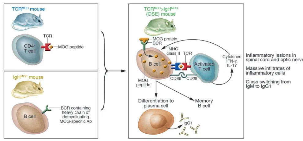
MS is an acquired inflammatory demy-elinating disorder of the human CNS, producing sensory, motor, and cognitive dysfunction in many affected individuals. For the past 75 years, much MS research has focused on EAE, an animal model of human MS. This useful model for scien-tific investigation (1) has been bedeviled by requirements for immunization, most often using myelin protein fragments along with powerful multicomponent adjuvants. The immunization protocols, especially the use of adjuvants, introduce complexity in data interpretation. Efforts to develop spontaneous EAE models have proceeded with ever-accelerating intensity in recent years. In the current issue of the JCI, 2 independent research groups report what I believe to be a new model of spon-taneous autoimmune demyelination (2, 3). Given its simplicity and predictability, this model holds great promise as a system for studying MS pathogenesis. Both groups emphasize that lesion distribution in the so-named opticospinal EAE (OSE) mice resembles that seen in patients with Devic disease (also referred to as neuromyelitis optica [NMO]). However, recent advances in NMO research are leading toward a new
nosology for human demyelinating dis- eases. Given this consideration and the dif-ferences in the neuropathology of murine OSE compared with human NMO, I sug-gest that this model will make its most salient contributions to MS rather than NMO research.
Important and largely harmonious findings of the current studies
Krishnamoorthy et al. (2) and Bettelli et al. (3) bred and studied essentially iden-tical strains of double-transgenic mice, derived from 2 previously described single-transgenic animals (4, 5) (Figure 1). The researchers performed overlapping analy-ses so that most findings (but not all) were instantly validated by independent evalua-tion. The first line of single-transgenic mice (4) was generated with knock-in technol-ogy, inserting the recombined heavy chain of a demyelinating anti–myelin oligoden-drocyte glycoprotein (anti-MOG) antibody (8.18C5) in the Ig J region, yielding mice in which 20–30% of circulating B cells pro-duced anti-MOG antibodies and expressed a B cell receptor (BCR) that recognized MOG protein. These MOG-specific Ig heavy chain knock-in mice on a C57BL/6 background were termed IgHMOG mice.
The second line, termed TCRMOG mice, was
generated by expressing a TCR that recog-nized an immunodominant determinant of MOG (5), and these mice spontaneously showed inflammation of the optic nerve, a typical early symptom of MS.
MOG is a stoichiometrically minor myelin protein but looms large in MS research: MOG immunization elicits autoimmune
demyelination of varying types in rodents and nonhuman primates, with histologi-cal findings that recapitulate the gamut of MS pathology (6). MOG is expressed virtually exclusively in the CNS (but not in thymus), and MOG-reactive lympho-cytes evade central thymic tolerance (7, 8). MOG is inserted into the external leaflet of the myelin membrane, so it is accessible to demyelinating antibodies, unlike other myelin components, which are buried deep within compact myelin (9, 10).
The 2 research groups reported exciting and concordant data (2, 3). First, approxi-mately 50% of the double-transgenic TCRMOG×IgHMOG offspring spontaneously
developed a severe form of EAE by 8 weeks of age and showed selective distribution of inflammatory lesions predominantly in their optic nerves and spinal cords, with cells in both meninges and parenchyma involved. The mouse model was descrip-tively termed OSE by Krishnamoorthy et al. (2). In the spinal cord, lesions were primar-ily subpial, as seen in active immunization– induced EAE. By contrast, spontaneous EAE occurred in most previously reported models only at low incidence or following exposure to microbes or by eliminating reg-ulatory cells (11–14). Both of the current reports found OSE severity and incidence unaffected by the usual strong influences such as gender and housing conditions (2, 3). Second, B cells in TCRMOG×IgHMOG
mice presented antigen and also received T cell help, reflected by B cell prolifera-tion and class switching to IgG1 (Figure 1). Crossing IgHMOG mice with transgenic
animals bearing non–MOG-specific TCRs (i.e., OVA-specific TCR transgenic mice on C57BL/6 background) did not induce EAE (2), indicating that spontaneous OSE was not equivalent to the opening of the blood-brain barrier to pathogenic antibodies by activated non–MOG-specific T cells (15). In vitro, activation of B cell proliferation was not dependent solely upon T cells that recognized MOG (3). Amazingly, Krish-namoorthy et al. found that class switching
from IgM to IgG1 MOG–specific antibod-Nonstandard abbreviations used: AQP4, aquaporin-4; BCR, B cell receptor; IgHMOG mice, MOG-specific Ig
heavy-chain knock-in mice on a C57BL/6 background; MOG, myelin oligodendrocyte glycoprotein; NMO, neuromyelitis optica; OSE, opticospinal EAE; TCRMOG
mice, MOG-specific TCR transgenic mice on a C57BL/6 background.
Conflict of interest: The author has declared that no conflict of interest exists.
commentaries
ies occurred, even in MOG–/– mice, raising
the question of whether a cross-reactive self antigen might be involved in the peripheral immune response that initiated spontane-ous OSE. Third, cytokine production in OSE mice resembled that seen in active immunization–induced EAE in the same background mouse strain as the transgen-ic animals in the current studies. Cytokine profiles obtained from cell cultures and inflamed CNS tissues were similar and were characterized by a mixture of Th1 and Th2 cytokines including IFN-γ, IL-5 (not typically detected in active immunization– induced EAE in this mouse strain), and lesser levels of IL-17, a cytokine of great interest in contemporary EAE research.
Immediate questions for a promising disease model
OSE holds enormous potential as a model for MS. The OSE mouse is a robust and convenient spontaneous EAE model and will support significant mechanistic stud-ies. So what are B cells doing in OSE? Findings from these 2 reports strongly implicate B cells as critical APCs in OSE. B cells have marked advantages as APCs:
antigen capture and internalization occurs at extraordinarily low antigen concentra-tions, mediated by the high-affinity B cell antigen receptor. Furthermore, signaling through the B cell antigen receptor follow- ing antigen engagement can promote anti-gen degradation to peptides and loading on MHC class II molecules (16) (Figure 1). The roles of B cells as APCs can be dissected in the OSE model. Current interest in anti–B cell therapeutics such as rituximab will be informed by this direction of research.
Another interesting question that remains to be answered concerns the abrupt onset of disease, which affords an opportunity to elucidate whether this pattern of disease evolution reflects the action of precipitat- ing factors or the accumulation of patho-genic elements that exceed some threshold. As another example of research that may be enabled by this model, the seeming paradox concerning IFN-γ as an inhibitory cytokine in EAE has been attributed to its interac- tion with adjuvants containing mycobac-terial components (17); if so, IFN-γ might function differently in OSE.
As noted above, Krishnamoorthy et al. (2) found evidence for IgHMOG B cell class
switching in MOG–/–
mice that lacked nom-inal antigen, suggesting that there may be another ligand for the transgenic IgHMOG
BCR and possibly for the TCRMOG TCR
as well. Clarifying this fascinating finding might identify mechanisms by which cross-reactivity to self antigens in the periphery leads to CNS autoimmunity.
It remains uncertain whether the mecha- nisms underlying the selective and pro-vocative occurrence of OSE lesions in the optic nerve and spinal cord will be suscep-tible to elucidation. It seems unlikely that this lesion distribution is determined by regional abundance of MOG, as suggested by Bettelli et al. (3), given the vast variety of CNS demyelinating syndromes medi-ated by autoimmunity to MOG (6, 18–20). It will be important to interrogate the local vascular anatomy and genetics of the model for answers.
Some discrepancies remain unresolved
[image:3.585.40.535.82.313.2]With regard to the presence of B cells in the lesions, Bettelli et al. reported aggregates of B220-immunoreactive leukocytes (3). In contrast, Krishnamoorthy et al. did not
Figure 1
detect B cells using immunohistochemistry in affected spinal cord and buttressed these negative findings by showing virtually no CD3–B220+ cells in lysates of the inflamed
CNS tissues (2). It is not likely that techni-cal differences underlie this incongruity, as tissue analysis in both cases was performed by very experienced research neuropathol-ogists. In addition, Krishnamoorthy et al. found a very impressive eosinophilic con-tribution to the leukocyte populations in lesions (2), while Bettelli et al. specifically reported the absence of eosinophils (3). These differences will need to be unraveled if the model is to bear utility for research, particularly for interventional studies to ameliorate or prevent disease.
What features distinguish OSE mice from NMO patients?
Despite the superficial resemblance con- ferred by lesion distribution of the phe-notype of the double-transgenic mice to human NMO, the present reports describe a pathology that much more resembles MS or EAE than NMO. It is unfortunate that we do not have an animal model of NMO, a variant and relatively uncommon form of human demyelinating disease that has presented a fertile field for research. Recent work on NMO provides hope that study-ing variant forms of CNS demyelinating disease might allow researchers to recapitu- late the remarkable progress made by inves-tigators into the inflammatory peripheral nerve syndromes (21–23).
Several key features distinguish human NMO pathology from that seen in OSE mice:
Lesion pathology. Human NMO possesses a distinct pathology characterized by inflam-
matory infiltrates that include polymor-phonuclear leukocytes and eosinophils, hyalinized small-caliber spinal arteries, and necrotic destruction of both gray and white matter within the affected spinal cord in addition to lymphocyte and macrophage infiltrates (24–26). Deposition of comple-ment C9 neoantigen and Ig bear testimony to the proposed humoral pathogenesis of NMO (26). In support of the concept of pathogenic humoral immunity in NMO, plasma exchange can be strikingly effective for the treatment of patients with severe attacks. Characteristics of NMO pathol-ogy that were specifically sought — but absent — in OSE mice included deposition of complement and Ig in the inflamed CNS as well as necrotic tissue damage and vas-cular hyalinization. Furthermore, there was no temporal relationship between Ig class switching and the presence or severity of EAE neurological deficits in OSE mice (2), arguing against a direct role for antibody in disease pathogenesis.
Lesion extent. Longitudinally extensive spinal lesions are reported to produce non-remitting paralysis in patients with NMO and constitute a major supporting diag-nostic criterion (27). These longitudinally extensive lesions, typical of NMO, were absent from the OSE mice described in these studies. By contrast, spinal lesions in OSE animals were relatively small.
Aquaporin-4 antibodies. About 55% of NMO patients harbor serum autoantibod-ies, initially characterized as NMO-Ig (28). Studies with NMO patients and appropri-ate controls, most importantly patients with classical MS and paraneoplastic syn-dromes, suggested that the detection of NMO-Ig was a specific indicator for the presence of disorders within the NMO spectrum (28). Further study identified an NMO-Ig antigen: aquaporin-4 (AQP4),
a water channel highly expressed on the astrocytic end-feet around CNS vessels (29). This serological reactivity represents the first potential biomarker of a subtype of human demyelinating disease and opens the possibility of a molecular nosology for these complex disorders. It remains uncer- tain whether AQP4 antibodies are patho-genic in NMO, although their presence in patients with spinal cord demyelination predicts relapse (25). There was no reason to suspect that OSE mice should possess AQP4 antibodies, and they did not (2). For now, NMO is pathologically and, at a molecular level, etiologically distinct from OSE (Table 1).
Summary
In this issue of the JCI, 2 groups of lead-ing scientists generated and characterized what I believe to be a new model of spon-taneous EAE (2, 3). Already, their work provides new insight into the potency of B cells as APCs. There are tantalizing hints that MOG-specific antigen receptors can also “see” a component distinct from MOG in MOG–/– mice. It is anticipated that full
exploitation of this spontaneous model will help resolve confusion that has arisen from the need to use immunization with adjuvants, as applied in most prior EAE studies. Prospects for new MS therapeutics are brighter due to the availability of this useful experimental system.
Address correspondence to: Richard M. Ransohoff, Neuroinflammation Research Center, Department of Neurosciences, Lerner Research Institute, Cleveland Clin-ic, Mail Code NC30, 9500 Euclid Avenue, Cleveland, Ohio 44195, USA. Phone: (216) 444-0627; Fax: (216) 444-7927; E-mail: [email protected].
Table 1
Few similarities and many differences between OSE mice and NMO patients
Characteristic OSE mice NMO patients
Pattern of neurobehavioral deficit evolution Abrupt, nonremitting Abrupt, often nonremitting
Spinal lesion pathology Subpial inflammation, demyelination; Necrotizing inflammation with eosinophils; eosinophils present (2) destruction of gray and white matter;
hyalinized small vessels
Evidence for humoral contribution to pathogenesis Burden of evidence against (no relation of Ig Preponderance of evidence favors (readily levels or presence to EAE severity; no detectable detectable Ig and complement in tissues; deposition of Ig or complement in tissues) clinical benefit with plasma exchange) Lesion location and extent Lesions restricted to 1 interspace Lesions extend for at least 2 interspaces
Presence of AQP4 antibodies No Yes
commentaries
1. Ransohoff, R.M. 2006. EAE: pitfalls outweigh vir-tues of screening potential treatments for multiple sclerosis. Trends Immunol. 27:167–168.
2. Krishnamoorthy, G., Lassmann, H., Wekerle, H., and Holz, A. 2006. Spontaneous opticospinal encepha-lomyelitis in a double-transgenic mouse model of autoimmune T cell/B cell cooperation. J. Clin. Invest. 116:2385–2392. doi:10.1172/JCI28330.
3. Bettelli, E., Baeten, D., Jäger, A., Sobel, R.A., and Kuchroo, V.K. 2006. Myelin oligodendrocyte glycoprotein–specific T and B cells cooperate to induce a Devic-like disease in mice. J. Clin. Invest. 116:2393–2402. doi:10.1172/JCI28334.
4. Litzenburger, T., et al. 1998. B lymphocytes produc-ing demyelinating autoantibodies: development and function in gene-targeted transgenic mice.
J. Exp. Med. 188:169–180.
5. Bettelli, E., et al. 2003. Myelin oligodendrocyte gly-coprotein-specific T cell receptor transgenic mice develop spontaneous optic neuritis. J. Exp. Med. 197:1073–1081.
6. Storch, M.K., et al. 1998. Autoimmunity to myelin oligodendrocyte glycoprotein in rats mimics the spectrum of multiple sclerosis pathology. Brain Pathol. 8:681–694.
7. Wekerle, H., and Linington, C. 2006. Organ specific autoantigens and the autoreactiveT cell repertoire: the case of myelin oligodendrocyte glycoprotein.
Eur. J. Immunol. 36:512–515.
8. Fazilleau, N., et al. 2006. Persistence of autoreac- tive myelin oligodendrocyte glycoprotein (MOG)-specific T cell repertoires in MOG-expressing mice.
Eur. J. Immunol. 36:533–543.
9. Kroepfl, J.F., Viise, L.R., Charron, A.J., Linington, C., and Gardinier, M.V. 1996. Investigation of myelin/oligodendrocyte glycoprotein membrane
topology. J. Neurochem. 67:2219–2222.
10. Mathey, E., Breithaupt, C., Schubart, A.S., and Lin-ington, C. 2004. Commentary: Sorting the wheat from the chaff: identifying demyelinating compo-nents of the myelin oligodendrocyte glycoprotein (MOG)-specific autoantibody repertoire. Eur. J. Immunol. 34:2065–2071.
11. Madsen, L.S., et al. 1999. A humanized model for multiple sclerosis using HLA-DR2 and a human T-cell receptor. Nat. Genet. 23:343–347.
12. Goverman, J. 1999. Tolerance and autoimmunity in TCR transgenic mice specific for myelin basic protein. Immunol. Rev. 169:147–159.
13. Furtado, G.C., et al. 2001. Regulatory T cells in spontaneous autoimmune encephalomyelitis.
Immunol. Rev. 182:122–134.
14. Waldner, H., Whitters, M.J., Sobel, R.A., Collins, M., and Kuchroo, V.K. 2000. Fulminant spontaneous autoimmunity of the central nervous system in mice transgenic for the myelin proteolipid protein-specific T cell receptor. Proc. Natl. Acad. Sci. U. S. A. 97:3412–3417.
15. Westland, K.W., et al. 1999. Activated non-neural specific T cells open the blood-brain barrier to cir-culating antibodies. Brain. 122:1283–1291.
16. Rodriguez-Pinto, D. 2005. B cells as antigen pre-senting cells. Cell. Immunol. 238:67–75.
17. Matthys, P., Vermeire, K., Heremans, H., and Billiau, A. 2000. The protective effect of IFN-gamma in experimental autoimmune diseases: a central role of mycobacterial adjuvant-induced myelopoiesis.
J. Leukoc. Biol. 68:447–454.
18. Kawakami, N., et al. 2004. The activation status of neuroantigen-specific T cells in the target organ determines the clinical outcome of autoimmune encephalomyelitis. J. Exp. Med. 199:185–197.
19. Linington, C., et al. 1993. T cells specific for the myelin oligodendrocyte glycoprotein mediate an unusual autoimmune inflammatory response in the central nervous system. Eur. J. Immunol. 23:1364–1372.
20. Berger, T., et al. 1997. Experimental autoimmune encephalomyelitis: the antigen specificity of T lymphocytes determines the topography of lesions in the central and peripheral nervous system. Lab. Invest. 76:355–364.
21. Stadelmann, C., and Bruck, W. 2004. Lessons from the neuropathology of atypical forms of multiple sclerosis. Neurol. Sci. 25(Suppl. 4):S319–S322.
22. Hughes, R.A., and Cornblath, D.R. 2005. Guillain-Barre syndrome. Lancet. 366:1653–1666. 23. Willison, H.J. 2006. Basic and clinical aspects of
autoimmune disorders in peripheral nerves. Acta Neurol. Scand. Suppl. 183:14–18.
24. Cree, B.A., Goodin, D.S., and Hauser, S.L. 2002. Neuromyelitis optica. Semin. Neurol. 22:105–122. 25. Giovannoni, G. 2006. Neuromyelitis optica and
anti-aquaporin-4 antibodies: widening the clini-cal phenotype. J. Neurol. Neurosurg. Psychiatry. doi:10.1136/jnnp.2006.090944
26. Lucchinetti, C.F., et al. 2002. A role for humoral mechanisms in the pathogenesis of Devic’s neuro-myelitis optica. Brain. 125:1450–1461.
27. Wingerchuk, D.M. 2006. Neuromyelitis optica. Int. MS J. 13:42–50.
28. Lennon, V.A., et al. 2004. A serum autoantibody marker of neuromyelitis optica: distinction from multiple sclerosis. Lancet. 364:2106–2112. 29. Lennon, V.A., Kryzer, T.J., Pittock, S.J., Verkman,
A.S., and Hinson, S.R. 2005. IgG marker of optic-spinal multiple sclerosis binds to the aquaporin-4 water channel. J. Exp. Med. 202:473–477.
Regeneration of the endothelium as a novel
therapeutic strategy for acute lung injury
Tohru Minamino and Issei Komuro
Department of Cardiovascular Science and Medicine, Chiba University Graduate School of Medicine, Chiba, Japan.
Acute lung injury (ALI) is characterized by the influx of protein-rich
edema-tous fluid into the airspaces due to increased permeability of the
alveolar-capillary barrier. Inflammatory mediators are thought to play a critical role
in the pathogenesis of this disorder. In this issue of the
JCI
, Zhao et al. report
that the forkhead box M1 (FoxM1) transcription factor induces endothelial
regeneration and thereby restores endothelial barrier function after ALI (see
the related article beginning on page 2333). Their findings raise the
intrigu-ing possibility that the promotion of endothelial regeneration may be a
novel therapeutic strategy for ALI.
Pathophysiology of acute lung injury
Acute lung injury (ALI) and its more severe form, acute respiratory distress syndrome
(ARDS), are characterized by an acute inflammatory process in the airspaces and lung parenchyma (1). These clinical syn-dromes are manifestations of the loss of barrier function of the alveolar epithelial and pulmonary capillary endothelial cells, resulting in respiratory failure. Epidemio- logical data suggest that the annual inci-dence of ALI/ARDS in the United States is 75 per 100,000 of the population. Although evidence exists that mortality in patients
with ALI/ARDS has declined over the last 10 years, it remains high at 30%–40% and is still an important cause of death in criti-cally ill patients.
The clinical course of ALI is a complex, variable process associated with severe lung dysfunction. The first stage, the exudative phase, is an acute inflammatory response accompanied by a marked influx of neu-trophils injuring epithelial and endothelial cells (Figure 1). The resulting death of type I epithelial cells invites a breakdown in the gas exchange and barrier function of the lung and is associated with the flood- ing of airspaces with protein-rich edema-tous fluid. Histological features are dense hyaline membranes and alveolar collapse. Injury to type II epithelial cells reduces sur-factant production and impairs the removal of edematous fluid from the alveolar space. Endothelial injury leads to a widening of
Nonstandard abbreviations used: ALI, acute lung injury; ARDS, acute respiratory distress syndrome; FoxM1, forkhead box M1; FoxM1 CKO, endothelial cell–restricted FoxM1-deficient (mice).
Conflict of interest: The authors have declared that no conflict of interest exists.