ISSN Print: 2165-3356
DOI: 10.4236/ojvm.2019.96006 Jun. 30, 2019 67 Open Journal of Veterinary Medicine
Double Blinded, Randomized and Controlled
Comparative Study Evaluating the Cleaning
Activity of Two Ear Cleaners in Client-Owned
Dogs with Spontaneous Otitis Externa
Geneviève Marignac
1, Jean Yanique Petit
2, Jean François Jamet
3, Loic Desquilbet
2,
Jean Luc Petit
4, Frédérique Woehrlé
5, Tessa Trouchon
5, Oscar Fantini
4*, Sébastien Perrot
21Dermatology Derpartment, Ecole Nationale Vétérinaire d’Alfort, Maisons-Alfort, France
2Institut of Alfort’s Clinical Research, IRCA, Ecole Nationale Vétérinaire d’Alfort, Maisons-Alfort, France 3Veterinary Clinic, Choisy le Roi, France
4Global Medical, Marketing & Communication, Vetoquinol SA, Magny-Vernois, Lure, France
5Preclinical & Clinical Development, Global Drug Development Division, Vétoquinol SA, Magny-Vernois, Lure, France
Abstract
Ear cleaning is a therapeutic component in the management of otitis externa in dogs. The objectives of this study were to evaluate the in vivo efficacy and safety of a new ear cleanser, Sonotix® against EpiOtic® Advanced for the management of canine otitis externa. Eighteen clients owned dogs with a di-agnosis of erythemato-ceruminous or purulent otitis externa were prospec-tively included and randomized to two treatment group: EpiOtic® Advanced and Sonotix®. Cytology and video-otoscopic examination (erythema, amount of cerumen and thickness and surface of ear canal covered by cerumen) of all affected ears were done at D0, both before (T0) and 30 minutes (T0 + 30 min) after ear cleaning. Then an ear medication was applied (Aurizon®, Vetoqui-nol). Owners were instructed to clean affected ears daily and apply the ear medication 30 minutes later for 5 days (D1-D5). Dogs were seen again at D6 for cytology and video-otoscopic examination. At T0, no significant differ-ences were found between both ear cleansers groups regarding macroscopic and microscopic scorings. At T0 + 30 min and D6 cytological and video-otoscopic scores were significantly decreased (Wilcoxon test; p < 0.01) compared to baseline in both groups. However, the cleaning activity of Sono-tix® was statistically superior to Epiotic® as evidenced by the median global scores of video-otoscopic examination at T0 + 30 min (Mann Whitney Test, p < 0.01). Effective ear cleaning is an essential part of any treatment scheme because it favours the contact between the ointment and the lining ear epi-How to cite this paper: Marignac, G.,
Petit, J.Y., Jamet, J.F., Desquilbet, L., Petit, J.L., Woehrlé, F., Trouchon, T., Fantini, O. and Perrot, S. (2019) Double Blinded, Randomized and Controlled Comparative Study Evaluating the Cleaning Activity of Two Ear Cleaners in Client-Owned Dogs with Spontaneous Otitis Externa. Open Journal of Veterinary Medicine, 9, 67-78.
https://doi.org/10.4236/ojvm.2019.96006
Received: May 20, 2019 Accepted: June 27, 2019 Published: June 30, 2019
Copyright © 2019 by author(s) and Scientific Research Publishing Inc. This work is licensed under the Creative Commons Attribution International License (CC BY 4.0).
DOI: 10.4236/ojvm.2019.96006 68 Open Journal of Veterinary Medicine dermis and speeds resolution by the removal of cerumen, microbial organ-isms and cellular debris. In our study, the important reductions in yeast, cocci, and rod-shaped organism counts were demonstrated in smears at T0 + 30 min and D6 in both groups. Video-otoscopic examination performed 30 minutes after ear cleaning suggests that Sonotix® seems to be more effective in removing cerumen than EpiOtic® Advanced.
Keywords
Ear Cleaning, Canine Ear Cleanser, Otitis Externa
1. Introduction
Otitis externa (OE) is a common pruritic and/or painful condition characterized by skin inflammation of the ear canal that can be related to a complex and mul-tifactorial aetiology [1] [2] [3]. Causes of otitis are various, involving primary factors such as hypersensitivities and ectoparasites, predisposing factors are re-lated to ear anatomy and environmental conditions, secondary factors that ex-acerbate inflammation such as bacterial/fungal infections and perpetuating fac-tors that prevent the resolution of otitis and lead to relapse [1] [3]. Reporting rate from veterinary clinics indicated that canine OE is a very common condi-tion (4% - 20%) [4] [5].
Chronic OE is associated with a thickening of the ear canal walls, stenosis, malfunctioning of secretory system and colonization by pathogenic microorgan-isms [2] [6].
Aggregated cerumen, debris and exudates are factors favouring the over-growth of pathogens such as Staphylococcus pseudintermedius, Pseudomonas aeruginosa and Malassezia pachydermatis [7] [8] [9] [10]. Therefore, the diag-nosis OE relies on a clinical history as well as a clinical examination of the ear canal and cytology of the ear exudates [1] [11] [12].
DOI: 10.4236/ojvm.2019.96006 69 Open Journal of Veterinary Medicine vitro trial whose aim was to compare the ceruminolytic activity of five commer-cial ear cleaners on synthetic canine cerumen, the ear solution (Sonotix®, Ve-toquinol, Lure, France) containing ethoxydyglycol, isopropyl alcohol, capric glycerides, lipacids, glycerin, thromethamine, polysorbate 80 and calendula had the most effective and rapid ceruminolytic activity of the products tested [17].
The purpose of this study is to investigate and to compare the safety and the efficacy of a new commercial ear cleaner (Sonotix®, Vetoquinol, Lure, France) in comparison to a commercial canine ear cleaner (EpiOtic® Advanced, Virbac, Carros France) to reduce cerumen accumulation and micro-organisms in the external ear canal of client-owned dogs with spontaneous otitis externa.
2. Materials & Methods
2.1. Overview
Dogs with a visible amount of cerumen and spontaneous otitis externa were prospectively included between December, 14th, 2014 and June, 12th, 2015. The study was conducted in compliance with applicable animal welfare regulations relating to the care and use of animals for scientific purposes, and the study protocol was approved by the relevant institutional Animal Ethics. The study was conducted in accordance with good clinical practice guidelines (GCP) and written informed consent was obtained from the owner of each participating dog. Owners and/or veterinarians were free to withdraw the dog from the study at any time. Owners were given a telephone number in case they had a question or observed an adverse effect. In compliance with GCP, clinicians either hold a French Certificate in Veterinary Dermatology or a Diploma of the European College of Veterinary Dermatology.
2.2. Study Design
The study was conducted as a double-blinded, randomized, controlled clinical trial in a teaching veterinary hospital. Each dog ear was considered as the ex-perimental unit and randomization was based on the order of enrolment. The animals were observed twice at one week interval (intended period of study for each enrolled dog was 7 ± 1 day).
2.3. Inclusion and Exclusion Criteria
DOI: 10.4236/ojvm.2019.96006 70 Open Journal of Veterinary Medicine
2.4. Prohibited Medications and Therapies
Withdrawal times for prohibited medications were as follows: long-acting corti-coids, 3 weeks; oral corticorti-coids, 8 days; antihistamines, 3 days; oral antibacte-rial/antifungal agents, 8 days. All current treatments, including those eventually administered during the study, were noted in the concomitant table in compli-ance with the guidelines.
2.5. Drug Administration
Ear cleaners tested
Dogs were blindly randomized to receive cleaning with either Sonotix® (Ve-toquinol, Lure France) or EpiOtic® Advanced (Virbac, Carros, France). The in-vestigators were blinded to the type of ear cleaner received by dogs as the prod-ucts were provided to investigators in identical bottles identified only by code numbers. The commercial ear cleaner EpiOtic® Advanced and the new formula-tion, Sonotix®, were packaged in a commercial Good Manufacturing Practices (GMP) facility into 80 ml polyethylene bottles with induction-nozzle caps. Bot-tles were labeled with the study reference, randomization number, batch num-ber, and expiration date and owner notice.
In 8 dogs, ears cleaning were performed with Sonotix® containing ethoxydyglycol, capric glycerides, isopropryl alcohol, undecylenoyl and capryloyl glycine, glyc-erin, thromethamine, polysorbate 80 and calendula.
In the other 10 dogs, ears were cleaned with EpiOtic® Advanced containing salicylic acid, PCMX, EDTA, sodium docusate, the monosaccharides D-galactose, D-mannose and L-rhamnose, and nonionic surfactant excipients. Ears were cleaned by the veterinarians, the day of inclusion study, and daily by the owner by introducing the cleaning fluid directly into the ear canal and massaging the ear for 1 minute. The new formulation had been already used in a preliminary study on laboratory beagle dogs (data not shown) done to explore the harmless-ness and tolerance of the formulation.
OE treatment
For the treatment of otitis, owners were instructed to administrate daily 10 drops of Aurizon® (Vetoquinol, Lure, France) 30 minutes after ear cleaning.
2.6. Study Schedule and Variable Measures
Following signed consent, baseline data (clinical history, concomitant medica-tions, body weight, physical examination) ear cytology and video-otoscopy were recorded for each dog enrolled in the study. The effect of ear cleanser was as-sessed by the median of the scores of four video-otoscopic features (erythema, cerumen amount and thickness and the surface of ear canal covered by ceru-men) and six cytological features (neutrophils, chromatin, yeast, cocci, rods and debris) Table 1 and Table 2. Scorings were adjusted during a preliminary study on Beagle dogs (data not shown). Each ear was scored separately.
DOI: 10.4236/ojvm.2019.96006 71 Open Journal of Veterinary Medicine was then rolled onto a clean microscope slide. After drying, the slide was stained with a quick staining kit (RAL 555) and scored following a semi-quantitative method (range 0 - 4) with a total score of 0 to 20.
The erythema, the amount and thickness of cerumen and surface of external ear canal covered by ear wax were assessed by a video-otoscopic examination of each ear canal with individual scores. A four-point-scale and a five-point-scale were used to score the erythema and the cerumen, respectively, with a total score of 0 to 11.
According to severity each video-otoscopic and cytological slides were evalu-ated blindly by one clinician. At the end of the study, each video-otoscopic and each cytological slide were blindly assessed by one clinician.
[image:5.595.210.538.331.504.2]Both ears video-otoscopy and cytology were performed prior (T0) and 30 minutes (T0 + 30 min) after the first ear cleaning at day 0 (D0), and during the follow-up visit scheduled on day 6. Ear cleaning was performed by the clinician at D0 and subsequently done daily by the owner until day 5 (D5).
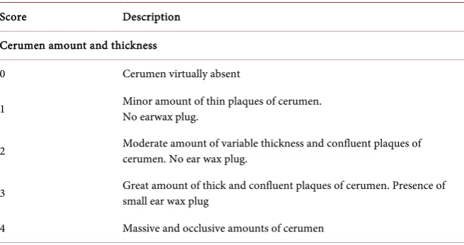
Table 1. Classification of the video-otoscopic scale of cerumen and erythema.
Score Description
Cerumen amount and thickness
0 Cerumen virtually absent
1 Minor amount of thin plaques of cerumen. No earwax plug.
2 Moderate amount of variable thickness and confluent plaques of cerumen. No ear wax plug.
3 Great amount of thick and confluent plaques of cerumen. Presence of small ear wax plug
4 Massive and occlusive amounts of cerumen
Surface of ear canal covered by cerumen
0 0
1 10% - 30%
2 30% - 50%
3 50% - 75%
4 75% - 100%
Erythema
0 No erythema
1 Very mild
2 Mild
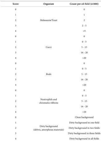
DOI: 10.4236/ojvm.2019.96006 72 Open Journal of Veterinary Medicine Table 2. Classification of the microbiologic semi quantitative scale.
Score Organism Count per oil field (x1000)
0
Malassezia/Yeast
0
1 1
2 2
3 2 - 5
4 >5
0
Cocci
0
1 0 - 5
2 5 - 15
3 16 - 20
4 >20
0
Rods
0
1 0 - 5
2 5 - 15
3 16 - 20
4 >20
0
Neutrophils and chromatin ribbons
0
1 0 - 5
2 5 - 15
3 16 - 20
4 >20
0
Dirty background (debris, amorphous materials)
Clean background
1 Dirty background in one field
2 Dirty background in two fields
3 Dirty background in three fields
4 Dirty background in all fields
2.7. Efficacy Outcome Measures
Video-otoscopic and cytological scores were used to assess the primary efficacy in this study: baseline score (T0 on D0) and scoring at each time point (T0 + 30 min on D0 and D6).
Any abnormal clinical signs reported by the owner or identified by the inves-tigator were recorded in the case report form.
2.8. Data Analysis
Dogs with one or major protocol deviations that affected the integrity of efficacy data were excluded from the analysis.
DOI: 10.4236/ojvm.2019.96006 73 Open Journal of Veterinary Medicine events) were compared using the fisher’s exact or a Chi-test.
Animal characteristics were compared at baseline (D0) to check comparability before treatment. A non-parametric analysis was used due to the low sample number. Intra-group comparison of scores over the study period was performed using a Friedman analysis of variance (ANOVA), if significant, a Wilcoxon rank sum test was used for individual comparisons between time points. Inter-group comparisons of scores were analysed using a Mann Whitney Test.
A value of p < 0.01 was considered statistically significant. All analyses were performed using statistical R software (R Foundation for Statistical Computing, Vienna, Austria, 2014).
3. Results
Population studyA total of eighteen dogs comprising 8 males (four castrated) and 10 females (2 spayed), aged between 5 months old and 14 years old were enrolled in the study, and randomisation resulted in 10 dogs in EpiOtic® Advanced and 8 dogs in Sonotix®. Breeds represented were 6 Cocker spaniels (3 from one breeder), 4 Labradors, 2 Golden Retrievers, 2 French Bulldogs, 1 Poodle and 3 cross breed dogs. The dogs weighed between 2.6 kg and 42.6 kg.
The descriptive characteristics of the dogs, the medical history and the scores were not significantly different on Day 0. Pendulous ears were overall overrep-resented but no significant differences were observed between groups. The sex ratio 1:1 was balanced and the majority of dogs had bilateral otitis in both groups (80% EpiOtic® Advanced, 87% Sonotix®). Fifteen dogs had bilateral and 3 had unilateral otitis externa on presentation, giving a total of 33 affected ears; 8 dogs presented as new cases of otitis, whereas the remaining 10 were undergoing recrudescence of a prior episode (no significant difference among groups).
Cytological and video-otoscopic clinical examination
[image:7.595.208.538.646.738.2]Cytological samples of the otic exudate at T0 revealed pure Malassezia yeast overgrowth in 17 of 33 ears (51.5%), rods and cocci in 8 of 33 ears (24.2%), cocci and Malassezia in 6 of 33 ears (18.2%) and rods, cocci and Malassezia in 2 of 33 ears (6.1%). Median of global cytological scores at T0 was 6 for Sonotix® and 5 for EpiOtic® Advanced (Table 3). The important reductions in yeast, cocci, and rod-shaped organism counts were demonstrated in smears 30 minutes after the cleansing in both groups and at D6 (Figure 1).
Table 3. Median global scores of the video-otoscopic and cytological examinations for EpiOtic® Advanced and Sonotix®. a significantly different from baseline value (p < 0.01).
Cytological global median score Video-otoscopic global median scores T0 T0 + 30 min D6 T0 T0 + 30 min D6
Sonotix® 5 2a 2a 6 3a 2a
EpiOtic®
DOI: 10.4236/ojvm.2019.96006 74 Open Journal of Veterinary Medicine Figure 1. Median global scores for EpiOtic® Advanced and
Sono-tix® before and after treatment.
Intra-group comparison shows a significant difference (p value < 0.01) be-tween the median of global cytological scores bebe-tween T0 (Sonotix®: 5, EpiOtic® Advanced: 6) and T0 + 30 min (Sonotix®: 2, EpiOtic® Advanced: 3) and T0 and D6 (Sonotix®: 2, EpiOtic® Advanced: 2). Inter-group comparison shows no sta-tistical difference at T0 + 30 min and D6 (P < 0.01).
Almost all ears (26 ears - 78.8%) had at T0 some degree of erythema on initial presentation, and erythema was rated as very mild in 17 ears (51.5%), mild in 7 ears (21.2%) and intense in 2 ears (6.1%).
All ears had a variable amount of cerumen: 18 (54.5%) external ear canals were entirely covered by confluent plaques of cerumen, 9 (27.3%) were partially covered by confluent plaques of cerumen, 5 (15.2%) were patchily covered by non-confluent plaques of cerumen and 1 (3%) was completely occluded by a massive amount of cerumen.
The median global scores of video-otoscopic examination, as shown in Table 3 and Figure 2, was 6 at T0 for both groups, 3 for Sonotix® and 4 for EpiOtic® Advanced at T0 + 30 min and 2 at D6 for both group.
30 minutes after the cleaning of ears there was a significant (P < 0.01) decrease of the amount of cerumen in vertical and horizontal ear canal from baseline. Intra-group comparison shows a significant difference (p value < 0.01) between T0 (Sonotix®: 6 EpiOtic® Advanced: 6) and T0 + 30 min (Sonotix®: 3, EpiOtic® Advanced: 4) and between T0 and D6 (Sonotix®: 2, EpiOtic® Advanced: 2).
Inter-group comparison shows a significant difference at T0 + 30 min (p < 0.01) between Sonotix® (median score: 3) and EpiOtic® Advanced (median score: 4) (Figure 3).
4. Discussion
In the present study, both ear cleansers reduced bacterial and yeast overgrowth and clinical signs (cerumen burden, erythema) in a population of client-owned dogs with spontaneous OE. The cleansers decreased ear wax, as early as 30 min-utes, as evidenced by marked reduction of amount and thickness of cerumen and surface of external ear canal covered by ear wax.
0 1 2 3 4 5 6 7
T0 T0 + 30 min D6
Cytological examination scores
DOI: 10.4236/ojvm.2019.96006 75 Open Journal of Veterinary Medicine Figure 2. Median global scores for EpiOtic® Advanced and
Sonotix® before and after treatment.
Figure 3. Video-otoscopy images showing the external auditory canals of dogs before and 30 minutes after receiving ear cleaning with either Sonotix® or Epi-Otic® Advanced. (a) External ear canal of a dog before receiving Sonotix® (T0) (b) The same external ear canal as showed in image a, 30 minutes after cleaning with Sonotix® (T0 + 30 min) (c) External ear canal of a dog before re-ceiving Epi-Otic® Advanced (T0) (d) The same external ear canal as showed in image c, 30 minutes after cleaning with Epi-Otic® Advanced (T0 + 30 min).
However, the cleaning activity of Sonotix® was statistically superior to Epi-Otic® Advanced as evidenced by the median global scores of video-otoscopic examination at T 30 min (p < 0.01).
This suggests that Sonotix® has a faster and more efficient action in cerumen removal. This enhanced ceruminolytic activity had also been demonstrated in an in vitro trial [17]. In that study, which the aim was to compare the ceruminolytic activity of five commercial ear cleaners on synthetic canine cerumen, the ear so-lution (Sonotix®, Vetoquinol, Lure, France) had the most effective and rapid ceruminolytic activity of the products tested. This fastest ceruminolytic activity could be in part explained by ingredient composition comprising three ceru-menolytic agents acting synergistically. Ethoxydiglycol (protic solvent) and
iso-0 1 2 3 4 5 6 7
T0 T0 + 30 min D6
Video-otoscopic examination scores
[image:9.595.259.487.258.451.2]DOI: 10.4236/ojvm.2019.96006 76 Open Journal of Veterinary Medicine propyl alcohol (organic solvent) disrupt the integrity of earwax by lysing the ag-glomerate and by breaking the bond between corneocytes whereas capric glyc-erides expedite the ceruminolytic and cleaning process by emulsifying debris. Cytological and clinical improvement, compared to T0, was also seen at D6 but no difference was seen between Sonotix® and EpiOtic® Advanced. As these dogs had clinical OE, for ethical reasons they were not treated with ear cleaner only. Both groups were prescribed daily application of Aurizon®, a product known to have strong anti-inflammatory, antibacterial and anti-yeast activity.
The video-otoscopic scoring used in this study was developed to assess the cerumen burden and erythema as opposed to other published scorings devel-oped to globally assess otitis externa severity [21]. In order to reduce inter clini-cian bias and before and after application bias, all video-otoscopy and cytology were evaluated by an experienced dermatologist (Dip. ECVD).
In healthy ear, the removal of cerumen and debris from the external ear canal is primarily achieved by epithelial migration. This physiologic process allows cerumen transport away from the tympanic membrane and towards the opening at the distal end of the ear canal. Recently this process has been demonstrated to follow both radial and centrifugal patterns in the dog [22]. While it is commonly accepted ear cleaning is not necessary for healthy ears, it has several benefits in many conditions as those in which epithelial migration is impaired. Failure of epithelial migration may result in the accumulation of flakes of skin in the ear canal and soft wax plugs. Furthermore, the migratory process is a key factor in the repair of spontaneous TM perforations and post-operative TM incisions
Effective ear cleaning is an essential part of any treatment scheme because it favours the contact between the ointment and the lining ear epidermis and speeds resolution by the removal of cerumen, microbial organisms, cellular de-bris and free fatty acids and prevents potential inactivation of topical therapy by purulent material [4] [13]. Our results comfort the common habit of prescribing a regular ear cleaner as a component in the management of OE in dogs.
A wide range of ear cleansing preparations and procedures aimed to remove exudates and ceruminous debris have become very popular in veterinary prac-tice. Knowledge of their properties helps to choose the best available product for specific clinical situations. Choosing the most appropriate product is akin to se-lecting a shampoo. It depends on the individual patient and it is of crucial im-portance to fully evaluate the primary, predisposing and perpetuating factors, the type otitis and the chosen topical therapy.
Oil-based ear cleaners, compared to those containing strong ceruminolytics, tend to be much gentler as they loosen wax, rather than disrupt it, but they do leave a residue in the canal. Moreover, it is questionable if they are of many benefits in ears with a purulent discharge. A cleaner with aqueous properties would be more indicated in a purulent otitis [23].
DOI: 10.4236/ojvm.2019.96006 77 Open Journal of Veterinary Medicine ear cleaners has been shown to be associated with a low pH, acid cleaners may inactive some antimicrobials (especially aminoglycosides and fluoroquinolones) [24]. In the present study, a complete clinical and microbiological resolution was observed at D6 suggesting that the concomitant use of Sonotix®, didn’t interfere with the activity of Aurizon® or didn’t inactivate the antimicrobial contained in it. Further studies to directly prove it (synergy in vitro test) are however neces-sary.
Funding Statement
This study was sponsored by Vetoquinol Lure, France
Conflicts of Interest
J. F. Jamet: None Declared; G. Marignac: None Declared; J. Y. Petit: None De-clared; J. L. Petit, F. Woehrlé, T. Trouchon and O. Fantini are employees of Ve-toquinol; S. Perrot: None Declared.
References
[1] Carlotti, D.N. (1991) Diagnosis and Medical Treatment of Otitis Externa in Dogs and Cats. Journal of Small Animal Practice, 32, 394-400.
https://doi.org/10.1111/j.1748-5827.1991.tb00963.x
[2] Huang, H.-P., Little, C.J.L. and McNeil, P.E. (2009) Histological Changes in the Ex-ternal Ear Canal of Dogs with Otitis Externa. Veterinary Dermatology, 20, 422-428.
https://doi.org/10.1111/j.1365-3164.2009.00853.x
[3] Radlinsky, M.G. and Mason, D.E. (2005) Diseases of the Ear. Textbook of Veteri-nary Internal Medicine, 2, 6.
[4] McKeever, P.J. and Torres, S.M.F. (1997) Ear Disease and Its Management. Veteri-nary Clinics of North America: Small Animal Practice, 27, 1523-1536.
https://doi.org/10.1016/S0195-5616(97)50137-9
[5] Gotthelf, L.N. (2005) Factors That Predispose the Ear to Otitis Externa. In: Small Animal Ear Diseases (An Illustrated Guide), 2nd Edition, Elsevier, Amsterdam, 141-171. https://doi.org/10.1016/B0-72-160137-5/50010-6
[6] Angus, J.C., Lichtensteiger, C., Campbell, K.L. and Schaeffer, D.J. (2002) Breed Variations in Histopathologic Features of Chronic Severe Otitis Externa in Dogs: 80 Cases (1995-2001). Journal of the American Veterinary Medical Association, 221, 1000-1006. https://doi.org/10.2460/javma.2002.221.1000
[7] Crespo, M.J., Abarca, M.L. and Cabanes, F.J. (2002) Occurrence of Malassezia Spp. in the External Ear Canals of Dogs and Cats with and without Otitis Externa. Medi-cal Mycology, 40, 115-121.
[8] Foster, A.P. and DeBoer, D.J. (1998) The Role of Pseudomonas in Canine Ear Dis-ease. Compendium of Continuing Education for the Practicing Veterinarian, 20, 909-919.
[9] Junco, M.T. and Barrasa, J.L. (2002) Identification and Antimicrobial Susceptibility of Coagulase Positive Staphylococci Isolated from Healthy Dogs and Dogs Suffering from Otitis Externa. Journal of Veterinary Medicine Series B, 49, 419-423.
https://doi.org/10.1046/j.1439-0450.2002.00571.x
DOI: 10.4236/ojvm.2019.96006 78 Open Journal of Veterinary Medicine America: Small Animal Practice, 29, 1303-1310.
https://doi.org/10.1016/S0195-5616(99)50128-9
[11] Ginel, P.J., Lucena, R., Rodriguez, J.C. and Ortega, J. (2002) A Semiquantitative Cy-tological Evaluation of Normal and Pathological Samples from the External Ear Canal of Dogs and Cats. Veterinary Dermatology, 13, 151-156.
https://doi.org/10.1046/j.1365-3164.2002.00288.x
[12] Tater, K.C., Scott, D.W., Miller, W.H. and Erb, H.N. (2003) The Cytology of the External Ear Canal in the Normal Dog and Cat. Journal of Veterinary Medicine Se-ries A, 50, 370-374. https://doi.org/10.1046/j.1439-0442.2003.00548.x
[13] Nuttall, T. and Cole, L.K. (2004) Ear Cleaning: The UK and US Perspective. Veteri-nary Dermatology, 15, 127-136. https://doi.org/10.1111/j.1365-3164.2004.00375.x
[14] Lloyd, D.H., Bond, R. and Lamport, I. (1998) Antimicrobial Activity in Vitro and in Vivo of a Canine Ear Cleanser. Veterinary Record, 143, 111-112.
https://doi.org/10.1136/vr.143.4.111
[15] Swinney, A., Fazakerley, J., McEwan, N. and Nuttall, T. (2008) Comparative in Vi-tro Antimicrobial Efficacy of Commercial Ear Cleaners. Veterinary Dermatology, 19, 373-379. https://doi.org/10.1111/j.1365-3164.2008.00713.x
[16] Rème, C.A., et al. (2006) The Efficacy of an Antiseptic and Microbial Anti-Adhesive Ear Cleanser in Dogs with Otitis Externa. Veterinary Therapeutics, 7, 15-26. [17] Brevet, A., Moreau, M. and Petit, J.L. (2015) Comparative in Vitro Ceruminolytic
Activity of 5 Commercialized Veterinary Ear Cleaners and a New Ear Cleaner Pro-totype. 21st FECAVA Eurocongress, Barcelona, Spain, 15-17 October 2015. [18] Mansfield, P.D., Steiss, J.E., Boosinger, T.R. and Marshall, A.E. (1996) The Effects of
Four, Commercial Ceruminolytic Agents on the Middle Ear. Journal of the Ameri-can Animal Hospital Association, 33, 479-486.
https://doi.org/10.5326/15473317-33-6-479
[19] Nielloud, F., et al. (2004) Development of an in Vitro Test to Evaluate Cerumen Dissolving Properties of Several Veterinary Ear Cleansing Solutions. Journal of Drug Delivery Science and Technology, 14, 235-238.
https://doi.org/10.1016/S1773-2247(04)50106-5
[20] Sánchez-Leal, J., Mayos, I., Homedes, J. and Ferrer, L. (2006) In Vitro Investigation of Ceruminolytic Activity of Various Otic Cleansers for Veterinary Use. Veterinary Dermatology, 17, 121-127. https://doi.org/10.1111/j.1365-3164.2006.00504.x
[21] Nuttall, T. and Bensignor, E. (2014) A Pilot Study to Develop an Objective Clinical Score for Canine Otitis Externa. Veterinary Dermatology, 25, 530-537.
https://doi.org/10.1111/vde.12163
[22] Tabacca, N.E., Cole, L.K., Hillier, A. and Rajala-Schultz, P.J. (2011) Epithelial Mi-gration on the Canine Tympanic Membrane. Veterinary Dermatology, 22, 502-510.
https://doi.org/10.1111/j.1365-3164.2011.00982.x
[23] Forsythe, P.J. (2016) Acute Otitis Externa: The Successful First-Opinion Ear Con-sultation. In Practice, 38, 2-6. https://doi.org/10.1136/inp.i412
[24] Nuttal, T. (2016) Successful Management of Otitis Externa. In Practice, 38, 17-21.