Testudines use water as a respiratory medium, exchanging O2 and CO2 across nonpulmonary surfaces (Belkin, 1968; Jackson et al., 1976; Gatten, 1980, 1984; Stone et al., 1992a). As such, many turtle species are bimodal gas exchangers, exchanging respiratory gases with both air and water. While the relative importance of aquatic gas exchange has been studied in only a handful of species, it has been documented that the utilization of water as a respiratory medium varies widely (Jackson et al., 1976; Stone et al., 1992a). For example, in members of the softshell family (Trionychidae), aquatic gas exchange accounts for approximately 38 % of the total V.O∑and 85 % of the total V.CO∑ (Gage and Gage, 1886; Stone et al., 1992a), whereas in members of the Emydidae, aquatic V.O∑ values range from 4 % (Belkin, 1968) to 11 % (Jackson et al., 1976). Additionally, the ability of these bimodal gas
exchangers to increase their reliance on aquatic gas exchange during periods of stress or increased metabolic rate has never been studied systematically.
One important physiological stressor is exercise, which can be utilized as a physiological correlate of survival (Bennett and Huey, 1990). Reptiles frequently undergo brief but sometimes intense periods of burst exercise in escaping from predators and/or capturing prey. During periods of increased anaerobic activity, an animal must cope with increased rates of CO2 production and the accumulation of lactic acid. The resultant respiratory and/or metabolic acidosis can be further complicated if exercise takes place while the animal is submerged and therefore unable to utilize aerial gas exchange. Exercise has been studied in sea turtles (Chelonia mydas), painted turtles (Chrysemys picta), pond sliders (Trachemys Printed in Great Britain © The Company of Biologists Limited 1999
JEB1655
The dynamics of bimodal respiration, diving behaviour and blood acid–base status in the softshell turtle Trachemys scripta and the pond slider Apalone ferox were investigated at rest and under conditions of stress induced by exercise and forced submergence. During periods of forced submergence, only A. ferox doubled its aquatic gas exchange rate. Both A. ferox and T. scripta increased their aerial gas exchange profoundly following exercise and forced submergence, a pattern indicative of increased anaerobic respiration. Emersion duration increased significantly in A. ferox following forced submergence, and mean apnoeic time decreased significantly in A. ferox following exercise, indicating that a larger proportion of time at the surface was spent ventilating. Also, A. ferox maintained a one-breath breathing bout regardless of treatment. Submergence produced a respiratory acidosis in the plasma of approximately 0.2 pH units in magnitude in T. scripta and a mixed respiratory/metabolic acidosis of 0.4 pH units in A. ferox. Exercise induced an acidosis of 0.2 pH units of primarily metabolic origin in both species. Intra-erythrocyte pH was also reduced in both species in
response to submergence and exercise. Both intracellular and extracellular acidoses were more severe and longer lasting in A. ferox after each treatment. Plasma [HCO3−] decreased by 25 % in both species following exercise, but only in A. ferox following submergence. Plasma lactate concentrations increased by equal amounts in each species following exercise; however, they returned to resting concentrations sooner in T. scripta than in A. ferox. A. ferox had significantly higher lactate levels than T. scripta following forced submergence as well as a slower recovery time. A. ferox, which is normally a good bimodal gas exchanger at rest, utilizes aerial respiration to a greater extent when under respiratory and/or metabolic stress. T. scripta, although almost entirely dependent on aerial respiration, is physiologically better able to deal with the respiratory and metabolic stresses associated with both forced submergence and exercise.
Key words: turtle, Trachemys scripta, softshell, Apalone ferox, bimodal breathing, aquatic gas exchange, exercise, forced submergence, blood, acid–base.
Summary
Introduction
EXERCISE AND FORCED SUBMERGENCE IN THE POND SLIDER (TRACHEMYS
SCRIPTA) AND SOFTSHELL TURTLE (APALONE FEROX): INFLUENCE ON BIMODAL
GAS EXCHANGE, DIVING BEHAVIOUR AND BLOOD ACID–BASE STATUS
B. BAGATTO* ANDR. P. HENRY
Department of Zoology and Wildlife, Auburn University, Auburn, AL 36849, USA
*Present address: Department of Biological Sciences, University of North Texas, Box 305220, Denton, TX 76203-5220, USA (e-mail: [email protected])
scripta) and snapping turtles (Chelydra serpentina); however, these studies have focused on the metabolic costs of and factors affecting aerobic exercise (Gatten, 1974, 1988; Butler et al., 1984; Lowell, 1990; Jackson and Prange, 1979; Zani and Claussen, 1994). The acid–base status during the recovery from exhaustive exercise has been well documented in fish (Wood and Perry, 1983; Boutilier et al., 1993) and in a few terrestrial reptiles (Seymour et al., 1985; Gleeson and Dalessio, 1989), but acid–base status and the way in which turtles deal with strenuous exercise have not yet been investigated in turtles, especially in those species that are aquatic and have varying abilities as bimodal gas exchangers.
Another significant physiological stressor is any period of submergence that is extended beyond a ‘routine’ dive duration. Turtles may extend dives beyond their aerobic capacity during predator/prey interactions and certainly during hibernation (Ultsch et al., 1985). Whether members of the genus Apalone are able to increase aquatic respiration significantly in response to prolonged submersion is not known.
Species within the Trionychidae are so highly aquatic that some have been reported to support resting metabolism solely via the aquatic medium (Dunson, 1960; Girgis, 1961). However, it is not known whether resting aquatic gas exchange is indicative of the maximal ability of the Trionychidae to respire aquatically. It is possible that members of this family could increase aquatic V.O∑/V
.
CO∑ during periods in which the
animal is effectively cut off from aerial respiration or when an increase in metabolic rate places a higher demand on total V.O∑.
Additionally, a comparison between Trionychidae and Emydidae, a family containing characteristically poor bimodal gas exchangers, will aid in determining the comparative importance of aquatic respiration under various physiologically stressful conditions. Pond sliders (Trachemys scripta), Florida softshells (Apalone ferox) and spiny softshells (Apalone spinifera) were used in a comparison of resting aquatic respiration to investigate inter-family differences as well as intra-genus differences. T. scripta and A. ferox were used to compare resting aquatic respiration versus maximal aquatic respiration to elucidate the degree of reliance on aerial versus aquatic gas exchange in freshwater turtles.
Materials and methods
Collection and maintenance of animals
Specimens of Apalone spinifera (Le Sueur) (N=7; 3 male, 4 female; mean mass 2196±616 g; range 237–4225 g), Apalone ferox (Schneider) (N=18; 17 male, 1 female; mean mass 1024±352 g; range 409–5900 g) and Trachemys scripta (Schoepff) (N=28; 15 male, 13 female; mean mass 987±53 g; range 545–1785 g) (means ± S.E.M.) were trapped from the Tallapoosa drainage in Alabama, USA, using baited hoop nets or purchased through a commercial supplier. All animals were housed in laboratory aquaria and were fed a combination of trout pellets and ReptoMin ad libitum. A. ferox and A. spinifera were housed individually, and T. scripta were housed in groups of a maximum of five for at least 2 weeks prior to
experimentation. Housing containers for both species were 4 m2in surface area and 1 m in height. Food was withheld for
at least 3 days prior to experimentation. Temperature remained between 22 and 25 °C, and the photoperiod was approximately 14 h:10 h L:D. All experiments began between 06:00 and 10:00 h and ended between 10:00 and 14:00 h.
Respiratory gas exchange
Turtles were placed in a Plexiglas chamber made up of a water-filled compartment (45 cm×45 cm×30 cm) and a smaller air-filled breathing hood (13 cm×13 cm×15 cm) built into the chamber lid. For T. scripta, the larger water compartment was reduced in size (25 cm×25 cm×25 cm) to measure changes in aquatic gas partial pressures more easily. The chamber structure was such that the turtle remained submerged in the water compartment but could raise its head into the air-breathing hood for pulmonary ventilation. Both compartments were fitted with ports from which gas and water samples were collected (Stone et al., 1992a) and subsequently analyzed for O2 partial pressure and CO2 concentration. To avoid behavioural acclimation (Stone et al., 1992b), each turtle was placed in the chamber for only one experiment.
Each turtle was maintained in the chamber for at least 12 h prior to an experiment. Air-equilibrated water and fresh air were supplied via a gas equilibrium column and air pump, respectively. This flow-through system was closed prior to the beginning of each experiment. Water was continuously stirred during the experiment by means of magnetic stir bars to prevent the occurrence of O2 and CO2 gradients. The small surface area of the air–water interface relative to the volume of water minimized movement of gases between phases. The negligible movement of gases was confirmed over 8 h periods. Either hypercapnic (10 % CO2) water or normoxic water was tested, each with normoxic air, hypercapnic air or anoxic air. Each air and water combination was replicated four times. There were no significant differences in either the air or water composition after the 8 h period.
refilled with air, and samples were withdrawn more frequently to prevent excessive O2depletion and CO2build-up in the air
chamber.
The exercise treatments utilized a similar protocol. In this case, the turtle was removed from the chamber following the rest period and exercised in a large plastic pool via caudal stimulation with tongs. A plastic screen was held over the anterior portion of the turtle to ensure that no air was inspired during the exercise period. Exercise proceeded to exhaustion, which was defined as the point at which a turtle no longer responded vigorously to caudal stimulation (i.e. a turtle no longer attempted to escape the grasp). Because exercise usually lasted 5–8 min and was performed in a large volume of water, aquatic gas exchange was not measured during this period. Immediately following exercise, the turtle was returned to the chamber in a closed, water-filled container to prevent it from breathing during the transfer. The metabolic chamber was then sealed, and samples were taken during a 2.0 h recovery period in the same manner as during the recovery period in the forced submergence protocol.
Oxygen partial pressures were measured using a Radiometer PHM72 blood/gas monitor equipped with an E5046 PO∑
electrode. Aquatic oxygen partial pressures were converted into molar concentrations using solubility coefficients from Dejours (1975). Carbon dioxide concentrations were measured using a Capni-con 5 total CO2analyzer (Cameron Instrument).
Mean aerial, aquatic and total V.O∑and V
.
CO∑were calculated for
each individual, and percentage aerial V.O∑ and V
.
CO∑ were
calculated from these means. Individual means were used to determine the mean ±S.E.M. of each variable for each species. Data analyses were performed on the means of individual animals, not on individual observations.
Ventilation and diving behaviour
The ventilation and diving behaviour of each turtle was monitored during all experiments (4 h duration) using a Dash IV oscillographic chart recorder (Astro-Med). Fine-gauge wires were fitted at opposite sides of the air–water interface and connected to a Colborne 2991 impedance converter (Morrow Bay, California, USA). Emersion, expiration, inspiration and immersion were monitored as a function of time by following the characteristic changes in impedance at the air–water interface. During preliminary experiments, a video camera was used to confirm behavioural measurements observed on the chart recorder. Variables measured included the duration of each immersion and emersion period, the duration of each period of emersion apnoea, the number of breaths per breathing bout and the number of breathing bouts per emersion period (Stone et al., 1992b). Incomplete immersion or emersion bouts in progress at the beginning or end of an experiment were excluded from the data.
Blood acid–base status Surgery
Trachemys scripta were anaesthetized using a pre-anaesthetic of NO2gas (10 % in air) followed by Halothane
(approximately 2 %). Surgical anaesthesia was characterized by the loss of a withdrawal reflex in response to pedal stimulation. A 2.5 cm diameter section of plastron was removed from the ventral side of T. scripta, exposing the right subclavian artery (Jackson et al., 1974). PE 90 tubing was used to catheterize this artery and was secured using silk ligatures. Heparinized reptilian Ringer’s solution (Lippe et al., 1966) was frequently flushed through the cannula to prevent clotting. The cannula was then routed posterior to the right limb and secured to the shell using rubber strips and superglue. The circular piece of plastron was replaced and sealed using plastic epoxy glue and covered with a thin acrylic sheet to prevent exchange of fluids in either direction.
In A. ferox, surgical anaesthesia was obtained using 3-aminobenzoic acid ethyl ester (MS-222, Sigma Chemical Co., St Louis, USA). A dosage of 500 mg kg−1 was injected
intracoelomically, after which the turtle was immersed in an aerated solution of 1 g l−1 MS-222 buffered to a pH of 7.0
(Bagatto et al., 1997a). After surgical anaesthesia had been achieved, a semicircular flap of skin was cut and folded back, exposing the left subclavian artery (Ultsch et al., 1984; Bagatto et al., 1997b). Cannulation proceeded in the same manner as for T. scripta, after which the flap of skin was sutured and sealed with superglue. Animals were allowed at least 24 h to recover from surgery.
Blood analyses
After a resting blood sample had been withdrawn, a turtle was given one of two treatments. Forced submergence of 1 h involved placing a turtle in aerated water and fitting the container with a plastic grate so that the turtle could not emerge. The exercise treatment was as described above. After a resting blood sample had been withdrawn, additional samples (800µl each) were withdrawn at 0, 30, 60, 120, 240 and 480 min following the forced submergence or exercise; these were replaced by equal volumes of heparinized reptile Ringer’s solution.
Each blood sample was withdrawn into a gas-tight Hamilton syringe and transferred into 0.5 ml polyethylene tubes, which were quickly sealed and were always full when sealed. Care was taken to minimize the exposure of the blood sample to environmental air. Plasma was then separated from the corpuscular component using a Fisher model 235B micro-centrifuge (3 min at 10 000 revs min−1), and the true plasma pH
(pHe) and plasma PCO∑ were immediately measured using a
Radiometer PHM 72 blood/gas monitor with associated G297 micro-pH unit/K497 reference electrode (Radiometer, Copenhagen) and associated E201 CO2 electrode (Cameron
Instrument), respectively. Simultaneously, a Corning model 965 CO2analyzer was then used to measure the total carbon
dioxide content (TCO∑) of the blood plasma. The red cell pellet
(1972). Measured values of true plasma TCO∑and PCO∑were
used to calculate true plasma bicarbonate concentration ([HCO3−]tpl) using the following rearrangement of the
Henderson–Hasselbach equation:
[HCO3−]tpl= TCO∑ −(αCO∑×PCO∑) ,
using values for αCO∑determined according to Boutilier et al. (1984).
Statistical analyses
The means of the respiration and behavioural data for both species at rest were compared using an analysis of variance (ANOVA) when assumptions of equal variance and normality were met. A Kruskal–Wallis ANOVA on ranks was used when either assumption failed. For percentage data, arcsine-square-root transformations were performed (Zar, 1984). The factors of mass and sex did not significantly affect the variables measured and were not therefore included as covariates. Comparisons among rest and recovery, species and treatment were calculated using a three-way ANOVA with repeated measures. For the blood characteristics, a two-way ANOVA with repeated measures was used to determine the differences among species, treatments and time intervals. All data were analyzed for significance at the P<0.05 level using SigmaStat for all procedures, except the three-way repeated-measures ANOVA, which was analyzed using SPSS. All values presented are means ±1S.E.M.
Results
Respiratory gas exchange
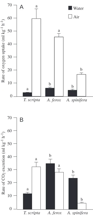
All three species relied primarily on air for O2 uptake;
however, both A. spinifera and A. ferox were able to acquire aquatic O2 at a significantly greater rate than T. scripta
(Fig. 1A). The total V.O∑of T. scripta was 62.7±7.8 ml kg−1h−1;
twice the value reported by Belkin (1968). The total V.O∑of A.
ferox was similar to that of T. scripta at 52.0±3.4 ml kg−1h−1,
and A. spinifera had a significantly lower total V.O∑ than the
other two species of 21.6±2.0 ml kg−1h−1(H=23.6; P<0.0001).
The total V.O∑of A. spinifera was similar to but slightly lower
than the value of 28.6 ml kg−1h−1 measured by Stone et al.
(1992a). The ratio of aquatic V.O∑ to total V
.
O∑, a standard
measure of an animal’s degree of reliance on water for gas exchange, was significantly different among the three species (H=35.1; P<0.0001). T. scripta relied the least on aquatic V.O∑,
exchanging 5.1 % of the total O2via water, while A. ferox was
intermediate (11.7 %) and A. spinifera wasthe most reliant on aquatic gas exchange (21.7 %).
Because of the greater solubility of CO2than O2in water,
rates of aquatic CO2excretion (V
.
CO∑) were approximately five
times greater than those for aquatic V.O∑. A. spinifera and A.
ferox had a significantly higher aquatic V.CO∑ than T. scripta
(H=30.6; P<0.0001), whereas T. scripta and A. ferox had a significantly higher aerial V.CO∑ than A. spinifera (H=23.3;
P<0.0001) (Fig. 1B). Therefore, A. ferox maintained a high CO2 excretion rate both aquatically and aerially, which
resulted in the highest total V.CO∑ of 64.1±6.0 ml kg−1h−1
compared with 45.0±4.6 ml kg−1h−1 for T. scripta (half the
value measured by Jackson et al., 1976) and 29.0±3.0 ml kg−1h−1 for A. spinifera (almost identical to the
value measured by Stone et al., 1992a). With aquatic and aerial CO2excretion partitioned almost equally in A. ferox, the ratio
of aquatic to total V.CO∑was 55.0 %. T. scripta relied the least
on water for CO2excretion (27.7 %), while A. spinifera was
the most reliant on water (81.8 %) (F=245.1; P<0.0001). Aquatic V.O∑and V
.
CO∑in T. scripta did not change significantly
in response to either exercise or forced submergence. Exercise did not have a significant effect on aquatic gas exchange in A. ferox; however, this was not true during forced submergence. A.
0 10 20 30 40 50 60 70
Rate of oxygen uptak
e (ml kg
-1 h -1)
Water
Air
T. scripta A. ferox A. spinifera
a a
b a
b b
Rate of CO
2
e
xcretion (ml kg
-1 h -1)
T. scripta A. ferox A. spinifera
a
b
b a
a
b
A
B
[image:4.609.335.525.69.505.2]0 10 20 30 40 50 60 70
Fig. 1. Resting mean aquatic and aerial V.O∑ (A) and V .
CO∑ (B) in Trachemys scripta (N=23), Apalone ferox (N=15) and Apalone spinifera (N=7). Letter groups indicate those species that are not
ferox demonstrated the ability to increase aquatic V.O∑(F=10.4;
P<0.0036) and V.CO∑significantly (F=19.8; P<0.0003), each by
twofold, during the submergence period.
Exercise and forced submergence both produced an increased aerial demand for O2 in T. scripta and A. ferox
(Fig. 2A). In T. scripta, aerial V.O∑ increased by fourfold in
response to exercise and forced submergence, and these values remained significantly higher than resting values until 30 min after each treatment (exercise F=76.82; P<0.0001; submergence F=188.9; P<0.0001). In A. ferox, aerial V.O∑
increased by fourfold following exercise and by fivefold following forced submergence. The increase following forced submergence in A. ferox was significantly greater than that exhibited by T. scripta following the same treatment (F=4.86; P<0.042). Aerial V.O∑in A. ferox returned to resting values by
60 min post-submergence, but aerial V.O∑remained significantly
higher than resting values even at 120 min post-exercise. In addition to increased aerial O2demand, the rate of aerial
CO2excretion increased in both species in response to exercise
and forced submergence (exercise F=47.09; P<0.0001; submergence F=111.3; P<0.0001) (Fig. 2B). In T. scripta, aerial V.CO∑increased by fivefold following both exercise and
forced submersion and returned to resting values by 60 min after each treatment. Aerial V.CO∑ in A. ferox increased by
sevenfold immediately after both exercise and forced submergence and returned to values for resting turtles by 60 min following each treatment. The increase following forced submergence in A. ferox was significantly greater than that exhibited by T. scripta following the same treatment (F=4.53; P<0.0036).
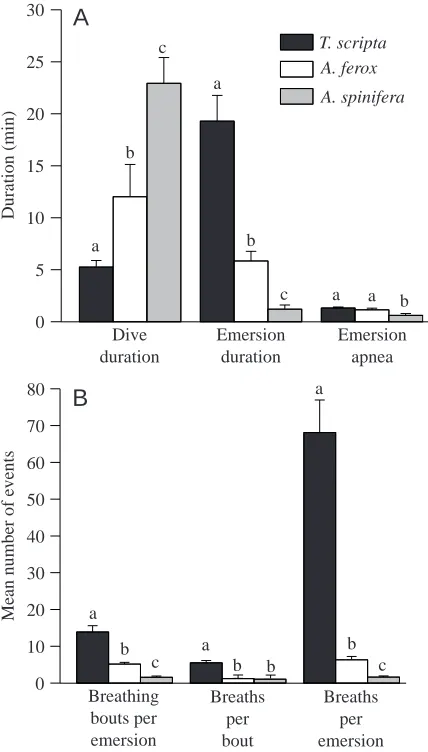
Ventilation and diving behaviour
At rest, each of the three species had a distinct ventilatory and diving pattern. Individual dives in T. scripta were short, with a mean duration of 5.3±0.6 min (Fig. 3A). In A. ferox,
0 70 140 210 280
T. scripta exercised T. scripta submerged A. ferox exercised A. ferox submerged
‡ ‡
*
*
*
*
*
Aerial
VO
2
(ml kg
-1 h -1)
Recovery time (min)
15 30 60 90 120
0 50 100 150 200 250
R
*
*
A
B
‡Aerial
VCO
2
(ml kg
-1 h -1) .
[image:5.609.332.546.290.666.2].
Fig. 2. Mean aerial V.O∑(A) and V
.
CO∑(B) in Trachemys scripta (N=8
exercise; N=8 forced submergence) and Apalone ferox (N=6 exercise; N=6 forced submergence) at rest (R) and after exercise and forced submergence. The dotted vertical line represents the time of application of the treatment. Asterisks indicate significant differences from corresponding resting values. The dotted brackets represent a grouping of data for the purpose of expressing significance. A double dagger (‡) denotes a significant difference between species at that corresponding time interval and treatment. Values are means ±S.E.M.
Duration (min)
0 5 10 15 20 25 30
T. scripta A. ferox
A. spinifera
Dive duration a
b c
Emersion duration a
b
Emersion apnea
a a b
0 10 20 30 40 50 60 70 80
Breathing bouts per emersion a
b c
Breaths per bout a
b b a
b
Mean number of events
c
c
A
B
Breaths per emersion
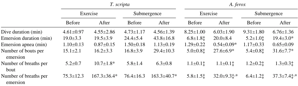
[image:5.609.62.284.311.636.2]mean dive duration was significantly longer (12.0±3.1 min) (H=26.7; P<0.0001), and mean dive duration was longer still in A. spinifera (22.9±2.5 min, a value twice that measured by Stone et al., 1992b). Dive duration was not significantly altered in T. scripta or in A. ferox by either exercise or submersion (Table 1).
While dive duration increased significantly from the least aquatically dependent species (T. scripta) to the most aquatically dependent species (A. spinifera), the reverse trend was true for emersion duration. The mean emersion duration of T. scripta was significantly longer than that for A. ferox, and both these species had significantly longer emersion times than A. spinifera (H=32.3; P<0.0001). Emersion duration in T. scripta was unaffected by exercise and, although an 83 % increase was noted after forced submergence, it was not significant (Table 1). Emersion duration in A. ferox increased fourfold after forced submergence (H=11.5; P<0.009), but did not increase significantly after exercise.
Emersion in all species was characterized by intermittent bouts of breathing separated by periods of apnoea. A. spinifera displayed significantly shorter apnoeic bouts than both T. scripta and A. ferox (F=5.76; P<0.006) (Fig. 3A). Emersion apnoea was not significantly altered by the treatments except in A. ferox after exercise, in which the mean apnoeic bout length decreased by 58 % compared with the corresponding resting value (F=10.8; P<0.02) (Table 1).
Another diving behaviour measured was the ratio of total apnoeic time during a given period of emersion to total emersion time. This conveyed the proportion of breathing time versus apnoeic time when the turtle was at the surface. A. spinifera spent a greater proportion of emersion time breathing compared with the other two species, with 73.0±7.8 % of the emersion time allotted to apnoea (F=4.01; P<0.025). T. scripta and A. ferox both spent less time at the surface breathing, with 86.4±1.6 % and 88.3±2.6 % of the emersion time spent in apnoea, respectively. However, when T. scripta and A. ferox
were subjected to exercise and forced submergence, the resultant increase in breathing frequency significantly reduced the ratio of apnoeic time to total emersion time.
The longer emersion durations in T. scripta allowed them to undergo a significantly greater number of breathing bouts per emersion period (13.9±1.7 versus 5.1±0.5 for A. ferox), and both performed significantly more breathing bouts per emersion period than A. spinifera (1.6±0.3) (H=32.0; P<0.0001) (Fig. 3B). The number of breathing bouts per emersion period did not change in T. scripta following either exercise or forced submersion (Table 1). However, A. ferox significantly increased the number of breathing bouts per emersion in response to exercise and forced submergence by factors of 5.5 and 5.8, respectively (H=17.4; P<0.0006).
At rest, A. spinifera and A. ferox were more similar in ventilatory characteristics compared to T. scripta (Fig. 3B). Both species retained a one-breath-per-bout behaviour that was unaltered by either submergence or exercise. T. scripta utilized a multi-breath bout, demonstrating a significantly greater number of breaths per bout than both A. spinifera and A. ferox (H=36.1; P<0.0001). In response to exercise, T. scripta significantly increased the number of breaths per bout by twofold (F=4.0; P<0.0172) (Table 1).
In T. scripta at rest, the number of breaths taken per emersion period was significantly greater than in A. ferox, and values for both these species were significantly greater than for A. spinifera (H=38.5; P<0.0001) (Fig. 3B). Following exercise and forced submersion, T. scripta increased the total number of breaths per emersion each by twofold (F=2.58; P<0.042) (Table 1). A. ferox, however, responded to exercise and forced submergence by increasing the number of breaths per emersion by factors of 5.5 and 5.8, respectively (F=7.58; P<0.0014). T. scripta exhibited significantly more breaths per emersion period than A. ferox for any given treatment.
The mean breath length (measured from the beginning of exhalation to the end of inhalation) of T. scripta (2.1±0.1 s) was
Table 1. Mean durations and numbers of ventilation behaviours in Trachemys scripta and Apalone ferox at rest, during and after forced submergence and after exercise
T. scripta A. ferox
Exercise Submergence Exercise Submergence
Before After Before After Before After Before After
Dive duration (min) 4.61±0.97 4.55±2.86 4.73±1.17 4.56±1.39 8.25±1.00 6.03±1.90 9.31±1.80 6.76±1.36 Emersion duration (min) 19.0±3.3 19.5±3.9 24.4±5.4 43.8±16.8 6.8±1.8‡ 20.0±8.4 5.2±1.0‡ 19.4±3.0* Emersion apnea (min) 1.10±0.13 0.87±0.15 1.50±0.18 1.13±0.19 1.29±0.22 0.54±0.09* 1.17±0.33 0.65±0.09 Number of bouts per 15.1±2.1 16.2±3.3 16.8±3.9 29.4±10.3 5.0±0.8‡ 27.6±6.9* 5.4±0.8‡ 31.6±7.7*
emersion
Number of breaths per 5.2±0.7 10.7±1.8* 5.8±1.4 6.3±0.8 1.1±0.1‡ 1.1±0.1‡ 1.2±0.2‡ 1.3±0.3‡ bout
Number of breaths per 75.3±12.3 167.3±36.4* 76.4±16.3 163.3±40.7* 5.8±1.5‡ 32.0±9.3‡,* 6.4±1.2‡ 37.3±7.4‡,*
emersion
Values are means ±S.E.M., N=8 for T. scripta; N=6 for A. ferox.
significantly shorter than that for A. ferox and A. spinifera (4.7±0.6 s and 5.1±0.3 s, respectively) (H=29.1; P<0.0001). Mean breath length was not significantly altered by either of the treatments in A. ferox or T. scripta.
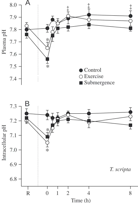
Blood acid–base status
In A. ferox and T. scripta, plasma pH (pHe) and intracellular pH (pHi) decreased significantly in response to exercise and forced submergence (Figs 4, 5). The acidosis produced by exercise was primarily metabolic in origin for both species, whereas the acidosis produced by forced submergence was almost completely of respiratory origin in T. scripta and mixed in A. ferox (see below).
Plasma pH in T. scripta decreased by 0.18 pH units in response to exercise, returning to resting values by 30 min (F=45.33; P<0.0001) (Fig. 4A). In A. ferox, pHe decreased by the same amount; however, resting values were not restored until 60 min post-exercise (Fig. 5A). Plasma pH in T. scripta and A. ferox decreased by 0.20 and 0.31 pH units, respectively,
in response to forced submergence, and resting values returned by 30 min in each species (F=45.52; P<0.0001).
Intracellular erythrocyte pH also exhibited a significant decline after exercise in each species (F=24.08; P<0.0001) (Figs 4B, 5B). In T. scripta, pHi decreased by 0.15 pH units in response to exercise, with resting values returning by 30 min. In A. ferox, a similar decrease was noted which recovered by 60 min. A. ferox pHi values remained significantly lower than the corresponding T. scripta values at each sampling interval until 120 min post-exercise (F=13.3; P<0.0026). Intracellular pH in T. scripta decreased by 0.13 pH units in response to forced submergence, returning to resting pHi values by 30 min (F=24.26; P<0.0001). In A. ferox, pHi decreased by 0.26 units immediately after forced submergence and had recovered by 60 min. A. ferox pHi values remained significantly lower than the corresponding T. scripta values at each sample time until 120 min post-submergence.
As pH declined following exercise, a concurrent decrease in plasma bicarbonate concentration was also noted in each species (F=16.63; P<0.0001) (Figs 6A, 7A). Plasma
Time (h)
Plasma pH
7.4 7.5 7.6 7.7 7.8 7.9 8.0
Control Exercise Submergence ‡
*
*
Intracellular pH
0 1 2 4 8
6.8 6.9 7.0 7.1 7.2 7.3
R
*
*
T. scripta
B
[image:7.609.332.553.70.384.2]A
‡ ‡Fig. 4. Mean blood plasma pH (A) and intra-erythrocyte pH (B) in
Trachemys scripta (N=7 control; N=9 exercise; N=10 forced
submergence) at rest (R) and after exercise and forced submergence. The dotted vertical line represents the time of application of either treatment. Asterisks denote a significant difference from both the resting value and the corresponding control value. Double daggers (‡) indicate a significant difference from the resting value only. Values are means ±S.E.M.
Time (h)
Plasma pH
7.4 7.5 7.6 7.7 7.8 7.9 8.0
Control Exercise Submergence
*
*
*
‡
§ §
Intracellular pH
*
0 1 2 4 8
6.8 6.9 7.0 7.1 7.2 7.3
R
*
*
§ §
§ §
A. ferox
B
A
[image:7.609.59.280.323.647.2]‡
Fig. 5. Mean blood plasma pH (A) and intra-erythrocyte pH (B) in
Apalone ferox (N=5 control; N=7 exercise; N=6 forced submergence)
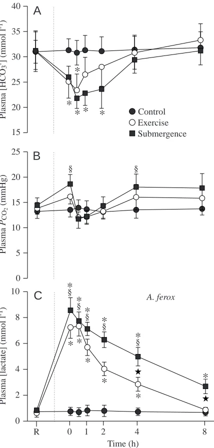
bicarbonate concentrations decreased by 25 % immediately following exercise in T. scripta, with resting values being achieved by 60 min post-exercise. In A. ferox, plasma bicarbonate concentration did not reach its maximal decrease of 25 % until 30 min post-exercise, but this also recovered by 60 min. Forced submergence had no effect on HCO3−
concentrations in T. scripta. Thirty minutes post-submergence, a maximal decrease of 30 % in [HCO3−] was exhibited in A.
ferox, and [HCO3−] did not return to resting levels until
240 min post-submergence (F=5.08; P<0.0002).
The partial pressure of CO2 (PCO∑) in the plasma did not
change in either species after exercise (Figs 6B, 7B). Plasma PCO∑ did not change in T. scripta after forced submergence;
however, there was a transient increase of 30 % in PCO∑in A.
ferox immediately after forced submergence.
Time (h) 15
20 25 30 35 40
Control Exercise Submergence
*
*
Plasma [HCO
3
-] (mmol l -1)
0 5 10 15 20 25
Plasma
PCO
2
(mmHg)
Plasma [lactate] (mmol l
-1)
0 1 2 4 8
0 2 4 6 8 10
R
*
*
*
‡
T. scripta
A
B
C
‡ ‡
‡ ‡ ★
[image:8.609.324.538.169.617.2]*
Fig. 6. Mean plasma bicarbonate concentration (A), plasma CO2 partial pressure (B) and plasma lactate concentration (C) in
Trachemys scripta (N=7 control; N=9 exercise; N=10 forced
submergence) at rest (R) and after exercise and forced submergence. The dotted vertical line represents the time of application of either treatment. Asterisks denote a significant difference from both the resting value and the corresponding control value. Double daggers (‡) indicate a significant difference from the resting value only. A star indicates a significant difference between treatments at a given time interval. Values are means ±S.E.M. 1 mmHg=0.133 kPa.
Time (h) Control Exercise Submergence 15
20 25 30 35 40
Plasma [HCO
3
-] (mmol l -1)
*
*
*
* *
0 5 10 15 20 25
§ §
Plasma
PCO
2
(mmHg)
Plasma [lactate] (mmol l
-1)
0 1 2 4 8
0 2 4 6 8 10
R §
* *
*
*
*
*
*
A. feroxC
B
A
★
★ §
*
§
*
§
*
§
*
Fig. 7. Mean plasma bicarbonate concentration (A), plasma CO2 partial pressure (B) and plasma lactate concentration (C) in Apalone
ferox (N=5 control; N=7 exercise; N=6 forced submergence) at rest
[image:8.609.54.276.179.624.2]With the significant decrease in blood pH, there was also a concurrent increase in plasma lactate concentration immediately following exercise (F=53.36; P<0.0001) (Figs 6C, 7C). In T. scripta, plasma lactate concentration increased by 6.21 mmol l−1.
Recovery in this species was variable after exercise; however, resting lactate concentrations returned by 240 min. An increase in lactate concentration of 6.61 mmol l−1 was observed in A.
ferox following exercise, with recovery apparent by 480 min. In T. scripta, forced submergence increased plasma lactate concentration by 5.05 mmol l−1. Significantly raised lactate
levels were maintained following the forced submergence treatment in T. scripta until 240 min post-treatment. A. ferox exhibited a remarkable trend in that forced submergence produced an increase in lactate concentration of 7.72 mmol l−1,
the largest increase of the study (F=90.83; P<0.0001). During forced submergence, lactate levels in A. ferox were significantly higher than the corresponding values for T. scripta at each sampling time until 480 min post-treatment (F=17.73; P<0.001). Lactate levels in A. ferox remained six times higher than at rest at 480 min post-submergence.
Discussion
Respiratory gas exchange Rest
The partitioning of oxygen uptake between air and water was unique for each species. These data, which were consistent with previous studies of T. scripta and A. spinifera, support the idea that among bimodal gas exchangers there is a spectrum of ability with regard to aquatic gas exchange that is related to overall reliance on water (Stone et al., 1992a). Absolute values for total and aquatic V.O∑in the present study were different from those
reported previously for the same species (Belkin, 1968; Stone et al., 1992a), but these differences did not change the general pattern of how gas exchange was partitioned between air and water. Although A. spinifera and A. ferox had a similar aquatic V.O∑, it accounted for a significantly larger proportion of aquatic
gas exchange in A. spinifera. A. ferox is generally found in sluggish streams, lakes and ponds and may not, therefore, be able to rely on aquatic O2as much as A. spinifera (Mount, 1975).
Slow-moving or stagnant water has a variable partial pressure of O2and is often hypoxic. It would not, therefore, be advantageous
for A. ferox to have a high aquatic V.O∑if this source of O2were
not constantly available. Furthermore, animals that have high aquatic V.O∑values could potentially lose O2from the blood if
the animal were exposed to a hypoxic environment (Belkin, 1968). Randall et al. (1981) documented such a transfer of oxygen from blood to water via the gills when Amia calva, a bimodally breathing fish, was exposed to hypoxic water.
Aquatic V.CO∑ has always been found to exceed V
.
O∑ in
bimodal gas exchangers, primarily because of the greater solubility of CO2in water (Wood and Lenfant, 1976). Again,
even though the absolute values of total and aquatic V.CO∑in
the present study differ from those reported previously, the partitioning pattern remains the same. Furthermore, the value of 27.7 % aquatic V.CO∑ for T. scripta, a poor aquatic gas
exchanger, may be more indicative of the minimal ability of aquatic turtles in general because this is consistent with aquatic V.CO∑ values from another poor aquatic gas exchanger,
Chelydra serpentina (30 %; B. Bagatto and R. P. Henry, unpublished observations). Jackson et al. (1976) reported an aquatic V.CO∑of 10.5 % for T. scripta; however, their total V
.
CO∑
was more than twice that of the present study and was probably not a true resting value, which may have led to an underestimation of the importance of aquatic V.CO∑.
Effects of submergence and exercise
In response to forced submersion, the first sign of respiratory plasticity was noted in A. ferox because this species increased both aquatic V.O∑and V
.
CO∑by twofold. Increasing aquatic gas
exchange may involve more frequent buccal pumping (Belkin, 1968) and/or increased capillary perfusion to the skin and buccal cavity, especially during exercise via increased cardiac output. The present study confirms that highly aquatic turtle species can alter their aquatic gas exchange and that this can be achieved more rapidly that previously reported (Belkin, 1968). Aquatic gas exchange remained minimal in T. scripta and did not change in response to either of the treatments. This confirms the aerial dependence of T. scripta and suggests that aquatic gas exchange is not plastic for every species; certainly not in species that are highly dependent on air.
Although A. ferox has the ability to increase cutaneous respiration during continued forced submergence, this probably did not occur during intense anaerobic exercise. Additionally, as A. ferox recovered from each of the treatments, direct access to air reduced the degree of utilization of aquatic gas exchange. This was especially apparent in A. ferox during recovery from forced submergence. During submergence, A. ferox increased both aquatic O2uptake and aquatic CO2excretion by twofold;
however, these rates decreased to resting levels once access was given to air during recovery.
Forced submergence had a greater impact on A. ferox than on T. scripta, an unexpected result (Fig. 2). Because A. ferox ventilate their lungs far less frequently than T. scripta, we hypothesized that A. ferox could easily withstand forced immersion for 1 h. Apparently the 22 breaths h−1 are vital to
resting metabolism; denying aerial respiration to A. ferox, even for 1 h, resulted in a large shift towards anaerobiosis as shown by the blood acidosis and increase in circulating lactic acid levels. Preliminary data using A. spinifera show the same trend even though this species only breathes approximately twice per hour. Apalone species appears to operate at the aerobic/anaerobic threshold, taking in enough O2to remain aerobic under resting
conditions. Thus, a disturbance in the natural rhythm of ventilatory and diving behaviour in these highly aquatic species seems to force them into anaerobic respiration.
Ventilation and diving behaviour Rest
metabolism via non-pulmonary avenues and have adopted a diving pattern similar to that of lungfish (Graham and Baird, 1982; Kramer, 1988), which come to the surface only for a single breath, then submerge again. A. spinifera have also evolved a one-breath bout, characteristic of highly aquatic species such as sea turtles Chelonia mydas (Butler et al., 1984) and sea snakes (Graham, 1974). This one-breath breathing pattern was also present in A. ferox; however, their increased reliance on aerial respiration was associated with increased numbers of breathing bouts per emersion and, thus, more breaths per emersion than A. spinifera. T. scripta was highly dependent on aerial respiration and, thus, displayed characteristics typical of terrestrial breathing patterns (a multi-breath bout).
Most reptiles undergo variable periods of apnoea (Wood and Lenfant, 1976). Mean lengths of apnoeic periods in T. scripta and A. ferox were 90 and 60 s, respectively. The mean emersion apnoea duration for A. spinifera was 36 s and was very similar to the value of 29 s measured by Stone et al. (1992b). As the capacity of a species for aquatic gas exchange increased, emersion times decreased (Stone et al., 1992b) and so did the duration of apnoea. It appears that the more efficient a species becomes at aquatic gas exchange, the more it approaches the condition observed in primitive lungfish; a one-breath bout and virtually no apnoeic period at the surface (Kramer et al., 1983; Kramer, 1988).
Agassiz (1857) noted that the more aquatic turtle species had significantly reduced lung volumes. In contrast, both A. spinifera and A. ferox had significantly longer breath durations than T. scripta, even after correction for differences in mass. Lung surface area and diffusion distance aspects aside, it seems that A. spinifera and A. ferox are able to ventilate more deeply as a result of having a less rigid outer shell structure and a reduced plastron. This would allow the visceral volume to vary to a greater extent, creating large intrapulmonary subatmospheric pressures, as found in Chelydra serpentina by Gaunt and Gans (1969). Although T. scripta have a larger lung volume per kilogram, their shell morphology allows them to produce only shallow breaths, requiring them to ventilate more quickly, as is the case for other strictly terrestrial species (Gans and Hughes, 1967). Thus, a one-breath bout is probably related to more than just a high level of aquatic gas exchange. Anatomical as well as other physiological variables may also relate to the existence of this seemingly unalterable phenomenon.
Another explanation of the utilization of the one-breath bout may involve the higher blood levels of CO2 in terrestrial
reptiles. Because A. spinifera and A. ferox are able to transfer a significant proportion of CO2into water, they may not need
to increase ventilation frequency in order to void CO2aerially.
T. scripta, as well as other terrestrial reptiles, may use many short breaths to reduce blood CO2levels, while concurrently
obtaining the required amount of O2(Rahn and Howell, 1976).
Effects of submergence and exercise
Because A. ferox did not alter their apnoeic period durations following forced submergence, compared with at rest, the emersion durations had to increase to allow for the increase in
the number of ventilation bouts per emersion (Table 1). However, A. ferox displayed a significant decrease in emersion apnoea duration after exercise, which may be the reason that the increase in emersion duration after exercise was not significant. Although there were no significant differences in diving behaviour after either treatment, T. scripta spent almost twice as much time at the surface after forced submergence, most likely to accommodate the increased number of bouts per emersion.
A. ferox retained a one-breath-per-bout pattern at all times; thus, the response of increasing breath frequency was simply a reflection of increased bout frequency (Table 1). This indicates that the characteristic of a one-breath bout is not plastic or reserved only for resting metabolism. These aquatic turtles have evolved a trait that is seemingly unalterable, even under extreme physiological conditions. This species-specific number of breaths per breathing bout was also documented for Kinosternon leucostomum and Staurotypus triporcatus following forced submergence (B. Bagatto, B. Hange and R. P. Henry, unpublished results). It was therefore interesting to note that exercise resulted in a significant increase in the number of breaths per bout in T. scripta. It is not known whether exercise would have a similar effect on breathing bouts in other species highly dependent on air. Perhaps exercise is so stressful that the increase in breathing frequency required for recovery causes breathing bouts to fuse, creating one long bout. This was certainly the trend in T. scripta immediately following exercise.
Blood acid–base status Exercise
The magnitudes of the decreases in pH and [HCO3−] were
similar in both T. scripta and A. ferox, indicating that brief intense anaerobic exercise had a similar physiological effect. It was apparent that the high aquatic to aerial gas exchange ratio in A. ferox neither delayed the onset of anaerobiosis nor aided the recovery from exhaustive exercise. In response to exercise, T. scripta was better able to cope with the metabolic acid load. Because of the compartmentalization of lactate within the body and long lag periods associated with intercompartmental transfers (especially from muscle to blood), blood lactate concentrations cannot be used to quantify the total amount of anaerobic metabolism (Bennett, 1994). However, relative levels of anaerobiosis can be compared between species and treatments using blood lactate level as an indicator, assuming equal perfusion and equal exchange rates between compartments. As with pH and [HCO3−], exercise
Forced submergence
Both A. ferox and T. scripta utilized anaerobic metabolism during the forced submergence period. It was surprising that A. ferox could not withstand the short duration of forced submergence, which produced the largest pH decrease of any treatment. Even though aquatic gas exchange was significantly increased during forced submergence, this was apparently not sufficient to sustain aerobic metabolism.
Plasma bicarbonate level decreased profoundly in A. ferox following forced submergence; however, T. scripta maintained resting levels of bicarbonate throughout recovery, further supporting the suggestion that this treatment did not create a long-lasting physiological disturbance. This indicates that T. scripta may have a larger reservoir of HCO3−with which to
buffer the by-products of anaerobic metabolism. Smith (1929) noted that freshwater turtles, such as T. scripta, possess a large volume of coelomic fluid with a pH more alkaline than plasma and having a HCO3−concentration three times that of plasma.
Because T. scripta have a higher volume to surface area ratio, this coelomic fluid may be present in greater quantity and may be utilized to a greater extent in buffering acidoses produced by anaerobic respiration. A. ferox, having a large surface to volume ratio, is equipped for increases in aquatic gas exchange, indicating that the role of coelomic fluid as a buffer may have been reduced or lost.
The hypothesized advantage that A. ferox had over T. scripta in avoiding anaerobic respiration via non-pulmonary respiration during forced submergence was not confirmed. Even though A. ferox doubled its rate of aquatic CO2excretion
during forced submergence, an increase in plasma PCO∑ was
nonetheless observed (Fig. 7). Furthermore, since the resting aerial CO2excretion rate in A. ferox was as high as that in T.
scripta, perhaps the increase in aquatic CO2excretion was not
adequate to compensate for internal CO2 produced by
metabolism during forced submergence.
Lactate concentrations in A. ferox were almost double those of T. scripta after forced submergence. This supports the observation that T. scripta is able to tolerate and recover from long bouts of anaerobiosis, even at warmer temperatures (Belkin, 1968). This also confirms the metabolic portion of the acidosis created in A. ferox during and after forced submergence. Perhaps resting lactate levels indicate the relative importance of anaerobiosis in each species. Because the resting levels of lactate in T. scripta are approximately four times those in A. ferox, shorter bouts of anaerobiosis may not affect T. scripta as they would A. ferox. Frequent periods of lactate production and catabolysis may allow T. scripta to tolerate and efficiently metabolize higher concentrations of lactate (Robin et al., 1964, 1981).
The marked differences in the effects of forced submergence between species may also be related to hibernation. It has been documented that Chrysemys picta, a close relative of T. scripta, undergoes bouts of severe anaerobiosis during hibernation that allow this species to remain submerged for periods of up to 6 months (Ultsch and Jackson, 1982). Although metabolic depression is critically involved in surviving hibernation
(Ultsch et al., 1985), massive amounts of lactic acid are nonetheless produced (Ultsch and Jackson, 1982, 1995). A. ferox, in contrast, has been documented to increase its aquatic oxygen uptake in response to decreasing water temperature and concurrent increasing oxygen solubility (S. Prassack, B. Bagatto and R. Henry, unpublished results). It is not known whether A. ferox undergoes anaerobic metabolism during the winter, but it is possible that gas exchange to support resting metabolism may be almost exclusively via the water. Given this, the severe anaerobic bouts that T. scripta experience during hibernation are likely to give them a physiological advantage when dealing with anaerobic respiration at any other time.
The findings of this study aid in defining more clearly the extent to which turtles rely on the aquatic environment for gas exchange. Even though the ability to increase aquatic gas exchange in response to forced submergence may be universal in species highly dependent on aquatic respiration, the resultant metabolic demands created by exercise and submergence force these species to revert to aerial gas exchange. Members of the family Trionychidae have evolved a remarkable ability to extract O2 from and void CO2 into the aquatic medium;
however, this appears to be an integral part of respiration only under resting conditions.
We would like to thank C. Guyer for his help with the statistical analyses and with editing the manuscript. We also thank M. Mendonça for her helpful comments on the manuscript. This research was supported by an Auburn University Graduate Student Research Award to B.B. and by NSF IBN 93-04844 to R.P.H.
References
Agassiz, L. D. (1857). Contributions to the Natural History of the United States, vol. 1. Boston: Little Brown. 452pp.
Bagatto, B., Blankenship, E. L. and Henry, R. P. (1997a).
Implanting an arterial catheter in softshell turtles. Herpetol. Rev.
28, 194–196.
Bagatto, B., Blankenship, E. L. and Henry, R. P. (1997b). Tricaine
methane sulfonate (MS-222) anesthesia in spiny and Florida soft-shell turtles, Apalone spinifera and Apalone ferox. Bull. Ass. Rep.
Amphib. Vet. 7, 9–11.
Belkin, D. A. (1968). Aquatic respiration and underwater survival of
two freshwater turtle species. Respir. Physiol. 4, 1–14.
Bennett, A. F. (1994). Exercise performance of reptiles. Adv. Vet. Sci. Comp. Med. 38B, 113–137.
Bennett, A. F. and Huey, R. B. (1990). Studying the evolution of
physiological performance. In Oxford Surveys in Evolutionary
Biology, vol. 7 (ed. D. J. Futuyma and J. Antonovics), pp. 251–284:
Oxford: Oxford University Press.
Boutilier, R. G., Ferguson, R. A., Henry, R. P. and Tufts, B. L.
(1993). Exhaustive exercise in the sea lamprey (Petromyzon
marinus): relationship between anaerobic metabolism and
intracellular acid–base balance. J. Exp. Biol. 178, 71–88.
Boutilier, R. G., Heming, T. A. and Iwama, G. K. (1984).
Butler, P. J., Milsom, W. K. and Woakes, A. J. (1984). Respiratory,
cardiovascular and metabolic adjustments during steady state swimming in the green turtle, Chelonia mydas. J. Comp. Physiol.
154B, 167–174.
Dejours, P. (1975). Principles of Comparative Respiratory
Physiology. Amsterdam: North-Holland/American Elsevier. 253pp. Dunson, W. A. (1960). Aquatic respiration in Trionyx spinifer asper.
Herpetologica 16, 277–283.
Gage, S. H. and Gage, S. P. (1886). Aquatic respiration in
soft-shelled turtles: A contribution to the physiology of respiration in vertebrates. Am. Nat. 20, 233–236.
Gans, C. and Hughes, G. M. (1967). The mechanism of lung
ventilation in the tortoise, Testudo graeca. J. Exp. Biol. 47, 1–20.
Gatten, R. E., Jr (1974). Effects of temperature and activity on
aerobic and anaerobic metabolism and heart rate in the turtles
Pseudemys scripta and Terrapene ornata. Comp. Biochem. Physiol. 48A, 619–648.
Gatten, R. E., Jr (1980). Aerial and aquatic oxygen uptake by
freely-diving snapping turtles (Chelydra serpentina). Oecologia 46, 266–271.
Gatten, R. E., Jr (1984). Aerobic and anaerobic metabolism of
freely-diving loggerhead musk turtles (Sternotherus minor).
Herpetologica 40, 1–7.
Gatten, R. E., Jr (1988). Cardiovascular correlates of exercise in
snapping turtles, Chelydra serpentina. Comp. Biochem. Physiol.
90A, 53–56.
Gaunt, A. S. and Gans, C. (1969). Mechanics of respiration in the
snapping turtle, Chelydra serpentina. J. Morph. 128, 195–218.
Girgis, S. (1961). Aquatic respiration in the common Nile turtle Trionyx triunguis (Forskal). Comp. Biochem. Physiol. 3, 206–217. Gleeson, T. T. and Dalessio, P. M. (1989). Lactate and glycogen
metabolism in the lizard Dipsosaurus dorsalis following exhaustive exercise. J. Exp. Biol. 144, 377–393.
Graham, J. B. (1974). Aquatic respiration in the sea snake. Respir. Physiol. 21, 1–7.
Graham, J. B. and Baird, T. A. (1982). The transition to air
breathing in fishes. I. Environmental effects on the facultative air breathing of Ancistrus chagresi and Hypostomus plecostomus (Loricariidae). J. Exp. Biol. 96, 53–67.
Jackson, D. C., Allen, J. and Strupp, P. K. (1976). The contribution
of non-pulmonary surfaces to CO2loss in 6 species of turtles at
20 °C. Comp. Biochem. Physiol. 55A, 243–246.
Jackson, D. C., Palmer, S. E. and Meadow, W. L. (1974). The
effects of temperature and carbon dioxide breathing on ventilation and acid–base status of turtles. Respir. Physiol. 20, 131–146.
Jackson, D. C. and Prange, H. D. (1979). Ventilation and gas
exchange during rest and exercise in adult green sea turtles. J.
Comp. Physiol. 134B, 315–319.
Kramer, D. L. (1988). The behavioural ecology of air breathing by
aquatic animals. Can. J. Zool. 66, 89–94.
Kramer, D. L., Manley, D. and Bourgeois, R. (1983). The effect of
respiratory mode and oxygen concentration on the risk of aerial predation in fishes. Can. J. Zool. 61, 653–665.
Lippe, C., Cremaschi, D. and Capraro, V. (1966). Solvent drag on
urea and thiourea across small intestine of Testudo hemanni and
Bufo bufo urinary bladder. Comp. Biochem. Physiol. 19, 179–186. Lowell, W. R. (1990). Aerobic metabolism and swimming energetics
of the painted turtle, Chrysemys picta. Exp. Biol. 48, 349–355.
Lowry, O. H. and Passonneau, J. V. (1972). A Flexible System of Enzymatic Analysis. New York: Academic Press.
Mount, R. H. (1975). The Reptiles and Amphibians of Alabama.
Auburn, Alabama: Auburn University, Agricultural Experiment Station. 347pp.
Rahn, H. and Howell, B. J. (1976). Bimodal gas exchange. In Respiration in Amphibious Vertebrates (ed. H. M. Hughes), pp.
271–285. London: Academic Press.
Randall, D. J., Cameron, J. N., Daxboeck, C. and Smatresk, N.
(1981). Aspects of bimodal gas exchange in the bowfin, Amia calva (Actinopterygii: Amiiformes). Respir. Physiol. 43, 339–348.
Robin, E. D., Robin, D. A., Ackerman, R., Lewiston, N., Hance, A. J., Caligiuri, M. and Theodore, J. (1981). Prolonged diving
and recovery in the freshwater turtle, Pseudemys scripta. I. Lung and blood gases, pH, lactate concentrations and ‘cation’ gap. Comp.
Biochem. Physiol. 70A, 359–364.
Robin, E. D., Vester, J. W., Murdaugh, H. V. J. and Millen, J. E.
(1964). Prolonged anaerobiosis in a vertebrate: anaerobic metabolism in the freshwater turtle. J. Cell. Comp. Physiol. 63, 287–297.
Seymour, R. S., Bennett, A. F. and Bradford, D. F. (1985). Blood
gas tensions and acid–base regulation in the salt-water crocodile,
Crocodylus porosus, at rest and after exhaustive exercise. J. Exp. Biol. 118, 143–159.
Smith, H. W. (1929). The inorganic composition of the body fluids
of the Chelonia. J. Biol. Chem. 2, 651–661.
Stone, P. A., Dobie, J. L. and Henry, R. P. (1992a). Cutaneous
surface area and bimodal respiration in soft-shelled (Trionyx
spiniferus), stinkpot (Sternotherus oderatus) and mud turtles
(Kinosternon subrubrum). Physiol. Zool. 65, 311–330.
Stone, P. A., Dobie, J. L. and Henry, R. P. (1992b). The effect of
aquatic O2 levels on diving and ventilatory behaviour in
soft-shelled (Trionyx spiniferus), stinkpot (Sternotherus odoratus) and mud turtles (Kinosternon subrubrum). Physiol. Zool. 65, 331–345.
Ultsch, G. R., Hanley, R. W. and Bauman, T. R. (1985). Responses
to anoxia during simulated hibernation in northern and southern painted turtles. Ecology 66, 388–395.
Ultsch, G. R., Herbert, C. V. and Jackson, D. C. (1984). The
comparative physiology of diving in North American freshwater turtles. I. Submergence tolerance, gas exchange and acid–base balance. Physiol. Zool. 57, 620–631.
Ultsch, G. R. and Jackson, D. C. (1982). Long-term submergence
at 3 °C of the turtle, Chrysemys picta bellii, in normoxic and severely hypoxic water. I. Survival, gas exchange and acid–base status. J. Exp. Biol. 96, 11–28.
Ultsch, G. R. and Jackson, D. C. (1995). Acid–base status and ion
balance during simulated hibernation in freshwater turtles from the northern portions of their ranges. J. Exp. Zool. 273, 482–493.
Wood, C. M. and Perry, S. F. (1983). Respiratory, circulatory and
metabolic adjustments to exercise in fish. In Circulation,
Respiration and Metabolism (ed. R. Gilles), pp. 1–22. Berlin:
Springer-Verlag.
Wood, S. C. and Lenfant, C. J. M. (1976). Respiration: mechanics,
control and gas exchange. In Biology of the Reptilia (ed. C. Gans and W. R. Dawson), pp. 225–274. London: Academic Press.
Zani, P. A. and Claussen, D. L. (1994). Voluntary and forced
terrestrial locomotion in juvenile and adult painted turtles,
Chrysemys picta. Copeia 1994, 466–471.
Zar, J. H. (1984). Biostatistical Analysis. Englewood Cliffs, NJ:
Prentice-Hall.
Zeidler, R. and Kim, H. D. (1977). Preferential hemolysis of
postnatal calf red cells induced by internal alkalinization. J. Gen.