1
Direct Thermal Desorption Gas Chromatographic Determination of
Toxicological Relevant Concentrations of Ethylene Glycol in Whole
Blood
James Robson, Stephen Townsend, Paul Bowdler and Kevin C. Honeychurch*
Department of Applied Sciences, Faculty of Health and Life Sciences, University of the West of England, Frenchay Campus, Coldharbour Lane, Bristol, BS16 1QY, UK.
*[email protected], Tel. +44 (0)117 3287357 Abstract
2 Introduction
Ethylene glycol is a common organic solvent that has a wide range of uses, such as antifreeze in vehicles, brake fluid, and as a chemical feedstock for polymer manufacture. Ethylene glycol is an odourless, colourless chemical with a sweet taste that produces effects similar to that of ethanol 1 with a dose of 1.4–1.6 mg/kg 2 considered a lethal dose in adults. Due to the sweet tasting nature of ethylene glycol and its ease of availability it is a commonly seen poison within emergency medicine in both humans and animals. Cases of ethylene glycol poisonings are rare within the United Kingdom, in the NPIS report of the year 2015/16 a total of 68 cases of poisoning were recorded. 3 However, cases are more prevalent within the United States of America; in their annual report of 2015, the American Association of Poison Control Centers reported a total of 6204 cases of ethylene glycol exposure resulting in 22 deaths and 178 major injuries. 4
Ethylene glycol is itself is sparingly toxic; once ingested into the body the minority (20 %) of ethylene glycol is excreted unchanged 5 and initial consumption results in symptoms of intoxication similar to that seen with ethanol. The remanding ethylene glycol is converted by the action of alcohol dehydrogenase into glycoaldehyde, which is then further metabolised to glycolic acid, glyoxylic acid and finally oxalic acid 5 resulting in acidosis and its associated adverse effects. Oxalic acid can then combine with calcium to form calcium oxalate crystals. These crystals precipitate in many organs, but are particularly toxic to the kidneys, resulting in renal failure. The average elimination half-life of ethylene glycol is about 3 hours, with metabolites having a biological half-life in the order of many days. 6
3 ethylene glycol concentrations fall below 20 mg/dL and, coupled with haemodialysis, generally provide a positive outcome in cases where diagnosis is made early enough. 7
Detection of ethylene glycol is very analytical challenging and possible poisoning is often diagnosed by analysis of serum osmol and anion gap. 5,8,9 However, these changes are not always easily detectable and as a result alternative methods for the determination of ethylene glycol poisoning have been sought. 8 Methods based on the detection of other common components of ethylene glycol antifreeze formulations, such as fluorescein, have been reported. 10 These offer a simple method for the detection of ethylene glycol consumption by measuring the fluorescence resulting from the dye in urine, but as a result can be non-specific. Enzyme-based assays have also been developed, but generally can only detect high concentrations (300 mg/dL). 11 The small molecular mass of ethylene glycol does not lend itself to LC/MS and other HPLC approaches suffer from the lack of a chromophore, requiring Refractive Index detection, 12 which lacks specificity and sensitivity.
4 However, studies have shown the possibility of determining other alcohols such as ethanol in blood by pyrolysis-gas chromatography. 16 We believe a similar approach such as thermal desorption-gas chromatography (TDC-GC) could be used to determine ethylene glycol. This would allow for the rapid, automated extraction, drying and introduction of sample overcoming the issues suffered by other GC methods. In this present investigation, we first explore this possibility and then optimise the conditions required for the TDC-GC determination of ethylene glycol in µL volumes of whole blood directly without the need for extraction or derivatization.
5
Technique Sample Detection limit, mg/L
Comment Ref.
GC- DMS Human saliva 100 Saliva volatiles collected on a polydimethylsiloxane coated titanium cylinder and thermally desorbed to the GC.
17
GC/MS Human plasma 5.0 De-proteinated with acetic acid; vortex; centrifugation; supernatant spiked with internal standard; reaction with butylboronic acid; neutralize with NH4OH, extraction with dichloromethane;
concentration.
18
GC/MS Human serum 10 Acetic acid/acetonitrile containing the internal standard, 1,3-propanediol add to the serum sample to precipitate proteins. Following centrifugation, 2,2-dimethoxypropane/dimethylformamide added to convert water to methanol, and the volume reduced to < 100 µL of dimethylformamide. Introduced to GC/MS after formation of tert-butyldimethylsilyl derivative.
19
GC/MS Human plasma 50 (limit of quantitation).
Samples (50 µL) vortex mixed with internal standard and centrifuged. Supernatant extracted with DMF and derivatization with BSTFA.
20
GC/MS Human blood 0.001
(estimated)
Whole blood was fortified with isotopically labelled ethylene glycol and extracted/deproteinated with acetonitrile and derivatised with heptafluorobutyric anhydride. The resulting derivatives extracted with n-hexane.
21
GC/MS Human serum and urine
50 Samples of urine or serum mixed with borate buffer and the internal standards: 1,3-propylene glycol, IS2: (3-(4-chlorophenyl) propionic acid added. Isobutanol and pyridine added and the mixture and derivatization phenylboronic acid undertaken.
22
GC/FID Human serum and urine
31 (limit of quantification )
Direct Injection of serum or urine samples diluted with water containing 2,3-butanediol as Internal standard.
23
GC/FID Human serum and urine
25 Samples of urine or serum mixed with borate buffer and the internal standards: 1,3-propylene glycol, IS2: (3-(4-chlorophenyl) propionic acid added. Isobutanol and pyridine added and the mixture and derivatization phenylboronic acid undertaken.
24
GC/FID Human serum Internal standard (in acetonitrile) added to sample; centrifugation to remove protein precipitate; esterification with butylboronic acid and 2,2-dimethoxypropane; neutralization with NH4OH in
acetonitrile.
25
GC/FID Human serum 25 Serum samples were deproteinated using ultrafiltration and 1 µL was injected directly to GC. 15 GC/FID Human plasma
and serum
10 Headspace GC using 1,3-propanediol as an internal standard following derivatization with phenylboronic acid.
26
GC/FID Human plasma 20.8 (lower limit of quantification )
Sample extracted with 2:1 (v:v) acetonitrile containing internal standard 1,2 butanediol. 27
6 phase and evaporation to dryness and derivatization with PNBDI.
TLC Urine Acidification; extraction with CHCl3; concentration; TLC. 29
HPLC/UV Human plasma 0.15 Heparinized blood deproteinated by addition of acetonitrile and phosphate buffer (pH 7), centrifugation, removal of solvent and evaporation to dryness.
30
HPLC/UV and LC/MS
Water 1.0 (UV), 0.03 (MS)
Following derivatization with benzoyl chloridebenzoyl esters of glycols are separated by microcolumn reversed-phase HPLC with either UV (237 nm) or electrospray ionization mass spectrometric (ESI-MS) detection using selected ion monitoring.
31
LC/MS Human Serum 1.1 Derivatization of ethylene glycol with benzoyl chloride in serum sample. Mixtures then extracted with pentane and blown down to dryness. Extract re-constituted in mobile phase.
32
GC/FTIR Water 120 Direct Injection. 33
GC-MS Human Serum 5.0 Derivatization with perfluorooctyl. 34
GC/ECD Human serum 0.38 Acetonitrile with internal standard added to sample; centrifugation; concentration; extraction with p-bromophenyl boric acid in ethyl acetate.
35
UV-Vis spectroscopy
Human serum 62.1 Colorimetric: precipitation of protein with trichloroacetic acid followed by centrifugation, addition of chromotropic acid, heating, and dilution. Absorbance at 580 nm.
36
GC/FID Air Sample adsorbed on Amberlite® XAD-2 with personal sampling pump; extraction with diethyl ether. 37 GC/FID Hydrocarbon
based or synthetic lubricants
1.0 Derivatization of ethylene glycol with phenyl boronic acid in lubricant sample followed by headspace GC analysis.
38
GC/FID Beer 0.73 Addition of ammonium sulfate and then extracted with ethyl acetate. 39
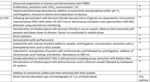
GC/FID Whole blood 3.11 Direct thermal desorption gas chromatography of 1 µL of whole blood. This method Gas chromatography/differential mobility spectrometry (GC/DMS); Gas chromatography/mass spectroscopy (GC/MS); Gas chromatography flame ionisation detection (GC/FID); High performance liquid chromatography UV detection (HPLC/UV); Gas chromatography/Fourier transformer infrared detection (GC/FTIR); Gas
[image:6.842.255.765.68.348.2]chromatography/electron capture detection (GC/ECD); Thin layer chromatography (TLC).
7 Experimental
Chemical and Reagents
All reagents were obtained from Sigma-Aldrich (Gillingham, UK) unless otherwise stated. Deionised water was obtained from a Purite RO200–Stillplus HP System, (Purite Oxon, UK). Defibrinated horse blood was obtained from Fisher Scientific (Loughborough, UK). Blood collection was carried out by the supplier in stress-free conditions by qualified, trained personnel in accordance with the Animals (Scientific Procedures) Act 1986.
Instrumentation
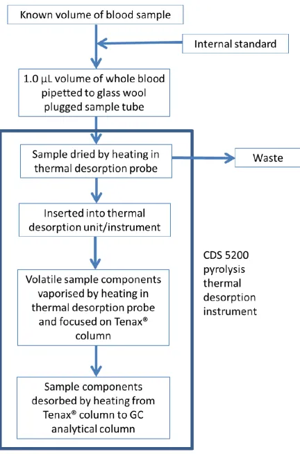
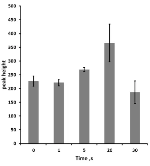
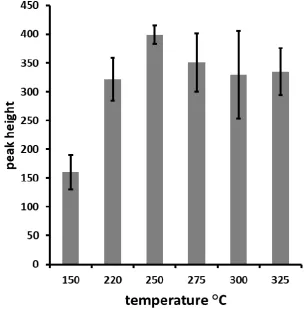
The CDS 5200 pyrolysis thermal desorption instrument conditions were as follows: Drying temperature 100 °C for 20 s; desorption temperature 250 °C for 2 min. Transfer line 250 °C, cleaning temperature, 1100 °C for 10 s. Gas chromatography analysis was carried out using an Agilent 6890 instrument with flame ionisation detection. Sample extracts were introduced via a CDS 5200 pyrolysis thermal desorption instrument (CDS Analytical, Oxford, PA) on to a HP-5 capillary column (15 m x 0.25 mm ID, 0.25 µm film thickness, 5 % diphenyl–95 % dimethylsiloxane phase). The GC oven temperature was maintained at 100 °C for 2 min and then programmed to 250 °C at 20 °C/min and finally held isothermally for 10.5 min at this temperature. The injector and flame ionisation detector were held at 250 °C and 260 °C respectively.
Sample Analysis
10 Results and Discussion
Proposed Analytical Procedure
Optimisation of Thermal Desorption Conditions
14 Precision, Calibration Plot, Limit of Detection and Quantification
Standard solutions containing ethylene glycol in the concentration range 0.05 - 500 mM were prepared in deionised water and determined by the optimized TD-GC procedure. This range was chosen as it covers the lower limit for antidote treatment through to severe toxic levels. 40 The calibration plot was found to be linear over the range 1.0 to 200 mM with an R2 value of 0.9997. The theoretical limit of detection, based on 3 σ, was calculated as 50.2 µM (3.11 mg/L) of ethylene glycol for a 1 µL sample and the limit of quantification was defined to be 1.0 mM (62.1 mg/L). It should be noted that larger sample volumes can be utilised and hence greater sensitivity can be obtained.
Studies of Possible Interferences
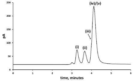
15 Compound RT, min RRT, min
methanol ND ND
ethanol ND ND
1,4-butandiol 4.19 1.16
1,2-butandiol 3.60 1.00
γ-butyrolactone 4.06 1.13
fomepizole 4.17 1.16
lactic acid 4.86 1.35
[image:15.595.190.407.71.213.2]ethylene glycol 3.20 0.89
Table 2. Retention times (RT) and relative retention times (RRT) (Analyte RT/Internal standard RT) for possible interfering compounds. ND = not detected.
Figure 5. Gas chromatogram showing the separation of ethylene glycol (i) and the internal standard, 1,2-butanediol (ii) in presence of methanol (ND), ethanol (ND), 1,4-butanediol (iii), ɣ- butyrolactone (iv) and fomepizole (v); 45 mM of each compound. No further peaks were detected after 6 minutes. ND = not detected.
Analytical Application
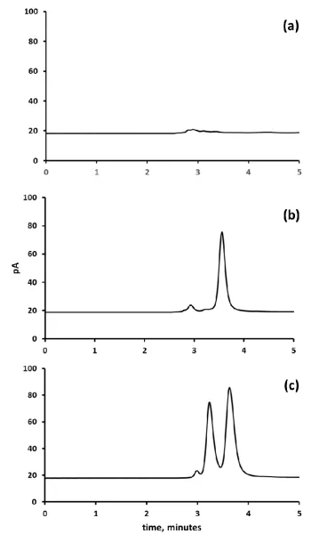
[image:15.595.77.521.297.568.2]16 external calibration. Figure 6 shows representative chromatograms for samples of whole blood (a) fortified with 31.2 mM ethylene glycol and (b) unadulterated whole blood.
Recoveries and precision data gained for the blood samples fortified with ethylene glycol are summarized in Table 3. These levels were selected as previous investigations have shown blood samples taken from human and animal victims to contain levels of ethylene glycol over this range.
13,22,23,41
18 Blood
Sample
Native Added, mM Mean Found, mM
% Mean Recovery
%CV
1 ND 12.5 10.5 84.8 4.4
2 ND 20.0 19.5 96.7 2.3
3 ND 32.2 29.4 94.3 5.8
4 ND 100 107 107 3.9
5 ND 200 209 105 1.7
[image:18.595.104.492.73.170.2]ND = not detected; %CV = percentage coefficient of variation
Table 3. Recovery and Precision Data for ethylene glycol obtained on whole blood
Conclusions
Our method requires very little sample preparation, requiring only the simple addition of internal standard to a whole blood sample. Other workers (table 1) have reported GC based assays for ethylene glycol, but these required rigorous extraction and derivatization procedures. Therefore, the overall time and skill required for these assays is much greater than our proposed assay, which is an important consideration in medical and forensic investigations. 7 In addition, a derivatization reagent itself and its reaction by-products might be expected to give responses with similar retention times to that of ethylene glycol itself.
19 Acknowledgements
We would like to thank the support of the Analytical Chemistry Trust Fund. Kevin Sudlow and Sarah Almond are thanked for their technical assistance.
Conflicts of Interest
There are no conflicts of interest to declare. References
[1] A. Vale, Medicine, 2012, 40, 89–93.
[2] R. C. Baselt, Disposition of Toxic Drugs and Chemicals in Man, 9th edition. Biomedical Publications, Seal Beach, CA, 2011, 637–640.
[3] National Poisons Information Service. National Poisons Information Service Report 2015/2016. National Poisons Information Service, 2016. 15 June 2017.
[4] J. B. Mowry, D. A. Spyker, D. E. Brooks, A. Zimmerman and J. L. Schauben, Clin. Toxicol. 2016, 54, 924-1109.
[5] J. Brent, Drugs, 2001, 61, 979-988.
[6] C. D. Peterson, A. J. Collins, J. M. Himes, M. L. Bullock and W. F. Keane, N. Engl. J. Med. 1981, 304, 21-23.
[7] J. Latus, M. Kimmel, M. Dominik Alscher and N. Braun, Clin. Kidney J. 2012, 5, 120–123. [8] J. A. Kraut, Clin. Toxicol. 2015, 53, 589–595.
[9] W. H. Porter, Clin. Chim. Acta 2012, 413, 365–377.
[10] K. L.Wallace, J. R. Suchard, S. C. Curry and C. Reagan, Ann. Emerg. Med. 2001, 38, 49–54. [11] J. B. Hack, W. K. Chiang, M. A. Howland and H. Patel, Acad. Emerg. Med. 2000, 7, 294–297. [12] Z. Siroka, R. Bily, V. Polacek, J. Dvorak and M. Svoboda, Vet. Med.-Czech. 2014, 59, 388–395. [13] T. G. Rosano, T. A. Swift, C. J. Kranick and M. Sikirica, J. Anal. Toxicol. 2009, 33, 508-513. [14] P. Van hee, H. Neels, M. De Doncker, K. E. Maudens, W. Lambert and L. Patteet, Clin. Chim.
Acta, 2013, 415, 107-108.
[15] R. H. Williams, S. M. Shah, J. A. Maggiore and T. B. Erickson, J. Anal. Toxicol. 2000, 24, 621-626.
[16] S. Yamamoto, K. Ueda, M. Noami and Y. Nokami, Japn. J. Sci. Tech. Identif. 2000, 5, 55-60. [17] L. Criado-García, D. M. Ruszkiewicz, G. A. Eiceman and C. L. P. Thomas, J. Breath Res. 2016,
10, 017101.
[18] C. Giachetti, G. Zanolo, A. Assandri and P. Poletti, Biomed. Environ. Mass Spectrom. 1989, 18, 592–597.
[19] W. H. Porter, P. W. Rutter and H. Ya, J. Anal. Toxicol. 1999, 23, 591-597.
[20] A. F. Robson, A. J. Lawson, L. Lewis, A. Jones and S. George, Ann. Clin. Biochem. 2017, 54, 481-489.
[21] A. Wurita, O. Suzuki, K. Hasegawa, K. Gonmori, K. Minakata, I. Yamagishi, H. Nozawa and K. Watanabe, Forensic Toxicol. 2013, 31, 272–280.
[22] T. Hložek, M. Bursová and R. Čabala, Clin. Biochem. 2015, 48, 189–191.
[23] K. Aarstad, O. Dale, O. Aakervik, S. Øvrebø and K. Zahlsen, J. Anal. Toxicol. 1993, 17, 218-221. [24] T. Hložek, M. Bursová and R. Čabala, Talanta, 2014, 130, 470-474.
[25] N. B. Smith, Clin. Chim. Acta, 1984, 162, 269–272.
20 [27] D. J. Orton, J. M. Boyd, D. Affleck, D. Duce, W. Walsh and I. Seiden-Long, Clin. Biochem. 2016,
49, 132–138.
[28] T. P. Hewlett, A. C. Ray and J. C. Reagor, J. Assoc. Off. Anal. Chem. 1983, 66, 276–283. [29] J. H. Riley, S. O’Brien and M. G. Riley, Vet. Hum. Toxicol. 1982, 24, 331–334.
[30] A. A. Brega, P. Quadri, P. Villa, P. Prandini, J. – Q. Wei, and C. Lucarelli, J. Liquid Chromatogr. 1992, 15, 501–511.
[31] M. Holcapek, H. Virelizier, J. Chamot-Rooke, P. Jandera and C. Moulin, Anal. Chem. 1999, 71, 2288-2293.
[32] L. Imbert, E. Saussereau and C. Lacroix, J. Anal. Toxicol. 2014, 38, 676–680.
[33] US EPA (1986) Standard scenarios for estimating exposure to chemical substances during use of consumer products. Volumes I and II. Prepared by Versar Inc. for Exposure Evaluation Division, Office of Toxic Substances, US Environmental Protection Agency ,Washington, DC (EPA Contract No. 68-02-3968).
[34] A. Dasgupt, W. Blackwell, J. Griego and S. Malikc, J. Chromatogr. B 1995, 666, 63-70.
[35] L. L. Needham, R. H. Hill, D. L. Orti, M. E. Felver and J. A. Liddle, J. Chromatogr. 1982, 233, 9– 17.
[36] A. D. Fraser and W. MacNeil, J. Toxicol. Clin. Toxicol. 1993, 31, 397-405.
[37] K. Andersson, J. – O. Levin, R. Lindahl and C. – A. Nilsson, Chemosphere 1982, 11, 1115-1119. [38] K. Gras, K. Luong, M. Lin, M. Gras and R. A. Shellie, Anal. Methods, 2015, 7, 5545–5550. [39] S. A. Williamson and W. G. Verson, J. Am. Soc. Brew. Chem. 1993, 51, 51:0114.