MEDICAL IMAGEFUSION USING DISCRETE WAVELET TRANSFORM (DWT) AND
DUAL TREE COMPLEX WAVELET TRANSFORM (DT
1,*
Sumit Narayan Jarholiya
1
Research Scholar,
2
Faculty of Engineering and Technology, JayotiVidhyapeeth Women’s University, Jaipur, India
ARTICLE INFO ABSTRACT
Image fusion is defined as the process of combing more than one image from various environments of same scene into a single image. Th
information contents are increased in the fused image. It helps to diagnose the diseases like tumor, cancer, fracture in bones, ulcer and stones in the body etc. effectively. Discrete Wavele
(DWT) is easy to implement, reduces the computation time, resources required and it also provides energy compaction, larger SNR and More accurate clinical information for medical diagnosis & evaluation. Dual Tree Complex Wavelet Transform (DT
and shift invariance feature. Hence medical image fusion with the combination of DWT and DT will provide very good quality of fused image and efficiency of fused image will increase significantly. Various fusi
evaluation of CT, MRI, and / or PET images.
Copyright©2017, Sumit Narayan Jarholiya and Dr. Shachi Awasthi
which permits unrestricted use, distribution, and reproduction in any medium, provided the original work is properly cited.
INTRODUCTION
Image fusion is defined as the process of combing more than one image from various environments of same scene into a single image (Roshan et al., 2015; Lakhwinder Singh 2015). The main objective of this technique is to obtain an image that is more suitable for visual perception
computer processing tasks (AvishekSen et al.,
and Meenakshi, 2015; RajkumarSoundrapandiyan
The fused image should have more complete information which is more useful for human or machine perception. The advantages of image fusion are (AvishekSen
Improving reliability Capability.
Improve the quality and increase the application of these data.
Image fusion techniques allow the integration of different information sources.
The goal of image fusion (IF) is to integrate complementary multisensor, multiview and multitemporal information into one
*Corresponding author: Sumit Narayan Jarholiya
Research Scholar, JayotiVidyapeeth Women’s University, Jaipur, India.
ISSN: 0975-833X
Vol.
Article History:
Received 03rd November, 2016
Received in revised form 10th December, 2016
Accepted 26th January, 2017
Published online 28th February,2017
Key words:
Discrete Wavelet Transform (DWT), Dual Tree Complex Wavelet Transform (DT-CWT) , CT, MRI and / or PET Image, Image registration, Image Resizing / Resampling, Image Enhancement, PSNR, MSE, SD and Mean.
Citation: Sumit Narayan Jarholiya and Dr. Shachi Awasthi
complex- wavelet transform (DT-CWT)”, International Journal of Current Research
RESEARCH ARTICLE
MEDICAL IMAGEFUSION USING DISCRETE WAVELET TRANSFORM (DWT) AND
DUAL TREE COMPLEX WAVELET TRANSFORM (DT-CWT)
Sumit Narayan Jarholiya and
2Dr. Shachi Awasthi
Research Scholar, JayotiVidyapeeth Women’s University, Jaipur, India
Faculty of Engineering and Technology, JayotiVidhyapeeth Women’s University, Jaipur, India
ABSTRACT
Image fusion is defined as the process of combing more than one image from various environments of same scene into a single image. The resultant fused image gives more accurate information therefore information contents are increased in the fused image. It helps to diagnose the diseases like tumor, cancer, fracture in bones, ulcer and stones in the body etc. effectively. Discrete Wavele
(DWT) is easy to implement, reduces the computation time, resources required and it also provides energy compaction, larger SNR and More accurate clinical information for medical diagnosis & evaluation. Dual Tree Complex Wavelet Transform (DT-CWT) provides better image visual eminence and shift invariance feature. Hence medical image fusion with the combination of DWT and DT will provide very good quality of fused image and efficiency of fused image will increase significantly. Various fusion applications have appeared in medical imaging like simultaneous evaluation of CT, MRI, and / or PET images.
Sumit Narayan Jarholiya and Dr. Shachi Awasthi. This is an open access article distributed under the Creative Commons Att use, distribution, and reproduction in any medium, provided the original work is properly cited.
Image fusion is defined as the process of combing more than one image from various environments of same scene into a Lakhwinder Singh et al.,
The main objective of this technique is to obtain an image that is more suitable for visual perceptionand the
et al., 2016; Nandeesh RajkumarSoundrapandiyan et al., 2016). The fused image should have more complete information which is more useful for human or machine perception. The
et al., 2016):
Improve the quality and increase the application of
Image fusion techniques allow the integration of
The goal of image fusion (IF) is to integrate complementary multisensor, multiview and multitemporal information into one
Sumit Narayan Jarholiya
Research Scholar, JayotiVidyapeeth Women’s University, Jaipur,
new image containing information the quality of which cannot be achieved otherwise. There are three types of fusion operators are used for wavelet image fusion: pixel, area and region. Pixel operators can quickly get a fused image, but the image can be blurred. Area operators consider the neighboring gray value, which can reduce the edge's sensitivity and the fused image has better vision characteristics. Region operators should operate image segment to source image. For fulfilling the aim of image fusion area operator will be used
et al., 2012). Image fusion has been used in many application areas like auto target recognition, computer vision, concealed weapon detection, remote sensing, astronomy, multisensory biometrics, automatic change detection,
navigation aid and medical imaging etc Lakhwinder Singh et al., 2015).
have appeared in medical imaging like simultaneous evaluation of CT, MRI, and/or PET images.
main focus of medical image fusion is on the anatomic image and the functional image, such as CT/PET, MRI/PET haiXu et al., 2012). Typically, the field of medical image analysis is divided into six categories:
Post-acquisition: Preprocessing techniques such
denoising and restoration are used to restore the images so that they can be used for diagnosis.
Available online at http://www.journalcra.com
International Journal of Current Research
Vol. 9, Issue, 02, pp.46224-46231, February, 2017
INTERNATIONAL
OF CURRENT RESEARCH
Jarholiya and Dr. Shachi Awasthi, 2017. “Medical image fusion using discrete wavelet tr International Journal of Current Research, 9, (02), 46224-46231.
z
MEDICAL IMAGEFUSION USING DISCRETE WAVELET TRANSFORM (DWT) AND
CWT)
Dr. Shachi Awasthi
JayotiVidyapeeth Women’s University, Jaipur, India
Faculty of Engineering and Technology, JayotiVidhyapeeth Women’s University, Jaipur, India
Image fusion is defined as the process of combing more than one image from various environments of e resultant fused image gives more accurate information therefore information contents are increased in the fused image. It helps to diagnose the diseases like tumor, cancer, fracture in bones, ulcer and stones in the body etc. effectively. Discrete Wavelet Transform (DWT) is easy to implement, reduces the computation time, resources required and it also provides energy compaction, larger SNR and More accurate clinical information for medical diagnosis &
CWT) provides better image visual eminence and shift invariance feature. Hence medical image fusion with the combination of DWT and DT-CWT will provide very good quality of fused image and efficiency of fused image will increase on applications have appeared in medical imaging like simultaneous
is an open access article distributed under the Creative Commons Attribution License, use, distribution, and reproduction in any medium, provided the original work is properly cited.
image containing information the quality of which cannot There are three types of fusion operators are used for wavelet image fusion: pixel, area and region. Pixel operators can quickly get a fused image, but the d. Area operators consider the neighboring gray value, which can reduce the edge's sensitivity and the fused image has better vision characteristics. Region operators should operate image segment to source image. For fulfilling operator will be used (Zhi-haiXu Image fusion has been used in many application target recognition, computer vision, concealed remote sensing, astronomy, multisensory biometrics, automatic change detection, machine vision, and medical imaging etc (Roshan et al., 2015; ). Numerous fusion applications have appeared in medical imaging like simultaneous evaluation of CT, MRI, and/or PET images. At present, the us of medical image fusion is on the anatomic image and the functional image, such as CT/PET, MRI/PET
(Zhi-Typically, the field of medical image analysis is divided into six categories:
Preprocessing techniques such as denoising and restoration are used to restore the images so that they can be used for diagnosis.
INTERNATIONAL JOURNAL OF CURRENT RESEARCH
Segmentation: Image segmentation is a process where the image can be partitioned into cluster of pixels which are similar based on some criteria. Different groups must not interact with each other, and neighboring cells can be compared. The result of segmentation is the splitting up of the image into connected areas. Thus segmentation is concerned with dividing an
meaningful regions (Sakthivel et al.,
such as brain MRI or abdomen CT scan contain multiple features (organs). Delineating features of interest is important for analysis and accurate diagnosis.
• Registration: In computer assisted
required to register or align the captured image with a model or a previously captured image.
Computation: Physical quantity derivation and other
computation such as fusion and compression are also required in several computer assisted thera
• Visualization: It is important to display medical images
on screen so that a medical professional can diagnose diseases.
• Security: Personal medical health-care information is
very sensitive and it is very important to secure it using techniques such as watermarking so that only legitimate user can access it and also accurately associate with the medical record with the correct patient.
The problems about comparison and synthesis between image CT, SPECT and MRI were frequently encountered. Doctor could easily find the position of illness after medical image fusion. Doctors manually combine the CT and MRI medical images of a patient with a tumor to make a more accurate diagnosis but it is very difficult task to complete this job and more important is doctors with different experience make inconsistent decisions. Manual process is subject to human error, and requires years of experience and time consuming. Thus, it is necessary to develop the efficiently automatic image fusion system to decrease doctor’s workload and improve the consistence of diagnosis (Lakhwinder Singh
Iterative image fusion technique is very useful in medical imaging (Lakhwinder Singh et al., 2015).
fusion is the idea to improve the image content by fusing images taken from different imaging tools like Computed Tomography (CT), Magnetic Resonance Imaging (MRI),Positron Emission Tomography (PET) and Single Photon Emission Computed Tomography (SPECT
Shanker Mishra and SmritiBhatnagar, 2014
approach of Medical imaging is Image guided interventions and therapy (IGIT) sometimes it is also called
therapy (IGT) or image-guided intervention (IGI) or image guided surgery (IGS) (Daniel Ruijters, 2010;
al.,). The minimally invasive nature of Image guided interventions and therapy (IGIT) is often preferred over open surgeries because less trauma to the patient’s body is caused, which generally is associated with easier and faster recovery (Daniel Ruijters, 2010).
Different types of medical images
In Medical field there are different types of medical scan available. Some of the examples of medical scan are image, MRI image, PET image and SPECT image.
Computed Tomography (CT) Image
CT images are used more often to as certain differences in tissue density depending upon their ability to block X
Image segmentation is a process where the image can be partitioned into cluster of pixels which similar based on some criteria. Different groups must not interact with each other, and neighboring cells segmentation is the splitting up of the image into connected areas. Thus segmentation is concerned with dividing an image into
et al., 2016). Images such as brain MRI or abdomen CT scan contain multiple features (organs). Delineating features of interest is important for analysis and accurate diagnosis. In computer assisted surgery, it is register or align the captured image with a model or a previously captured image.
Physical quantity derivation and other computation such as fusion and compression are also required in several computer assisted therapy.
It is important to display medical images on screen so that a medical professional can diagnose
care information is very sensitive and it is very important to secure it using watermarking so that only legitimate can access it and also accurately associate with the medical record with the correct patient.
The problems about comparison and synthesis between image CT, SPECT and MRI were frequently encountered. Doctor easily find the position of illness after medical image fusion. Doctors manually combine the CT and MRI medical images of a patient with a tumor to make a more accurate diagnosis but it is very difficult task to complete this job and ors with different experience make inconsistent decisions. Manual process is subject to human error, and requires years of experience and time consuming. Thus, it is necessary to develop the efficiently automatic image rkload and improve the Lakhwinder Singh et al., 2015). Iterative image fusion technique is very useful in medical 2015). Medical image fusion is the idea to improve the image content by fusing ges taken from different imaging tools like Computed Tomography (CT), Magnetic Resonance Imaging (MRI),Positron Emission Tomography (PET) and Single Photon Emission Computed Tomography (SPECT) (Hari Om , 2014). Another approach of Medical imaging is Image guided interventions and therapy (IGIT) sometimes it is also called image-guided guided intervention (IGI) or
image-, 2010; Simon DiMaio et
minimally invasive nature of Image guided interventions and therapy (IGIT) is often preferred over open surgeries because less trauma to the patient’s body is caused, ith easier and faster recovery
In Medical field there are different types of medical scan are available. Some of the examples of medical scan are CT image, MRI image, PET image and SPECT image.
often to as certain differences in density depending upon their ability to block X-rays
(Roshan et al., 2015). CT can clearly reflect the anatomical structure of bone tissues. It provides detailed cross sectional views of all types of tissues (Lakhw
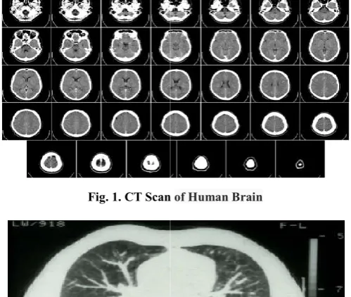
[image:2.595.311.559.128.636.2]CT Image provides the best information on denser / hard tissue with less distortion (Hari Om Shanker Mishra and Smriti Bhatnagar, 2014; Swathi et al.,
Fig. 1. CT Scan
[image:2.595.309.559.142.353.2]Fig. 2. CT Scan of Chest
Fig. 3 CT Scan of Abdomen
Magnetic Resonance Imaging (MRI)
MRI provides good contrast between the different soft tissues with more distortion of the body, which make it especially useful in detecting brain tissues, and cancer
2015). MRI can clearly reflect the anatomical structure of soft tissues, organs and blood vessels. And with high spatial resolution can provide anatomical structure information of organs (Lakhwinder Singh et al.,
Positron Emission Tomography (PET)
A PET image shows chemical and other on comparing to that of CT and MRI images 2016).
CT can clearly reflect the anatomical structure of bone tissues. It provides detailed cross sectional Lakhwinder Singh et al., 2015). A CT Image provides the best information on denser / hard tissue Hari Om Shanker Mishra and Smriti
2016).
CT Scan of Human Brain
[image:2.595.312.557.342.594.2]CT Scan of Chest
Fig. 3 CT Scan of Abdomen
Magnetic Resonance Imaging (MRI) Image
MRI provides good contrast between the different soft tissues with more distortion of the body, which make it especially detecting brain tissues, and cancer (Roshan et al.,
MRI can clearly reflect the anatomical structure of soft tissues, organs and blood vessels. And with high spatial resolution can provide anatomical structure information of
et al., 2015)
Positron Emission Tomography (PET) Image
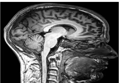
Fig. 4. MRI Scan of Normal Human Brain
Fig. 5. Full Body MRI Scan
Fig. 6. PET scan of Brain
Single Photon Emission Computed Tomography (SPECT) Image
[image:3.595.39.290.257.431.2]A Single Photon Emission Computed Tomography (SPECT) scan is a type of nuclear test that shows how blood flow changes in brain, tissues and organs.
Fig. 7. SPECT Image
Preprocessing for image fusion
Preprocessing and improvement techniques are used to improve the detection of the suspicious region. The preprocessing and enhancement method consists of two steps; first the removal of film vestiges such as labels and X-ray marks. Second the removal of high frequency components (Arnavi et al., 2016). Two images taken at different angles of the same scene or different times from different sensors, or from different viewpoints sometimes cause distortion. So before fusing the images, we have to make sure that both the images are spatially aligned and have the same dimensions (MedhaBalachandraMule and Padmavathi, 2015). The pre-processing can be the best solution of it. When MRI images are viewed on computer screen, theylook like black and white but in actual they contain someprimary colors (RGB) content. So, for further processing of MRI brain image, it must be converted to perfect grayscaleimage in which the red, green and blue components all haveequal intensity in RGB space.
[image:3.595.308.558.480.559.2]The original MRI brain image has properties 320x320x3 and conversion to grayscale image makes the properties 320x320. This step is carried out to improve the quality of the imageto make it ready for further processing. This improved andenhanced image will help in detecting edges and improving the quality of the overall image (Sakthivel et al., 2016). Image pre-processing is used to reduce noise and prepare the images for further steps such as segmentation. It diminishes distortion in image and enhances the relevant features. Thus a rectified image is obtained. For this purpose, MATLAB software has been used (Neelima Singh and Asuntha, 2016; Vaishnavi and Uma, 2016). The basic steps used in image fusion are, given in Fig.8 below (Gopal Kumar and Manish Trivedi, 2016; Medha Balachandra Mule and Padmavathi, 2015). The pre-processing includes imageregistration, image resizing and image enhancement (Deron Rodrigues et al., 2014).
Fig. 8. Pre-processing of image fusion
Image Registration
The images which are obtained by different modalities might be of different orientations and hence are needed to be registered before they are fused (Deron Rodrigues et al., 2014). Image registration transforms different set of data into one coordinate system. Image registration or image alignment algorithms can be classified into intensity-based and feature based registration. One of the images is referred to as the source image and the others are respectively referred to as the target images (Medha Balachandra Mule and Padmavathi, 2015). Intensity-based method uses correlation metrics to compare intensity patterns in the images. While feature-based method compare the features such as points, lines, and contours in the images to be registered (Medha Balachandra Mule and Padmavathi, 2015). Image registration will repair /remove the problem of distortion (Gopal Kumar and Manish Trivedi, 2016).
[image:3.595.35.294.671.783.2]Image Resizing / Resampling
Also the sizes of the images might vary so before fusion, the images are needed to be resized so that both the images are of the same size. This is done by interpolating the smaller size image by rows and columns duplication (Deron Rodrigues et al., 2014). Image resampling is the process of changing the pixel dimension of an image. For performing image fusion images should have same pixel dimensions. Resampling changes the image dimension, so that the images to be fused will be of same dimension. Down sampling an image means that information is deleted from the image. Similarly up sampling an image means that new pixels are added based on color and intensity values of existing pixels. Image resampling can be performed by using the methods such as nearest neighbor, bilinear, and bicubic (cubic convolution) (Medha Balachandra Mule and Padmavathi, 2015). Re-sampling in order to assure that the image coordinate system is correct (ShahidEqbal and Ansari, 2015).
Image Enhancement
The principal objective of image enhancement is to modify attributes of an image to make it more suitable for a given task and a specific observer. During this process, one or more attributes of the image are modified. Filtering is technique for enhancing the image (Sakthivel et al., 2016). If both or any of the images are not of grayscale then it is desired that it is converted to grayscale. The next step which follows this is equalization of thehistograms of the images so that the contrast of the image is enhanced and that both the images have similar range of values for wavelet coefficients (Deron Rodrigues et al., 2014). The various image enhancement techniques can be categorized as spatial domain methods and Frequency domain methods. Different image enhancement techniques are used for all the different images. This includes smoothing of image and removal of noises, blurring etc. Gabor filter was found to be suitable for both the CT and MRI images. The filtering of image proves to be useful for further steps (Neelima Singh and Asuntha, 2016). Contrast enhancement to assure that relevant information can be detected (ShahidEqbal and Ansari, 2015).
Performance evaluation parameters
The measurement and analysis on the fused images are done both objective as well as subjective quality measures. This effectively helps in better assessment of the information in the images (Rajkumar Soundrapandiyan et al., 2016). Even though wavelets sharing same properties in nature, the fusion result are different. This is because of their uniqueness in image decomposition as well as the image reconstruction characteristics. Entropy (H), Root Mean Square Error (RMSE), Peak Signal to Noise Ratio (PSNR), Mean Square Error (MSE), Standard Deviation (SD) and Mean ( ) are used as performance evaluation parameters (Rajesh Bhandari and Shivakumar, 2016).
Entropy (H)
Entropy indicates the amount of information contents present in the image. If the entropy is higher in the fused imagethen it indicates that fused image carrying greater information compare to the source image. The larger the value of entropy, better the fusion results (Nandeesh and Meenakshi, 2015; Rajesh Bhandari and Shivakumar, 2016). Entropy is a measure
of the uncertainty in a random variable. Higher entropy values are extracted from homogeneous scenes, and lower ones are from inhomogeneous scenes (Vaishnavi and Uma, 2016). Entropyof an image designates the information content of the merged image and hence its value must be high (Indira et al.,
2016). The fusion quality entropy is, the moreabundant of information the fusion image contains.The entropy is defined as follows (Zhi-haiXu et al., 2012; Nandeesh and Meenakshi, 2015),
L-1
H= −∑ Pl log2P l ………..…… (1)
l=0
Pl = Probability of fused image pixels, l = 0 to 255 (Asokan et
al., 2016)
Root Mean Square Error (RMSE)
It is a method to measure the differences between values predicted by an ideal (reference) image and the fused images. It is calculated as
w h
RMSE= √ (1/w x h )∑ ∑ (Rij – Fij )2 ……….… (2)
i=1 j=1
RMSE = 1/( × ℎ) ∑ ∑ ( − ) …….… (3)
Here, Rij = Intensity value of the reference (original) image at
coordinate i,j, Fij= Intensity value of fused image at
coordinates i, j, w =Width of the image and h= Height of the image.
It also presents the amount of deviation present in the fused image (Fij) compared to source image (Rij).RMSE value should
aslow as possible for the better quality image (Rajesh Bhandari and Shivakumar, 2016). RMSE for the reference and fused images will increase with decrease in similarity, and approaches zero whenever they are similar (Rajkumar Soundrapandiyan et al., 2016).
Peak Signal to Noise Ratio (PSNR)
PSNR is the measure of ratio between the maximum value of an image and the magnitude of the noise present in the image background. Normally PSNR is used to measure the reconstruction quality of fused image. It indicates the similarity between two images. The higher PSNR value gives the better fused image (Nandeesh and Meenakshi, 2015; Rajesh Bhandari and Shivakumar, 2016; Indira et al., 2016).
It is a method used to measure the quality of the fused image with respect to the reference image. It is defined as (Roshan et al., 2015):
PSNR = 10 log 10(MAX2 / MSE) ………..……..…….… (4)
Mean Square Error (MSE)
MSE is a frequently used measure of the difference between original image and fused image pixels. MSE is defined as image with size of given by below equation (Asokan et al.,
2016),
MSE = 1/( × ℎ) ∑ ∑ ( − ) ..…..… (5)
Here, Rij = Intensity value of the reference (original) image,
Fij= Intensity value of fused image, w =Width of the image and
h= Height of the image (Nayera Nahvi and Onkar Chand Sharma, 2014).
Standard Deviation (SD)
It shows the extent of variation or dispersion from the average or mean. Standard deviation takes into account the original image and the acquired transmission noise. Absence of any noise in the transmitted image increases its effectiveness and portraits the image’s contrast (Rajkumar Soundrapandiyan et al., 2016). The Standard deviation is a measure that is used to quantify the amount of variation of a fused image values. SD can be calculated as (Asokan et al., 2016).
SD = 1/( × ℎ) ∑ ∑ ( − ) ...…….… (6)
Where Fij = Intensity value of fused image,μ = Mean,
w =Width of the image and h= Height of the image.
Mean ( )
The mean intensity estimates the luminance of afused image. This is deduced by (Rajkumar Soundrapandiyan et al., 2016)
MEAN ( ) = 1/( × ℎ) ∑ ∑ | | ...…….… (7)
The large value of Mean (μ) means that thepixel values in the reconstructed image aremore deviated from actual pixel value. Larger value of Mean (μ) indicates imageis of poor quality (Roshan et al., 2015).
IMAGE FUSION METHODS
There are many image fusion methods that can be used to produce resolution multispectral images from a high-resolution panchromatic image and low-resolution multispectral images (ShahidEqbal and Ansari, 2015). The term CBIR (content-based image retrieval) designates the retrieval of images based on their visual similarity to a user-supplied query image or to a set of user-specified image features. CBIR can be widely used in many areas, such as photograph archives, medical diagnosis, the military, retail catalogs, and remote sensing systems. There are so many image fusion methods are available. Most of these image fusion methods are as follows:
Wavelet Transform Fusion
The Discrete Wavelet Transform ( DWT)
The Dual Tree Complex Wavelet Transform (DTCWT) Pixel Based Wavelet Transform Fusion
Edge Based Wavelet Transform Fusion Operator Based Image Fusion Method
Fusion of Multimodal Brain Images using Redundant Discrete Wavelet Transform (RDWT)
Image Fusion Using The Convolution Of Meridian Distributions
Image Fusion Method Based On Principal Component Analysis (PCA)
Image Fusion Method Based On Brovey Transform Image Fusion Method Based On Ehlers Transform Simple Average Based Image Fusion
Image Fusion Based On Radon Transform
Shift Invariant Discrete Wavelet Transform For Image Fusion (SIDWT)
Fusion Of Images In Multi Resolution Domain Complex Wavelet Image Fusion
Morphological Methods Knowledge Based Methods
Neural Network Based Image Fusion Methods Fuzzy Logic Based Image Fusion Methods Discrete Cosine Transform (DCT)
Daubechies Discrete Wavelet Transform Based Image Fusion
Symlet Based Image Fusion
Image Fusion Technique Using SVD
Image Fusion For Radiotherapy, Prostate Biopsy And Lesion Targeted Prostate Intervention and many more
Wavelet Transform Fusion
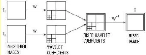
Wavelet transforms are linear transforms whose basis functions are called wavelets (AvishekSen et al., 2016). In all wavelet based image fusion schemes the wavelet transforms W of the two registered input images I1(x,y) and I2(x,y) are
computed and these transforms are combined using some kind of fusion rule∅ (Fig 5). Then the inverse wavelet transform W−1is computed and the fused image I(x,y)is reconstructed (Lakhwinder Singh et al., 2015; NayeraNahvi and Onkar Chand Sharma, 2014).
I (x,y) = W−1 {∅ (W (I1(x,y)), W (I2))) } ………....…..… (8)
[image:5.595.309.557.613.706.2]Where x= Scaling parameter and y= Location parameter. Wavelet means “small wave” so wavelet analysis is about analyzing signal with short duration finite energy functions (Hari Om Shanker Mishra and SmritiBhatnagar, 2014). Wavelet Transform is reduces the storage cost but it is not able to maintain edge information efficiently.
Fig. 9.Fusion of the wavelet transforms of two images
For a given scaling parameter x, we translate the wavelet by varying the parameter y. we define the wavelet transform as
W(x,y)= ∫ f(t) { 1/ √|x| }∅ ((t-y)/x) ..…………....…..… (9)
According equation (9), for every (x, y), we have a wavelet transform co-efficient, representing how much the scaled wavelet is similar to the function at location, t = y/x. “If scale(x) and position(y) is varied very smoothly, then transform is called continuous wavelet transform” (Hari Om Shanker Mishra and SmritiBhatnagar, 2014). “If scale(x) and position(y) are changed in discrete steps, the transform is calleddiscrete wavelet transform” (Hari Om Shanker Mishra and SmritiBhatnagar, 2014).
Discrete Wavelet Transform (DWT)
The Discrete Wavelet Transform (DWT), which is based on sub-band coding is found to yield a fast computation of Wavelet Transform. It is easy to implement and reduces the computation time and resources required. The Discrete Wavelet Transform (DWT) also converts the image from the spatial domain to frequency domain (Roshan et al., 2015). It provides some other advantages over other simple methods, like: energy compaction, larger SNR, reduced features, etc. The discrete wavelet transform DWT is a spatial frequency decomposition that provides a flexible multi resolution analysis of an image in one dimension. The aim of the wavelet transform is to represent the signal as a superposition of wavelets, If a discrete signal is represented by f(t) its wavelet decomposition is then given by
f( t) = ∑ , , , ( ), ……..…………....…..… (10)
Where m, n( t) is the dilated and or translated version of the mother wavelet given by the equation
, (t) = 2 –m /2 [2 -m t –n ] ……..…………....…..… (11)
[image:6.595.309.559.50.190.2]Where m and n are integers (NayeraNahvi and Deep Mittal, 2014). This ensures that the signal is decomposed into normalized wavelets at octave scales (NayeraNahvi and Deep Mittal, 2014). It also provide More accurate clinical information for medical diagnosis & evaluation but it offers Poor directionality, Shift sensitivity, Absence of phase information (Swathi et al., 2016). The DWT decomposition works on the cascade of special low pass and high-pass filters and a sub-sampling operation (Swathi et al., 2016). Fig. 10 shows image fusion using DWT (Nalini et al., 2016).
Fig.10. Image fusion using DWT
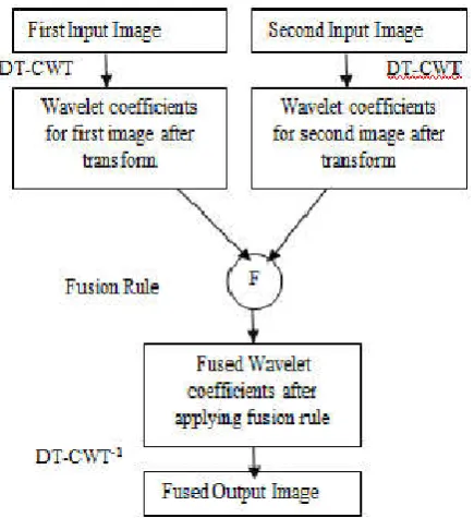
The Dual Tree Complex Wavelet Transform (DT-CWT)
In this method, fusion is executed using the masks to remove information from the decomposed structure of DT-CWT. Fig11 demonstrates the complex transform of a signal using two split DWT decompositions: Tree a and Tree b (VinaySahu and Dinesh Sahu, 2014).
Fig. 11. DT-CWT Structure
[image:6.595.325.543.466.704.2]It can be observed that the DT-CWT structure, involves both real and complex coefficients. It is known that DT-CWT is relevant to visual sensitivity. Fusion procedure involves the formation of a fused pyramid using the DT-CWT coefficients which are obtained from the decomposed pyramids of the source images. The used image is obtained through conventional inverse dual tree complex wavelet transform or reconstruction process. This results show a significant reduction of distortion (VinaySahu and Dinesh Sahu, 2014). The dual tree complex wavelet transform (DT-CWT) iteratively applies separable spatial filters to produce frequency sub bands in a similar way to the classic discrete wavelet transform. The prime motivation for producing the dual tree complex wavelet transform was shift invariance. In normal wavelet decomposition small shifts of the input signal are able to move energy between output sub bands. This is a result of the sub sampling necessary for critical decimation. Its advantage is to provide better image visual eminence and shift invariance feature. its disadvantage is that it has limited directionality but better than DWT (Swathi et al., 2016). Fig 12 shows the image fusion process using the DT-CWT.
Fig. 12. The image fusion process using the DTCWT
RESULTS AND DISCUSSION
[image:6.595.44.283.567.694.2]46230 International Journal of Current Research, Vol. 9, Issue, 02, pp.46224-46231, February, 2017
Table 1. Comparison of different image fusion methods
S. No. Name of Method Advantages Disadvantages
1. Wavelet Transform It Reduces the storage cost. It is not able to maintain edge information efficiently.
2. Discrete Wavelet
Transform (DWT )
1. It is easy to implement, reduces the computation time and resources required.
2 It provides energy compaction, larger SNR andMore accurate clinical information for medical diagnosis & evaluation.
It offers Poor directionality, Shift sensitivity, Absence of phase information and presence of spectral degradation.
3. Dual Tree Complex
Wavelet Transform (DT-CWT)
Itprovides better image visual eminence and shift invariance feature.
It has limited directionality and It introducesaliasing.
4. Pixel Based Wavelet
Transform
It gives contrast sensitivity of the human visual system. It is not suitable for Medical diagnosis since image can be blurred.
5. Edge Based Wavelet
Transform (I)Discrete Dyadic Wavelet Transform DDWT
It is applicable on two dimensional images and produces a hierarchy of edge features indexed by scale.
It can not be applicable for 3D images andorthogonal spline wavelets.
(II) Fusion of Images Using their Multiscale Edges
1. It reconstructs a very close and visually approximation of the input image.
2. It also stable for slightly modified of input image.
3. It is possible to suppress the nonlinear noise in the image as well as some light textures.
It does not provide the exact approximation of the input image.
6. Operator Based Image Fusion
Area operator is the best image fusion operator since it reduces the edge's sensitivity and the fused image has better vision characteristics.
Computation time is moderate.
7. Redundant Discrete
Wavelet
Transform(RDWT)
It provides better shift invariance rather than of DWT RDWT is a pixel based fusion techniques hence image can be blurred
8 Convolution Of Meridian Distributions
1. It provides improved fusion resultswhen compared to the Weighted Average (WA), FLOM, Flom Cauchy and CauchyConvolution fusion methods.
2. It also works like a feature detector,retaining thesalient features,well edges preservation and obtains higher metric values .
Its approach intends to maximize the cost function derived from the convolution of Meridiandistributions.
9. Principal Component
Analysis (PCA)
1.Arbitrary number of bands can be used 2. Simple algorithm.
3. It reduces the dimensionality of large data sets. 4.PCA shows fair amount of entropy.
PCA is a pixel based fusion techniques hence image can be blurred
and contrast of fused image is not good
10. Brovey transform It avoids the drawbacks of the multiplicative method. It is a pixel based fusion techniques hence image can be blurred and not suitable for Medical Image Fusion
11. Ehlers Transform This replaces the high frequencypart of the intensity component with that from the Pan image.
It is a pixel based fusion techniques hence image can be blurred and not suitable for Medical Image Fusion
12. Simple Average
BasedImage Fusion
It is very easy to understand and implement. High information loss
13. Radon Transform It is good to find linear features and able to transform two dimensional images with lines and it provide more information regarding medical image.
It is not able to transform three dimensional images and it is also not clear that the information is provided by this transform for two dimensional medical images are sufficient.
14. Shift Invariant Discrete
Wavelet Transform
(SIDWT)
It provides the better Shift Invariant feature of Image. Since its entropy is average hence information content present in the fused images are limited.
15. Image Fusion Based on Multi Resolution Domain
The fusion of images makes it possible to enrich information to be treated and fusion can be effective for certain system of imagery.
It can be proves to be ineffective fusion techniques for others system of imagery.
16. Complex Wavelet
Transform (CWT)
It provides magnitude or phase, shift invariant and free from aliasing.
It is Most expensive and computational intensive.
17. Morphological Image
Fusion Methods
It is used for noise removal and image smoothing without altering shapes and locations of objects in the image.
This method is highly sensitive to the inter-image variability.
18. Knowledge Based
Methods
The advantage is the ability to benchmark the images with the known human vision standards.
The drawback is the limitations imposed by human judgment in images that are prone to large pixel intensity variability.
19. Neural Network Based Image Fusion
It reduces development time and cost while improving performance
Neurons should be trained.
20. Fuzzy Logic Based Image Fusion Method
It provides advantage of efficient disease diagnoses, retrieval ofimages, undergo surgery treatment, tumoridentification etc.
The selection of membership functions and fuzzy sets are difficult.
21. Discrete Cosine Transform 1. It is Simple, fast & energyefficient multifocus image fusion scheme
2. It provides better result for JPEG Images.
It consumes more time than wavelet based method as two diff multiscale decomposition process are applied.
22. Daubechies Discrete
(db2)Wavelet Transform
It is able to manage different images resolution and provide better results than Haar Transform.
It considers only wavelet coefficient value and unable give better results than SIDWT and RDWT.
23. Symlet based image
fusion
Symlet Wavelets are very much similar to the Daubechies wavelets except the only difference being the vanishing moments of the wavelets function. Thus the wavelet coefficients differ than that of the daubechies.
Same as db2. Poor regularity is the only concern in case of Symlets.
24. Image Fusion Technique Using SVD
It is robust, simple andfast to implement. Efficiency is still improved if noise is reduced.
25. Image Fusion For
Radiotherapy, Prostate Biopsy And Lesion
Targeted Prostate
Intervention
1. Improved visualization and dexterity.
2. Reduction of radiation exposure to surgeon, e.g., by removing the surgeon’s hand fromthe fluoroscope field of view,
3. Provision of a “third hand”, e.g., to hold cameras, retractors, etc, 4. Increased accuracy in carrying out a surgical plan.
5. Improved safety via the use of virtual fixtures (“no fly” zones).
For evaluating the outcome various performance metrics were like Entropy(H), Root Mean Square Error (RMSE), Peak Signal to Noise Ratio(PSNR), Mean Square Error (MSE), Standard Deviation (SD) and Mean ( ). It is used to how much information carry from input images to fused image.
Conclusion
Finally this paper concludes that a image fusion based on combination of DWT and DT-CWT will provides better solution for medical diagnosis and clinical purpose.
REFERENCES
Arnavi A. Patil, S. S. Badhe and A. A. Mulajkar, 2016. “Brain Tumor Detection and its Area Measurement using K-Means Clustering baesd on Genetic Algorithm”,
International Journal of Science and Research (IJSR),
Volume 5 Issue 8, August, ISSN (Online): 2319-7064, pp 26-31.
Asokan, R., T.C. Kalaiselvi and M. Tamilarasi, 2016. “MedicalImage Fusion Using Stationary Wavelet Transform With Different Wavelet Families”, Pak. J. Biotechnol. Vol. 13 (special issue on Innovations in information Embedded and Communication Systems) Pp. 10 –14, (2016).
AvishekSen, SouvikChatterjee, MainakBiswas, SoumiBiswas and Pallab Kr Ghosh , 2016. “Robust Image Fusion Using Single Level DWT And SSIM”, International Journal of Scientific & Engineering Research, Volume 7, Issue 4, April, ISSN 2229-5518, pp 152-156.
Daniel Ruijters, 2010. “Multi-modal image fusion during minimally invasive treatment”, TechnischeUniversiteit Eindhoven, ISBN: 978-94-6018-174-0.
Deron Rodrigues, Hasan Ali Virani and ShajahanKutty, 2014. “Multimodal Image Fusion Techniques for MedicalImages using Wavelets”, International Journal of Research in Advent Technology, Vol.2, No.3, March, E-ISSN: 2321-9637, pp 310-313.
Dr. Sakthivel, K., B.R. Swathi, S.VishnuPriyan and C. Yokesh, 2016. “Analysis of Medical Image Processing and itsApplication in Healthcare”, International Journal of Advanced Engineering Research and Science (IJAERS)
Vol-3, Issue-2 , Feb, ISSN: 2349-6495,pp 25-29.
Gopal Kumar and Manish Trivedi, 2016. “Medical Image Fusion based on DWT and SPIHT Techniques with Quantitative Analysis”, International Journal of Computer Applications (0975 – 8887) Volume 147 – No.12, August, pp 5-8.
Hari Om Shanker Mishra and SmritiBhatnagar, 2014. “MRI and CT Image Fusion Based on Wavelet Transform”,
International Journal of Information and Computation Technology., ISSN 0974-2239 Volume 4, Number 1, pp. 47-52
Indira, K.P., R. Rani Hemamalini and N.M. Nandhitha, 2016. “Performance evaluation of DWT, SWT and NSCT for fusion of PET and CT Images using different fusion rules”,
Biomedical Research, 27 (1): 123-131, ISSN 0970-938X, pp 123-131.
Lakhwinder Singh, Dr. Sunil Agrawaland Mrs. Preeti Gupta, 2015. “Review On Medical Image Fusion Based On Neuro-Fuzzy Approach”, International Journal of Scientific Research Engineering & Technology (IJSRET),
Volume 4, Issue 7, July, ISSN 2278 – 0882 , pp 777-781.
MedhaBalachandraMule and Padmavathi, N.B. 2015. “Basic Medical Image Fusion Methods”, International Journal of Advanced Research in Computer Engineering & Technology (IJARCET), Volume 4 Issue 3, March, ISSN: 2278 – 1323, pp 1046-1049.
Nalini B. Kolekar and Prof. R.P. Shelkikar, 2016. “A Review on Wavelet transform based image fusion and classification”, International Journal of Application or Innovation in Engineering & Management (IJAIEM),
Volume 5, Issue 3, March, ISSN 2319 – 4847, pp 111-115. Nandeesh, M.D. and Dr. M. Meenakshi, 2015. “Image Fusion
Algorithms for Medical Images-A Comparison “, Bonfring International Journal of Advances in Image Processing, Vol. 5, No. 3, July, pp 23-26.
NayeraNahvi and Deep Mittal, 2014. “Medical Image Fusion Using Discrete Wavelet Transform”, Int. Journal of Engineering Research and Applications, ISSN : 2248-9622, Vol. 4, Issue 9( Version 5), September, pp.165-170 NayeraNahvi and Onkar Chand Sharma, 2014.
“Implementation of Discrete Wavelet Transform ForMultimodal Medical Image Fusion”, International Journal of Emerging Technology and Advanced Engineering, Volume 4, Issue 7, July, ISSN 2250-2459, pp 312-317.
Neelima Singh and A. Asuntha, 2016. “Image Processing used for Lung Cancer Detection in Medical Imaging”, Journal of Chemical and Pharmaceutical Research, 8(4), ISSN : 0975-7384,pp 1044-1049.
Rajesh Bhandari and Shivakumar B. R. 2016. “Wavelet based Analysis of Medical Image Fusion using MATLAB GUI”, International Journal of Innovative Research in Science, Engineering and Technology, ISSN(Online) : 2319- 8753, ISSN (Print) : 2347-6710, pp 512-517.
Rajkumar Soundrapandiyan, RishinHaldar, Swarnalatha Purushotham and ArvindPillai, 2016. “Multimodality Medical Image Fusion Using Block Based Intuitionistic Fuzzy Sets”, SPECIAL ISSUE (SCMDSA), IIOABJ, Vol.7, 5, ISSN: 0976-3104, pp85-94.
Roshan P. Helonde and Prof. M.R. Joshi , 2015. “Image Fusion Based on Medical Images Using DWT andPCA Methods”, International Journal of Computer Techniques, Volume 2 Issue 1, ISSN: 2394-2231 , pp 75-79.
ShahidEqbal and Dr. M. A. Ansari , 2015. “Medical Image Feature Extraction for Computer Aided Diagnosis of Lung Cancer”, International Journal of Advanced Research in Computer Science and Software Engineering, Volume 5, Issue 6, June, ISSN: 2277 128X, pp 193-197.
Simon DiMaio et al. “Challenges in Image-Guided Therapy System Design”, pp 1-28.
Swathi, P.S, Sheethal, M.S, Vince Paul, “Survey on Multimodal Medical Image Fusion Techniques”, IJCSET , January 2016 , Vol 6, Issue 1, ISSN:2231-0711, pp 33-39 Vaishnavi, B and Uma R. 2016. “A Hierarchical Model to
Classify Brain Cancer using GLCM”, International Journal of Science and Research (IJSR), ISSN (Online): 2319- VinaySahu and Dinesh Sahu, 2014. “Image Fusion using
Wavelet Transform: A Review”, Global Journal of Computer Science and Technology: F Graphics & Vision, Volume 14 Issue 5 Version 1.0, Year, Online ISSN: 0975-4172 & Print ISSN: 0975-4350, pp 20-28.
Zhi-haiXu, Ling-xiang Liu, Lei Tong, Lin-hong Zhou, and Chao-min Chen, 2012. “Wavelet medical image fusion algorithm based on local area feature”, Research Article/Biological and Biomedical Reports, (ISSN: 2162-4186), 2(1), 25-31.