Received 19 February 2017 Accepted 1 March 2017
Edited by C. Rizzoli, Universita degli Studi di Parma, Italy
Keywords:crystal structure; chalcone thiophene derivative; Hirshfeld surface analysis;in silico evaluation.
CCDC reference:1535563
Supporting information:this article has supporting information at journals.iucr.org/e
Crystal structure of (2
E
)-3-[4-(dimethylamino)-phenyl]-1-(thiophen-2-yl)prop-2-en-1-one
Gabriela Porto de Oliveira,aLeandro Bresolin,a* Darlene Correia Flores,a Renan Lira de Fariasband Adriano Bof de Oliveirac
aUniversidade Federal do Rio Grande (FURG), Escola de Quı´mica e Alimentos, Rio Grande, Brazil,bUniversidade
Estadual Paulista (UNESP), Instituto de Quı´mica, Araraquara, Brazil, andcUniversidade Federal de Sergipe (UFS),
Departamento de Quı´mica, Sa˜o Cristo´va˜o, Brazil. *Correspondence e-mail: [email protected]
The equimolar reaction between 4-(dimethylamino)benzaldehyde and
2-acetylthiophene in basic ethanolic solution yields the title compound, C15H15NOS, whose molecular structure matches the asymmetric unit. The
molecule is not planar, the dihedral angle between the aromatic and the thiophene rings being 11.4 (2). In the crystal, molecules are linked by C—
H O and weak C—H S interactions along [100], formingR22(8) rings, and by weak C—H O interactions along [010], forming chains with aC(6) graph-set motif. In addition, molecules are connected into centrosymmetric dimers by weak C—H interactions, as indicated by the Hirshfeld surface analysis. The most important contributions for the crystal structure are the H H (46.50%) and H C (23.40%) interactions. The crystal packing resembles a herringbone arrangement when viewed along [100]. A molecular docking calculation of the title compound with the neuraminidase enzyme was carried out. The enzyme
shows (ASN263)N—H O, (PRO245)C—H Cg(thiophene ring) and
(AGR287)C—H N intermolecular interactions with the title compound. The crystal structure was refined as a two-component twin with a fractional contribution to the minor domain of 0.0181 (8).
1. Chemical context
Chalcone derivatives are compounds with an aromatic conjugated enone as the main fragment and are synthesized by hydroxide-catalysed aldol condensation between an aromatic aldehyde and a ketone. Some of the first preparative methods of the aldol condensation were reported in the second half of the 19th Century (Claisen & Clapare`de, 1881; Schmidt, 1881) and the experimental procedure remains the same to the present time. Chalcone compounds can be obtained from a great number of starting materials, resulting in a class of compounds with a wide range of properties and applications, specially in the medicinal chemistry. Several 4-dialkylamino-chalcones have shown antiproliferative activity on cancer cell lines and one method to monitor the chalcone–protein inter-action, e.g. tubulin proteins, is the chalcone’s fluorescence (Zhouet al., 2016). Another example of the pharmacological background for the title compound and its derivatives is the anti-influenza viral activity through the neuraminidase enzy-matic inhibitionin vitro(Kingeret al., 2012). Thus, the crystal structure determination of chalcone-based molecules is an intensive research area, in particular for its contributions in medicinal chemistry. As part of our studies in this field, we
describe herein the crystal structure, the Hirshfeld surface analysis and the molecular docking evaluation of the title compound.
2. Structural commentary
In the crystal structure of the title compound, a chalcone-thiophene derivative, the asymmetric unit contains one crys-tallographically independent molecule (Fig. 1). The molecule is not planar: the r.m.s deviations from the mean plane of the non–H atoms range from0.158 (3) A˚ for C3 to 0.1318 (15) A˚ for S1 and the dihedral angle between the benzene and thio-phene rings amounts to 11.4 (2). In addition, the plane
through the amino group atoms (C7/C8/N1) is rotated by 9.7 (6)with respect to the plane of the aromatic ring. Finally,
the molecule shows the Econfiguration about the C9—C10 bond.
3. Supramolecular features
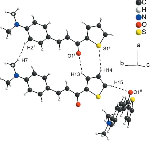
In the crystal, the molecules are connected by very weak C13—H13 O1i and C14—H14 S1i hydrogen-bonding interactions (see Table 1 for symmetry codes), forming rings with an R22(8) graph-set motif. The R22(8) rings are the subunits of the periodic arrangement along [100] and one very weak H7 H2icontact is also observed [H H = 2.26 A˚ ]. The molecular units are also linked by very weak C15—H15 O1ii links into chains along [010] with aC(6) graph-set motif (Fig. 2; Table 1). Additionally, the molecules are connected into centrosymmetric dimers by very weak C—H interactions involving the thiophene ring (Fig. 3; Table 1). The inter-molecular contacts are slightly longer than the sum of the van der Waals radii for the respective atoms (Bondi, 1964; Rowland & Taylor, 1996) and suggest weak interactions only.
4. Hirshfeld surface analysis
[image:2.610.313.565.108.168.2] [image:2.610.81.257.111.202.2]The Hirshfeld surface analysis (Hirshfeld, 1977) of the crystal structure suggests that the contribution of the H H inter-molecular interactions to the crystal packing amounts to 46.50% and the contribution of the H C interactions Table 1
Hydrogen-bond geometry (A˚ ,).
Cgis the centroid of the S1/C12–C15 thiophene ring.
D—H A D—H H A D A D—H A
C13—H13 O1i 0.95 2.65 3.451 (4) 142 C14—H14 S1i 0.95 3.00 3.779 (3) 141
C15—H15 O1ii 0.95 2.57 3.291 (4) 133 C8—H8 Cgiii 0.98 2.64 3.457 (4) 141
Symmetry codes: (i)xþ1;y;z; (ii)x1 2;y
1 2;zþ
3
[image:2.610.315.565.295.531.2]2; (iii)x;y;zþ1.
Figure 2
Graphical representation of the weak intermolecular C—H O, C— H S and H H interactions (dashed lines) in the crystal structure of the title compound. [Symmetry codes: (i)x+ 1,y,z; (ii)x1
2,y 1 2,z+
3 2.]
Figure 1
[image:2.610.316.565.598.707.2]The molecular structure of the title compound, showing displacement ellipsoids drawn at the 40% probability level.
Figure 3
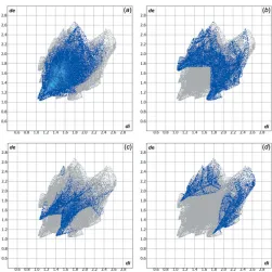
[image:2.610.46.293.604.710.2]amounts to 23.40%. Other important intermolecular contacts for the cohesion of the structure are (values given in %): H O = 10.80 and H S = 10.00. Graphical representations of the Hirshfeld surface with transparency and labelled atoms (Figs. 4 and 5) indicate, in a magenta colour, the locations of the strongest intermolecular contacts, e.g. the H2, H7, H13, H15 and O1 atoms. The C—H interaction is also well represented in the Hirshfeld surface (for details, compare Figs. 3 and 5). The H H, H C, H O and H S contributions to the crystal packing are shown as a Hirshfeld surface two-dimensional fingerprint plot with cyan dots. Thede(yaxis) and
di(xaxis) values are the closest external and internal distances
(values given in A˚ ) from given points on the Hirshfeld surface contacts (Fig. 6; Wolffet al., 2012).
5. Molecular docking evaluation
In addition, a lock-and-key supramolecular analysis between the neuraminidase enzyme, whose inhibition is believed to be a key point to block the influenza viral infection (Kingeret al., 2012), and the title compound was performed. The semi-empirical equilibrium energy of the title compound was obtained using the PM6 Hamiltonian and the experimental bond lengths were conserved. The calculated parameters were: heat of formation = 139.28 kJ mol1, gradient normal = 0.62031, HOMO =8.96 eV, LUMO =0.866 eV and energy gap = 7.421 eV (Stewart, 2013). The rigid molecular docking was carried out with theGOLDsoftware (Joneset al., 1997)
using the ChemPLP score function (Chen, 2015). The chal-cone thiophene derivative and the active site of the neur-aminidase match (Fig. 7) and the structure–activity relationship can be assumed by the following observed inter-molecular interactions (H A distance values given in A˚ ):
(ASN263)N—H O1 (d = 1.796), (PRO245)C—
H Cg(thiophene ring) (d= 2.829) and (AGR287)C—H N1 (d= 2.620) (Fig. 8). More details about thein silicoevaluation, with additional references, can be found in the Supporting Information. For the intermolecular interactions, it is impor-tant to report that the H Cg(thiophene ring) contact is observed in the structure interpretation, by the centrosym-metric dimeric arrangement of the molecules (Figs. 3 and 9), in the Hirshfeld surface analysis (Fig. 5) and in the molecular docking evaluation (Fig. 8).
6. Database survey
[image:3.610.314.565.67.318.2]Chalcone-thiophene derivatives have some molecular struc-tural features in common, namely the nearly planar geometry, as a result of thesp2-hybridized C atoms of the main fragment, and the weak intermolecular interactions,e.g. H H, H C or– contacts. One example for comparison with the title compound is the crystal structure of the compound 3-(4-methylphenyl)-1-(3-thienyl)-2-propen-1-one (Li & Su, 1993). In both of the structures, the molecules are linked by weak interactions into centrosymmetric dimers and the crystal Figure 6
Hirshfeld surface two-dimensional fingerprint plots for the title compound showing the (a) H H, (b) H C, (c) O H and (d) H S contacts in detail (cyan dots). The contributions of the interactions to the crystal packing amount to 46.50, 23.40, 10.80 and 10.00%, respectively. Thede(yaxis) anddi(xaxis) values are the closest external and internal
[image:3.610.46.295.75.170.2]distances (values in A˚ ) from given points on the Hirshfeld surface contacts.
Figure 4
A graphical representation of the Hirshfeld surface (dnorm) for the title
[image:3.610.45.296.598.688.2]compound. The surface is drawn with transparency and all atoms are labelled. The surface regions with strongest intermolecular interactions for atoms H2, H15 and O1 are shown in magenta.
Figure 5
A graphical representation of the Hirshfeld surface (dnorm) for the title
packing shows a herringbone motif: for the title compound this molecular arrangement is clear when looking along the [100] direction (Fig. 9a) and for the above-mentioned 3-thienyl derivative, along [001] (Fig. 9b).
7. Synthesis and crystallization
All starting materials are commercially available and were used without further purification. The synthesis of the title compound was adapted from a previously reported procedure (Claisen & Clapare`de, 1881; Schmidt, 1881; Zhouet al., 2016). In a hydroxide-catalysed reaction, a mixture of
4-(dimethyl-amino)benzaldehyde (10 mmol) and 2-acetylthiophene
(10 mmol) in ethanol (80 mL) was stirred under room temperature for 4 h. After cooling in an ice bath and filtering,
an orange solid was obtained. Orange crystals were grown from the solution after 24 h.
8. Refinement
Crystal data, data collection and structure refinement details are summarized in Table 2. H atoms were located in a differ-ence-Fourier map but were positioned with idealized geometry and were refined with isotropic displacement para-meters using a riding model (HFIX command) withUiso(H) =
1.2Ueq(C) or 1.5Ueq(C) for methyl H atoms. A rotating model
was used for the methyl groups. The crystal was refined as a two-component twin {twin law: two-axis (001) [105], BASF = 0.0181 (8)}.
Acknowledgements
[image:4.610.44.296.67.247.2]ABO is an associate researcher in the project ‘Dinitrosyl complexes containing thiol and/or thiosemicarbazone: synth-esis, characterization and treatment against cancer’, founded by FAPESP, Proc. 2015/12098–0, and acknowledges Professor Jose´ C. M. Pereira (UNESP, Brazil) for his support. GPO thanks CNPq for the MSc scholarship and RLF thanks the CAPES foundation for the PhD scholarship. The authors acknowledge Professor Manfredo Ho¨rner for access to Figure 7
[image:4.610.317.562.72.395.2]Graphical representation of the lock-and-key model for the title compound, with the molecular main fragment in green, and the neuraminidase structure, with selected amino acids residues, in stick model. The structure of the enzyme is simplified for clarity.
Figure 8
Intermolecular interactions between the title compound and the neuraminidase enzyme. The interactions are shown as dashed lines and the structure of the enzyme is simplified for clarity.
Figure 9
[image:4.610.46.298.537.708.2]experimental facilities and Guilherme Alves de Moraes for the data collection (Federal University of Santa Maria, Brazil).
References
Allen, F. H., Johnson, O., Shields, G. P., Smith, B. R. & Towler, M. (2004).J. Appl. Cryst.37, 335–338.
Bondi, A. (1964).J. Phys. Chem.68, 441–451.
Brandenburg, K. (2006).DIAMOND. Crystal Impact GbR, Bonn, Germany.
Bruker (2014). APEX2, SADABS and SAINT. Bruker AXS Inc., Madison, Wisconsin, USA.
Chen, Y.-C. (2015).Trends Pharmacol. Sci.36, 78–95. Claisen, L. & Clapare`de, A. (1881).Chem. Ber.14, 2460–2468. Farrugia, L. J. (2012).J. Appl. Cryst.45, 849–854.
Hirshfeld, H. L. (1977).Theor. Chim. Acta,44, 129–138.
Jones, G., Willett, P., Glen, R. C., Leach, A. R. & Taylor, R. (1997).J. Mol. Biol.267, 727–748.
Kinger, M., Park, Y. D., Park, J. H., Hur, M. G., Jeong, H. J., Park, S. J., Lee, W. S., Kim, S. W. & Yang, S. D. (2012).Arch. Pharm. Res.35, 633–638.
Li, Z. & Su, G. (1993).Acta Cryst.C49, 1075–1077.
Rowland, R. S. & Taylor, R. (1996).J. Phys. Chem.100, 7384–7391. Schmidt, J. G. (1881).Chem. Ber.14, 1459–1461.
Sheldrick, G. M. (2008).Acta Cryst.A64, 112–122. Sheldrick, G. M. (2015).Acta Cryst.C71, 3–8. Stewart, J. J. (2013).J. Mol. Model.19, 1–32. Westrip, S. P. (2010).J. Appl. Cryst.43, 920–925.
Wolff, S. K., Grimwood, D. J., McKinnon, J. J., Turner, M. J., Jayatilaka, D. & Spackman, M. A. (2012). Crystal Explorer. University of Western Australia, Perth, Australia.
[image:5.610.44.294.90.365.2]Zhou, B., Jiang, P., Lu, J. & Xing, C. (2016).Arch. Pharm. Chem. Life Sci.349, 539–552.
Table 2
Experimental details.
Crystal data
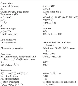
Chemical formula C15H15NOS
Mr 257.34
Crystal system, space group Monoclinic,P21/n
Temperature (K) 120
a,b,c(A˚ ) 6.2405 (4), 9.9975 (6), 20.7815 (13)
() 93.097 (2)
V(A˚3) 1294.65 (14)
Z 4
Radiation type MoK
(mm1) 0.24
Crystal size (mm) 0.530.160.09
Data collection
Diffractometer Bruker APEXII CCD area detector
Absorption correction Multi-scan (SADABS; Bruker, 2014)
Tmin,Tmax 0.885, 0.979
No. of measured, independent and observed [I> 2(I)] reflections
50820, 3381, 3116
Rint 0.031
(sin/)max(A˚
1
) 0.677
Refinement
R[F2> 2(F2)],wR(F2),S 0.068, 0.182, 1.14
No. of reflections 3381
No. of parameters 166
H-atom treatment H-atom parameters constrained
max, min(e A˚
3
) 1.16,0.81
Computer programs:APEX2andSAINT(Bruker, 2014),SHELXS97(Sheldrick, 2008),
sup-1 Acta Cryst. (2017). E73, 476-480
supporting information
Acta Cryst. (2017). E73, 476-480 [https://doi.org/10.1107/S2056989017003437]
Crystal structure of (2
E
)-3-[4-(dimethylamino)phenyl]-1-(thiophen-2-yl)prop-2-en-1-one
Gabriela Porto de Oliveira, Leandro Bresolin, Darlene Correia Flores, Renan Lira de Farias and
Adriano Bof de Oliveira
Computing details
Data collection: APEX2 (Bruker, 2014); cell refinement: SAINT (Bruker, 2014); data reduction: SAINT (Bruker, 2014);
program(s) used to solve structure: SHELXS97 (Sheldrick, 2008); program(s) used to refine structure: SHELXL2016
(Sheldrick, 2015) and WinGX (Farrugia, 2012); molecular graphics: DIAMOND (Brandenburg, 2006), GOLD (Chen et
al., 2015) and Crystal Explorer (Wolff et al., 2012); software used to prepare material for publication: publCIF (Westrip,
2010) and enCIFer (Allen et al., 2004).
(2E)-3-[4-(Dimethylamino)phenyl]-1-(thiophen-2-yl)prop-2-en-1-one
Crystal data
C15H15NOS
Mr = 257.34
Monoclinic, P21/n
a = 6.2405 (4) Å
b = 9.9975 (6) Å
c = 20.7815 (13) Å
β = 93.097 (2)°
V = 1294.65 (14) Å3
Z = 4
F(000) = 544
Dx = 1.320 Mg m−3
Mo Kα radiation, λ = 0.71073 Å Cell parameters from 9620 reflections
θ = 2.3–28.7°
µ = 0.24 mm−1
T = 120 K Block, orange
0.53 × 0.16 × 0.09 mm
Data collection
Bruker APEXII CCD area detector diffractometer
Radiation source: fine-focus sealed tube, Bruker APEXII CCD area detector
φ and ω scans
Absorption correction: multi-scan (SADABS; Bruker, 2014)
Tmin = 0.885, Tmax = 0.979
50820 measured reflections 3381 independent reflections 3116 reflections with I > 2σ(I)
Rint = 0.031
θmax = 28.8°, θmin = 2.3°
h = −7→8
k = −13→13
l = −28→28
Refinement
Refinement on F2 Least-squares matrix: full
R[F2 > 2σ(F2)] = 0.068
wR(F2) = 0.182
S = 1.14 3381 reflections 166 parameters
0 restraints
Primary atom site location: structure-invariant direct methods
Secondary atom site location: difference Fourier map
sup-2 Acta Cryst. (2017). E73, 476-480
H-atom parameters constrained
w = 1/[σ2(F
o2) + 7.019P] where P = (Fo2 + 2Fc2)/3
(Δ/σ)max < 0.001 Δρmax = 1.16 e Å−3 Δρmin = −0.81 e Å−3
Special details
Geometry. All esds (except the esd in the dihedral angle between two l.s. planes) are estimated using the full covariance matrix. The cell esds are taken into account individually in the estimation of esds in distances, angles and torsion angles; correlations between esds in cell parameters are only used when they are defined by crystal symmetry. An approximate (isotropic) treatment of cell esds is used for estimating esds involving l.s. planes.
Refinement. Refined as a 2-component twin.
Fractional atomic coordinates and isotropic or equivalent isotropic displacement parameters (Å2)
x y z Uiso*/Ueq
C1 −0.1108 (5) 0.1473 (3) 0.52979 (15) 0.0159 (6) C2 −0.2422 (5) 0.1940 (4) 0.47768 (16) 0.0189 (6)
H1 −0.382513 0.157938 0.471036 0.023*
C3 −0.1753 (5) 0.2904 (4) 0.43572 (15) 0.0193 (6)
H2 −0.269640 0.319201 0.401045 0.023*
C4 0.0319 (5) 0.3467 (3) 0.44378 (15) 0.0178 (6)
C5 0.1659 (5) 0.2992 (3) 0.49602 (16) 0.0188 (6)
H3 0.306383 0.334799 0.502939 0.023*
C6 0.0956 (5) 0.2024 (3) 0.53690 (15) 0.0181 (6)
H4 0.190279 0.171734 0.571104 0.022*
C7 0.3103 (6) 0.5007 (4) 0.4112 (2) 0.0295 (8)
H5 0.318786 0.553158 0.451058 0.044*
H6 0.336378 0.559200 0.374572 0.044*
H7 0.418747 0.429736 0.413870 0.044*
C8 −0.0527 (6) 0.5041 (4) 0.35627 (17) 0.0241 (7)
H8 −0.117365 0.435643 0.327624 0.036*
H9 0.021999 0.570086 0.330728 0.036*
H10 −0.165267 0.548805 0.379384 0.036*
C9 −0.1918 (5) 0.0467 (3) 0.57189 (15) 0.0168 (6)
H11 −0.336808 0.020903 0.562907 0.020*
C10 −0.0899 (5) −0.0158 (3) 0.62222 (15) 0.0163 (6)
H12 0.056053 0.004730 0.633416 0.020*
C11 −0.2021 (5) −0.1147 (3) 0.65969 (15) 0.0164 (6) C12 −0.0788 (5) −0.1847 (3) 0.71167 (15) 0.0155 (6) C13 0.1366 (5) −0.1816 (3) 0.72937 (15) 0.0174 (6)
H13 0.237533 −0.126760 0.709136 0.021*
C14 0.1911 (5) −0.2697 (3) 0.78141 (16) 0.0194 (6)
H14 0.333099 −0.281313 0.799380 0.023*
C15 0.0180 (5) −0.3357 (3) 0.80267 (15) 0.0180 (6)
H15 0.024715 −0.397159 0.837576 0.022*
N1 0.0994 (5) 0.4417 (3) 0.40233 (14) 0.0232 (6)
sup-3 Acta Cryst. (2017). E73, 476-480
Atomic displacement parameters (Å2)
U11 U22 U33 U12 U13 U23
C1 0.0188 (14) 0.0149 (14) 0.0140 (14) 0.0025 (12) 0.0012 (11) −0.0014 (11) C2 0.0160 (14) 0.0211 (16) 0.0195 (15) 0.0028 (12) 0.0003 (12) −0.0010 (13) C3 0.0187 (15) 0.0221 (16) 0.0167 (14) 0.0051 (13) −0.0014 (12) 0.0004 (13) C4 0.0226 (16) 0.0152 (14) 0.0159 (14) 0.0039 (12) 0.0030 (12) −0.0011 (12) C5 0.0200 (15) 0.0170 (15) 0.0191 (15) −0.0006 (12) −0.0011 (12) −0.0016 (12) C6 0.0211 (15) 0.0179 (15) 0.0148 (14) 0.0020 (13) −0.0029 (11) −0.0011 (12) C7 0.0287 (19) 0.0276 (19) 0.032 (2) −0.0026 (16) 0.0024 (15) 0.0082 (16) C8 0.0300 (18) 0.0202 (16) 0.0220 (16) 0.0055 (14) −0.0009 (14) 0.0054 (13) C9 0.0190 (15) 0.0161 (14) 0.0156 (14) 0.0017 (12) 0.0027 (11) −0.0033 (12) C10 0.0172 (14) 0.0158 (14) 0.0159 (14) −0.0027 (12) 0.0011 (11) −0.0025 (12) C11 0.0183 (14) 0.0159 (14) 0.0150 (14) 0.0004 (12) 0.0023 (11) −0.0018 (11) C12 0.0168 (14) 0.0146 (14) 0.0154 (14) −0.0017 (11) 0.0030 (11) −0.0020 (11) C13 0.0165 (14) 0.0174 (15) 0.0182 (15) −0.0029 (12) 0.0004 (11) 0.0020 (12) C14 0.0176 (14) 0.0202 (16) 0.0203 (15) −0.0023 (12) 0.0011 (12) 0.0047 (13) C15 0.0184 (15) 0.0199 (15) 0.0157 (14) −0.0009 (12) 0.0001 (11) 0.0010 (12) N1 0.0267 (15) 0.0216 (14) 0.0211 (14) 0.0009 (12) −0.0012 (12) 0.0062 (12) O1 0.0145 (11) 0.0256 (13) 0.0258 (12) −0.0025 (10) −0.0014 (9) 0.0034 (10) S1 0.0137 (4) 0.0218 (4) 0.0210 (4) −0.0023 (3) 0.0030 (3) 0.0038 (3)
Geometric parameters (Å, º)
C1—C6 1.401 (5) C8—H8 0.9800
C1—C2 1.403 (4) C8—H9 0.9800
C1—C9 1.442 (4) C8—H10 0.9800
C2—C3 1.379 (5) C9—C10 1.348 (5)
C2—H1 0.9500 C9—H11 0.9500
C3—C4 1.412 (5) C10—C11 1.461 (4)
C3—H2 0.9500 C10—H12 0.9500
C4—N1 1.364 (4) C11—O1 1.240 (4)
C4—C5 1.416 (5) C11—C12 1.469 (4)
C5—C6 1.375 (5) C12—C13 1.375 (4)
C5—H3 0.9500 C12—S1 1.728 (3)
C6—H4 0.9500 C13—C14 1.422 (4)
C7—N1 1.445 (5) C13—H13 0.9500
C7—H5 0.9800 C14—C15 1.359 (4)
C7—H6 0.9800 C14—H14 0.9500
C7—H7 0.9800 C15—S1 1.709 (3)
C8—N1 1.452 (4) C15—H15 0.9500
C6—C1—C2 116.5 (3) H8—C8—H10 109.5
C6—C1—C9 124.1 (3) H9—C8—H10 109.5
C2—C1—C9 119.4 (3) C10—C9—C1 128.9 (3)
C3—C2—C1 122.4 (3) C10—C9—H11 115.6
C3—C2—H1 118.8 C1—C9—H11 115.6
sup-4 Acta Cryst. (2017). E73, 476-480
C2—C3—C4 120.7 (3) C9—C10—H12 119.7
C2—C3—H2 119.7 C11—C10—H12 119.7
C4—C3—H2 119.7 O1—C11—C10 122.8 (3)
N1—C4—C3 121.0 (3) O1—C11—C12 119.3 (3)
N1—C4—C5 121.9 (3) C10—C11—C12 117.9 (3)
C3—C4—C5 117.1 (3) C13—C12—C11 130.7 (3)
C6—C5—C4 121.0 (3) C13—C12—S1 111.0 (2)
C6—C5—H3 119.5 C11—C12—S1 118.3 (2)
C4—C5—H3 119.5 C12—C13—C14 112.3 (3)
C5—C6—C1 122.2 (3) C12—C13—H13 123.8
C5—C6—H4 118.9 C14—C13—H13 123.8
C1—C6—H4 118.9 C15—C14—C13 112.7 (3)
N1—C7—H5 109.5 C15—C14—H14 123.6
N1—C7—H6 109.5 C13—C14—H14 123.6
H5—C7—H6 109.5 C14—C15—S1 112.1 (3)
N1—C7—H7 109.5 C14—C15—H15 124.0
H5—C7—H7 109.5 S1—C15—H15 124.0
H6—C7—H7 109.5 C4—N1—C7 120.9 (3)
N1—C8—H8 109.5 C4—N1—C8 120.1 (3)
N1—C8—H9 109.5 C7—N1—C8 117.8 (3)
H8—C8—H9 109.5 C15—S1—C12 91.87 (16)
N1—C8—H10 109.5
C6—C1—C2—C3 −0.9 (5) O1—C11—C12—C13 175.7 (3)
C9—C1—C2—C3 179.7 (3) C10—C11—C12—C13 −5.9 (5)
C1—C2—C3—C4 0.0 (5) O1—C11—C12—S1 −2.0 (4)
C2—C3—C4—N1 179.7 (3) C10—C11—C12—S1 176.4 (2)
C2—C3—C4—C5 0.5 (5) C11—C12—C13—C14 −177.5 (3)
N1—C4—C5—C6 −179.3 (3) S1—C12—C13—C14 0.3 (4)
C3—C4—C5—C6 0.0 (5) C12—C13—C14—C15 −1.0 (4)
C4—C5—C6—C1 −0.9 (5) C13—C14—C15—S1 1.2 (4)
C2—C1—C6—C5 1.3 (5) C3—C4—N1—C7 179.1 (3)
C9—C1—C6—C5 −179.3 (3) C5—C4—N1—C7 −1.7 (5)
C6—C1—C9—C10 −2.8 (5) C3—C4—N1—C8 11.2 (5)
C2—C1—C9—C10 176.5 (3) C5—C4—N1—C8 −169.6 (3)
C1—C9—C10—C11 179.2 (3) C14—C15—S1—C12 −0.9 (3)
C9—C10—C11—O1 −4.9 (5) C13—C12—S1—C15 0.3 (3)
C9—C10—C11—C12 176.7 (3) C11—C12—S1—C15 178.4 (3)
Hydrogen-bond geometry (Å, º)
Cg is the centroid of the S1/C12–C15 thiophene ring.
D—H···A D—H H···A D···A D—H···A
C13—H13···O1i 0.95 2.65 3.451 (4) 142
sup-5 Acta Cryst. (2017). E73, 476-480
C15—H15···O1ii 0.95 2.57 3.291 (4) 133
C8—H8···Cgiii 0.98 2.64 3.457 (4) 141
![Figure 9Section of the crystal structures of (a) the title compound viewed along[100],and(b)the3-(4-methylphenyl)-1-(3-thienyl)-2-propen-1-onecompound (Li & Su, 1993) viewed along [001], showing the herringbonemotif.](https://thumb-us.123doks.com/thumbv2/123dok_us/409965.538530/4.610.317.562.72.395/section-structures-compound-methylphenyl-thienyl-onecompound-showing-herringbonemotif.webp)