Original Article
miRNA-15b-5p promotes expression of osteoblast
differentiation-associated markers via targeting SMAD7
He Hu1, Guowei Zhang2, Geng Tian1, Guang Lv1, Yongli Jin3
1Medical College, 2College of Nursing, Inner Mongolia University for The Nationalities, Tongliao City 028041, Inner Mongolia, China; 3The Affiliated Hospital to Inner Mongolia University for The Nationalities, Tongliao, China
Received April 21, 2017; Accepted January 7, 2018; Epub April 15, 2018; Published April 30, 2018
Abstract: Recent studies have recognized the involvement of microRNAs (miRNAs) in the development of osteoporosis, which regulates the balance between osteogenesis and osteoclasis. The present study was to investigate the regulation of miRNA-15b-5p on the osteoblast differentiation-associated markers in the mouse osteoblast-like MC3T3-E1 cells, and to identify the targeting regulation by miRNA-15b-5p on the 3’ untranslated region (UTR) of SMAD7, which was a well-recognized inhibition to osteoblast differentiation. Firstly, quantitative real-time polymerase chain reaction (qRT-PCR) was performed to examine the expression of Osteocalcin, Procollagen type I N-terminal propeptide (P1NP) and Runt-related transcription factor 2 (RUNX2) in the MC3T3-E1 cells, which were transfected with 30 nM miR-15b-5p mimics or scramble miRNA (as control). Then the extracellular matrix (ECM) mineralization and the Alkaline Phosphatase (ALP) activity were also examined. Finally, we investigated the regulation by miRNA-15b-5p on the expression of SMAD7, with luciferase reporter. Results demonstrated that the transfection with miRNA-15b-5p mimics significantly promoted the miRNA-15b-5p level, and then upregulated the expression of Osteocalcin, P1NP and RUNX2 in MC3T3-E1 cells; promoted the ECM mineralization and the ALP activity. The alignment analysis indicated that miRNA-15b-5p was homologous with the 3’UTR of SMAD7. Moreover, the luciferase reporter assay demonstrated that miRNA-15b-5p targeted the 3’UTR of SMAD7, and inhibited the expression of SMAD7 in both mRNA and protein levels. In conclusion, we found the promotive effects of miRNA-15b-5p on the expression of osteoblast differentiation-associated markers, the ECM mineralization and the ALP activity in mouse osteoblast-like MC3T3-E1 cells, via targeting the 3’UTR of SMAD7. Our study suggests that miRNA-15b-5p might be an important target to promote osteoblast differentiation and to prevent osteoporosis.
Keywords: Differentiation, miRNA-15b-5p, osteoblast, SMAD7
Introduction
Osteoporosis is a common disease, which could increase bone weakness and the risk of fracture, especially at hip, forearm, spine and wrist [1-3]. The main cause of osteoporosis is bone loss and bone fragility, and the incidence of osteoporosis is high in elderly people, espe-cially in females [4]. In addition, osteoporosis is typically with no clinical symptoms, it has a long latent period until a bone broken. A person with fracture, more often than not, suffering from severe osteoporosis [5, 6]. Therefore, the pre-diction and diagnosis of osteoporosis is even more important. The balance between osteo-genesis and osteoclasis affects the bone for-mation and resorption, which is associated
with many factors, such as SMAD4, CACNG1 and TRIM63 [7, 8]. It also has reported that microRNAs (miRNAs) play important roles in osteoporosis development [4].
pro-miRNA-15b-5p inhibits SMAD7 and promotes EMT in osteoblasts
gression of many diseases, such as carcinoma
[15, 16], chronic hepatitis B [17] and Alzheimer’s disease [18]. Recently, miRNAs have been rec-ognized to involve in osteoporosis develop-ment, by regulating the balance between osteo-genesis and osteoclasis [19, 20]. The expres-sion level of miRNA-133a was up-regulated in the patients with postmenopausal osteoporo-sis, negatively correlating with CXCL11, CXCR3 and SLC39A1 genes [21]. In addition, miRNA-422a and miRNA-502 are potential biomarker for osteoblast differentiation [22, 23]. MiRNA-15 family is a highly conserved miRNA family containing six highly conserved miRNAs (miR-15a, miR-15b, miR-16-1, miR-16-2, miR-195, miRNA-497). miRNA-15 family could modulate cell apoptosis, differentiation, vascular remod-eling and insulin synthesis [15, 24]. miRNA-15b-5p is processed from the 5’ of the pri-miR-NA-15b. The function role of miRNA-15b-5p in osteoblast differentiation still remains to be elucidated.
In this study, we investigated the function role of miRNA-15b-5p in osteoblast differentiation in the osteoblast-like MC3T3-E1 cells. miRNA-15b-5p mimics was utilized to upregulate miR-NA-15b-5p level, and then the mRNA expres-sion level of the osteoblast-differentiation mar- kers, such as osteocalcin, procollagen type I N-terminal propeptide (P1NP) and runt-related transcription factor 2 (RUNX2) were examined. In addition, the extracellular matrix (ECM) min-eralization and the alkaline phosphatase (ALP) activity were also examined in the miR-15b-5p-manipulated MC3T3-E1 cells. We confirmed the regulatory role of miRNA-15b-5p on osteo-blast differentiation in MC3T3-E1 cells. Taken together, miRNA-15b-5p might be a rational biomarker for diagnostic and therapeutic appli-cations in osteoporosis.
Materials and methods
Cell culture and transfection
The MC3T3-E1 cells (mouse embryo osteoblast precursor cells) were used in this study, which were obtained from ATCC (Rockville, MD, USA). The base medium for MC3T3-E1 cells is formu-lated Dulbecco’s Modified Eagle’s Medium (Gibco, Rockville, MD, USA). The MC3T3-E1 cells were cultured in the complete growth medium supplemented with 10% fetal bovine serum (FBS; Gibco, Rockville, MD, USA) based on the base medium. The culture condition of
the cells was 37°C 5% CO2 in an incubator. The MC3T3-E1 cells were seeded into a 12-well (3.8 cm2) plate and cultured for 24 h, then the cells were transfected with miRNA-15b-5p mimics or recombinant plasmid by Lipofecta- mine® 2000 Transfection Reagent (Invitrogen, Carlsbad, CA, USA) according to the manufac-turer’s instructions.
mRNA extraction and quantitative real-time polymerase chain reaction assay
The relative mRNA level of the osteoblast differ-entiation-associated markers (osteocalcin, P1- NP, RUNX2, SMAD7) was utilized to represent the change of the expression level of each marker. The total RNA was extracted from the MC3T3-E1 cells transfected with miRNA-15b-5p mimics or scramble by the Tri-Reagent (Ambion, Huntingdon UK) following the user’s specification. The RNase-free water was used to dissolve the total RNA. The quantitative anal-ysis of mRNA was determined by qRT-PCR. The mRNA sequence of each marker was obtained from NCBI, and the primers for qRT-PCR were designed by Primer premier 5. One Step SYBR PrimeScript PLUS RT-PCT Kit (Takara, Tokyo, Japan) was used to examine the relative level of each sample according to the manufacturer’s manual. After the reaction, the mRNA level of each marker was calculated and presented as mean ± SEM for triple independently-performed experiments, with β-actin as internal control. Construction of the recombinant plasmid and the luciferase reporter assay
exam-ined the relative luciferase activity assayed with the Dual-Luciferase Assay kit (Promega, Madison, WI, USA) by a GLOMAX (Promega, Madison, WI, USA).
ECM mineralization assay and alkaline phos-phatase activity
The ECM mineralization assay was performed after 24 h transfection. The transfected cells were collected and washed with PBS (phos-phate-buffered saline) for three times. Then the cells were fixed with 70% ethanol on ice for 60 min. After fixation and several washes in PBS, the cell layers were stained for 10 min at room temperature with 2% (w/v) Alizarin red solution (Sigma-Aldrich, St. Louis, MO, USA) at pH 4.2. After staining, the cell layers were washed to
remove the unbound stain. The distribution of mineral staining was observed from image of the culture well. To further investigate the rela-tive level of the ECM mineralization, therefore, we solubilized the bound stain in 10% cetylpyri-dinium chloride (Sigma-Aldrich, St. Louis, MO, USA) and the optical density value of the resul-tant solution determined at 450 nm by spectro-photometry (Crystaleye, Olympus, Tokyo, Japan)
[image:3.612.94.302.67.516.2][25]. The relative ALP activity in the miR-15b-5p mimics or scramble miRNA-transfected (30 or 60 nM; 24 hours) MC3T3-E1 cells, was per-formed by the Alkaline Phosphatase Dieth- anolamine Activity Kit (Sigma-Aldrich, St. Louis, MO, USA) according to the manufacturer’s instructions. And the ALP activity was quanti-fied at 450 nm by spectrophotometry.
miRNA-15b-5p inhibits SMAD7 and promotes EMT in osteoblasts
Western blot assay
The expression of SMAD7 on protein level were determined by Western blotting. To harveste the MC3T3-E1 cells transfected with or without miRNA-15b-5p mimics, the cells were lysed into cell lysis buffer (Bio-Rad, Hercules, CA, USA). The protein supernatant was extracted from the cell lysis after centrifuging at 12,000×g at 4°C for 15 min. the protein extract was sepa-rated by SDS-PAGE gel. Then the protein bands were transferred onto the PVDF membranes (Millipore, Bedford, MA, USA), and the mem-brane was blocked with 5% skimmed milk pow-der at 4°C overnight. The membrane was incu-bated with SMAD-specific antibody (1:1000, mouse monoclonal antibody, Abcam, Cambri- dge, UK) or β-actin-specific antibody (1:3000, mouse monoclonal antibody, Abcam, Cambri- dge, UK) in TBST at 37°C for 1 h, washed the membrane with TBST, subsequently incubated with the secondary antibody, anti-mouse IgG conjugated with HRP (1:1000, New England Biolab, Ipswich, UK) at 37°C for 30 min. After washing with TBST, the membrane was treated with the ECL kit (Thermo Scientific, Rockford, IL, USA) and scanned by a Smart ChemiTM lamp Analysis System (Life Technologies, Grand Island, NY, USA). The protein level of the SMAD7 was quantified according to the band density by Quantity One software based on the western blot assay, with β-actin as loading control. Each value was averaged for triple independent results.
Statistical analysis
Data were expressed as mean ± SEM for triple independently-performed experiments. The dif-ference between two groups was analyzed by Student’s t test using the GraphPad Prism (GraphPad Software, La Jolla, CA, USA). A p value <0.05 or less was considered to be significant (*p<0.05, **p<0.01, ***p<0.001, ****p<0.0001, ns: no significance).
Results
The osteoblast differentiation-associated markers were up-regulated in mouse osteo-blast-like MC3T3-E1 cells post transfection of miRNA-15b-5p
To investigate the function of miRNA-15b-5p in osteoblast, we examined the osteoblast
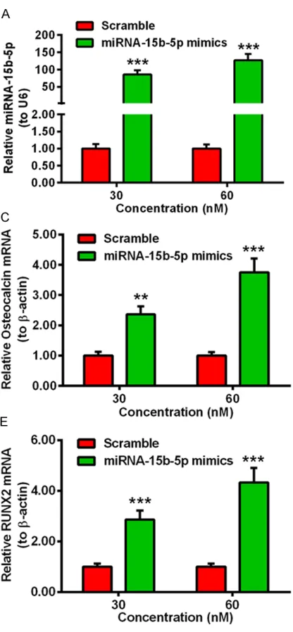
differ-entiation-associated markers in MV3T3-E1 cells with or without the transfection of miRNA-15b-5p. The miRNA-15b-5p mimics was used to increase the level of miRNA-15b-5p in vitro. The level of miRNA-15b-5p was significantly up-regulated in MC3T3-E1 cells transfected with 30 or 60 nM miRNA-15b-5p mimics (Figure 1A, ***p<0.001) by qRT-PCR with U6 as internal standard template. The relative level of miRNA-15b-5p in MC3T3-E1 cells post transfection of 30 nM miRNA-15b-5p mimics or scramble miRNA for 0, 1, 3, 6, 12 or 24 hours, the trend of the change of miRNA-15b-5p was depicted in Figure 1B, which showed that the level of miR-NA-15b-5p was significantly increased at 3 h post transfection, moreover the level was sta-ble within 3 h to 24 h, compared to the control group (**p<0.01, ****p<0.0001). Then we investigate the mRNA level of the differentia-tion-associated markers in MC3T3-E1 cells, such like osteocalcin, P1NP and RUNX2, with β-actin as internal control. The results was depicted in Figure 1, the mRNA level of osteo-calcin was significantly increased in the MC3T3-E1 cells post transfection with miRNA-15b-5p mimics for 12 hours, compared to the scramble groups. Moreover, the mRNA level of osteocalcin was higher in the cells transfected with 60 nM miRNA-15b-5p mimics than those transfected with 30 nM miRNA-15b-5p mimics (Figure 1C, **p<0.01, ***p<0.001). Beyond that, Figure 1D and 1E also showed that, trans-fected with miRNA-15b-5p mimics also incre- ased the mRNA level of P1NP and RUNX2 in the MC3T3-E1 cells (*p<0.05, **p<0.01), espe-cially the mRNA level of RUNX2 (***p<0.001). All the above data implied that the level of the differentiation-associated markers (osteocal-cin, P1NP, RUNX2) was positive correlated with the miRNA-15b-5p in MC3T3-E1 cells.
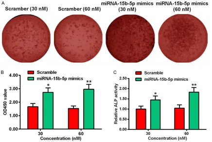
miRNA-15b-5p upregulates the extracellular matrix (ECM) mineralization in MC3T3-E1 cells
transfect-ed with 30 or 60 nM miR-15b-5p mimics was much increased than the cells transfected with 30 or 60 nM scramble. Besides the image, we also quantified the OD450 value of the cells. The OD450 value was increased in the cells transfected with 30 nM (*p<0.05) or 60 nM (**p<0.01) miRNA-15b-5p mimics, as com-pared with the scramble group (Figure 2B). Alkaline Phosphatase (ALP) is a marker in the early stage of osteogenesis, the relative ALP activity in the miR-15b-5p mimics-transfected MC3T3-E1 cells was much higher than the scramble cells, especially at the concentration of 60 nM miRNA-15b-5p mimics (Figure 2C, *p<0.05, **p<0.01).
miRNA-15b-5p targets the 3’UTR of SMAD7 in the MC3T3-E1cells
Based on the above finding, miRNA-15b-5p could increase the level of osteoblast differen-tiation-associated markers and promote the osteogenesis in the mouse osteoblast-like
[image:5.612.91.527.72.366.2]miRNA-15b-5p inhibits SMAD7 and promotes EMT in osteoblasts
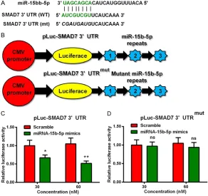
activity in the pLuc-SMAD7 3’UTRmut-transfe- cted MC3T3-E1 cells, there was no significant difference between the cells co-transfected with 30 nM or 60 nM miRNA-15b-5p mimics and scramble (Figure 3D, ns: no significance). The results demonstrated that the miRNA-15b-5p mimics reduced the relative luciferase activity by targeted the 3’UTR of SMAD7. The expression level of SMAD7 was down-regulated by miRNA-15b-5p
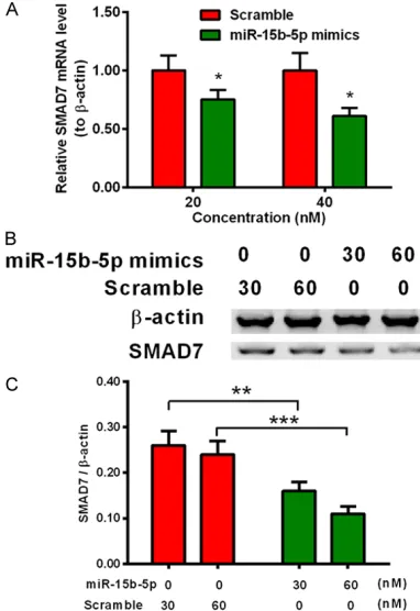
The relative luciferase activity was reduced by the targeting of the miRNA-15b-5p and 3’UTR SMAD7 in the luciferase-15b-5p-mim-ics co-transfection system, therefore, miRNA-15b-5p may play a role in modulating the expression level of SMAD7 in the MC3T3-E1 cells. Then, the relative mRNA level of SMAD7 in MC3T3-E1 cells transfected with or without miRNA-15b-5p mimics were examined by qRT-PCR assay. Figure 4A showed that transfecting
transfecting groups. Furthermore, the higher concentration of miRNA-15b-5p mimics led to the lower expression of SMAD7 (Figure 4C, ** p<0.01, ***p<0.0001).
Discussion
[image:6.612.92.384.75.350.2]miRNAs have been shown to regulate osteo-blast differentiation. And miRNA-15 family has been showed to participate in various cellular processes especially the differentiation pro-cess. But there is no report on the regulator role of miRNA-15b-5p in osteoblast differentia-tion. In the present study, the miRNA-15b-5p mimics was used to increase the level of miR-NA-15b-5p in the MC3T3-E1 cells by cell trans-fection. To investigate the regulation role of the miRNA-15b-5p in osteoblast differentiation, the typical osteoblast-differentiation markers were examined in this parts. Osteocalcin is an extracellular matrix protein, which is expressed in the later stage of osteogenic induction. The Figure 3. miRNA-15b-5p targets the 3’UTR of SMAD7 in MC3T3-E1 cells. (A)
Alignment of murine mature miRNA-15b-5p and the 3’UTR of SMAD7 sequenc-es; (B) Sketch of the luciferase reporter plasmid with the wild type or mutant type 3’UTR of SMAD7; (C) and (D) Relative luciferase activity in the MC3T3-E1 cells, which were transfected with miR-15b-5p mimics or scramble miRNA, by the reporter plasmid with the 3’UTR of SMAD7 (C) or with the mutant 3’UTR of SMAD7 (D). All experiments were independently performed in triplicate. *p<0.05, **p<0.01 or ns: no significance.
mimics-expression level of osteocalcin is positively cor-related with the mineralized deposition, it is a key index to indicate the osteoblast activity
[26]. Procollagen type I N-terminal propeptide is an osteoblast-derived protein [27, 28]. The osteoblast-related gene RUNX2 is a bone tran-scription factor, which could regulate the diff- erentiation of mesenchymal stem cells into osteoblasts. RUNX2 plays an important role in the differentiation and function of osteoblasts, it could promote the bone matrix deposition rate [29]. These markers are indexes of the osteoblast, the expression level of these mark-ers are positively related with the osteoblast differentiation. The effect of miRNA-15b-5p on osteoblast differentiation was represented by these markers indirectly. We found that the mRNA level of osteocalcin, P1NP, RUNX2 were both up-regulated in the MC3T3-E1 cells trans-fected with miRNA-15b-5p mimics, compared
to the scramble groups. The results demon-strated that the miRNA-15b-5p promoted the expression of the osteoblast-differentiation markers.
[image:7.612.90.281.73.351.2]ALP activity is an early marker of bone forma-tion, the activity is the precondition of cells (or tissues) mineralization, and affecting the formation of mineralized tissues [30]. The MC3T3-E1 cells transfected with miRNA-15b- 5p mimics showed a higher activity than the control group. In addition, from the staining of the extracellular matrix (ECM) mineralization, the stained mineralized nodes was more in the cells transfected with 30 nM or 60 nM miRNA-15b-5p mimics than the cells transfected wi- th same concentration scramble. The relative level of mineralization represented by OD450 nm was also showed the enhancement of min-eralization. As is known to all, the mineralized nodes is the index for the formation of mineral-ized matrix, it is the final signal of the osteo-genic phenotype in vitro, which means the maturity of the osteoblast [31-34]. The differen-tiation makers and the mineralization assay implied that miRNA-15b-5p could promote the osteoblast differentiation in MC3T3-E1 cells. The mechanism of miRNA-15b-5p in modulat-ing the differentiation of osteoblast was the next issue that we were going to solve. SMAD7 is a well-recognized inhibitor to osteoblast dif-ferentiation. Many studies had reported that SMAD7 plays an important role in regulating cell cycle, it also modulates cancer growth and progression [35-37]. SMAD7 promotes and enhances skeletal muscle differentiation by inhibiting TGF-b/activin signaling and bone morphogenetic protein (BMP) pathways [38]. Therefore, we suspected that miRNA-15b-5p may target the mRNA of SMAD7 to modulate the cell differentiation. The sequence of miR-NA-15b-5p and the 3’UTR of SMAD7 sequenc-es was complementary. To verify this hypothe-sis, we designed a recombinant plasmid con-tained the luciferase reporter and the wild type 3’UTR of SMAD7 (or mutant type 3’UTR of SMAD7). The relative luciferase activity was decreased in the miRNA-15b-5p-transfected cells, which demonstrated that the miRNA-15b-5p could target the wild type 3’UTR of SMAD7, and further terminate the expression of luciferase reporter. On the other hand, the protein level of SMAD7 was also reduced in the Figure 4. miRNA-15b-5p downregulates the
miRNA-15b-5p inhibits SMAD7 and promotes EMT in osteoblasts
cells transfected with miRNA-15b-5p by west-ern blot.
In conclusion, there are two major outcomes in this study. Firstly, miRNA-15b-5p improved the expression level of the osteoblast-differentia-tion markers (osteocalcin, P1NP, RUNX2, ALP activity), and promoted the formation of ECM mineralization. Therefore, the miRNA-15b-5p could promote the osteoblast differentiation. Secondly, miRNA-15b-5p could modulate the cell differentiation via targeting the 3’UTR of SMAD7. Our study suggests that miRNA-15b-5p might be an important target to promote osteo-blast differentiation and to prevent osteopo- rosis.
Disclosure of conflict of interest
None.
Address correspondence to: He Hu, Medical College, Inner Mongolia University for The Nationalities, 996 West Lamulun Street, Tongliao City 028041, Inner Mongolia, China. Tel: 00864758314234; Fax: 00864758314234; E-mail: [email protected]
References
[1] Maeda SS and Lazaretti-Castro M. An overview on the treatment of postmenopausal osteopo-rosis. Arq Bras Endocrinol Metabol 2014; 58: 162-171.
[2] Li XS, Zhang JR, Meng SY, Li Y and Wang RT. Mean platelet volume is negatively associated with bone mineral density in postmenopausal women. J Bone Miner Metab 2012; 30: 660-665.
[3] Tella SH and Gallagher JC. Prevention and treatment of postmenopausal osteoporosis. J Steroid Biochem Mol Biol 2014; 142: 155-170.
[4] Meng J, Zhang D, Pan N, Sun N, Wang Q, Fan J, Zhou P, Zhu W and Jiang L. Identification of miR-194-5p as a potential biomarker for post-menopausal osteoporosis. Peer J 2015; 3: e971.
[5] Tian J, Xu XJ, Shen L, Yang YP, Zhu R, Shuai B, Zhu XW, Li CG, Ma C and Lv L. Association of serum Dkk-1 levels with beta-catenin in pa-tients with postmenopausal osteoporosis. J Huazhong Univ Sci Technolog Med Sci 2015; 35: 212-218.
[6] Diab DL and Watts NB. Postmenopausal osteo-porosis. Curr Opin Endocrinol Diabetes Obes 2013; 20: 501-9.
[7] Ozmen B, Kirmaz C, Aydin K, Kafesciler SO, Guclu F and Hekimsoy Z. Influence of the se-lective oestrogen receptor modulator
(raloxi-fene hydrochloride) on IL-6, TNF-alpha, TGF-beta1 and bone turnover markers in the treat-ment of postmenopausal osteoporosis. Eur Cytokine Netw 2007; 18: 148-153.
[8] Liu Y, Wang Y, Yang N, Wu S, Lv Y and Xu L. In silico analysis of the molecular mechanism of postmenopausal osteoporosis. Mol Med Rep 2015; 12: 6584-6590.
[9] Hu J, Xu JF and Ge WL. MiR-497 enhances metastasis of oral squamous cell carcinoma through SMAD7 suppression. Am J Transl Res 2016; 8: 3023-3031.
[10] Pereira DM, Rodrigues PM, Borralho PM and Rodrigues CM. Delivering the promise of miR-NA cancer therapeutics. Drug Discov Today 2013; 18: 282-289.
[11] Gyvyte U, Juzenas S, Salteniene V, Kupcinskas J, Poskiene L, Kucinskas L, Jarmalaite S, Stuo- pelyte K, Steponaitiene R, Hemmrich-Stanisak G, Hubenthal M, Link A, Franke S, Franke A, Pangonyte D, Lesauskaite V, Kupcinskas L and Skieceviciene J. MiRNA profiling of gastrointes-tinal stromal tumors by next-generation se-quencing. Oncotarget 2017; 8: 37225-37238. [12] Di Leva G and Croce CM. miRNA profiling of
cancer. Curr Opin Genet Dev 2013; 23: 3-11. [13] Elton TS, Selemon H, Elton SM and Parinan-
di NL. Regulation of the MIR155 host gene in physiological and pathological processes. Gene 2013; 532: 1-12.
[14] Kim VN, Han J and Siomi MC. Biogenesis of small RNAs in animals. Nat Rev Mol Cell Biol 2009; 10: 126-139.
[15] Janaki Ramaiah M, Lavanya A, Honarpisheh M, Zarea M, Bhadra U and Bhadra MP. miR-15/16 complex targets p70S6 kinase1 and controls cell proliferation in MDA-MB-231 breast can-cer cells. Gene 2014; 552: 255-264.
[16] Zhao S, Sun H, Jiang W, Mi Y, Zhang D, Wen Y, Cheng D, Tang H, Wu S, Yu Y, Liu X, Cui W, Zhang M, Sun X, Zhou Z, Peng Z and Yan D. miR-4775 promotes colorectal cancer invasion and metastasis via the Smad7/TGFβ-mediated epithelial to mesenchymal transition. Mol Can- cer 2017; 16.
[17] Tan Y, Ge G, Pan T, Wen D and Gan J. Serum MiRNA panel as potential biomarkers for chronic hepatitis B with persistently normal al-anine aminotransferase. Clin Chim Acta 2015; 451: 232-239.
[18] Bekris LM and Leverenz JB. The biomarker and therapeutic potential of miRNA in Alzheimer’s disease. Neurodegener Dis Manag 2015; 5: 61-74.
[19] Chen J, Qiu M, Dou C, Cao Z and Dong S. MicroRNAs in bone balance and osteoporosis. Drug Dev Res 2015; 76: 235-245.
[21] Wang Y, Li L, Moore BT, Peng XH, Fang X, Lappe JM, Recker RR and Xiao P. MiR-133a in human circulating monocytes: a potential biomarker associated with postmenopausal osteoporo-sis. PLoS One 2012; 7: e34641.
[22] Cao Z, Moore BT, Wang Y, Peng XH and Lappe JM. MiR-422a as a potential cellular microRNA biomarker for postmenopausal osteoporosis. PLoS One 2014; 9: e97098.
[23] Chen C, Cheng P, Xie H, Zhou H, Wu X, Liao E and Luo X. MiR-503 regulates osteoclasto- genesis via targeting rank. J Bone Miner Res 2014; 29: 338-347.
[24] Sullivan RP, Leong JW, Schneider SE, Ireland AR, Berrien-Elliott MM, Singh A, Schappe T, Jewell BA, Sexl V and Fehniger TA. MicroRNA-15/16 AntagonizesMyb to control NK cell maturation. J Immunol 2015; 195: 2806-2817.
[25] Vijayakumar P, Laize V, Cardeira J, Trindade M and Cancela ML. Development of an in vitro cell system from zebrafish suitable to study bone cell differentiation and extracellular ma-trix mineralization. Zebrafish 2013; 10: 500-509.
[26] Enomoto H, Furuichi T, Zanma A, Yamana K, Yoshida C, Sumitani S, Yamamoto H, Enomoto-Iwamoto M, Enomoto-Iwamoto M and Komori T. Runx2 deficiency in chondrocytes causes adipogenic changes in vitro. J Cell Sci 2004; 117: 417-425.
[27] Lee J and Vasikaran S. Current recommenda-tions for laboratory testing and use of bone turnover markers in management of osteopo-rosis. Ann Lab Med 2012; 32: 105-112. [28] Krege JH, Lane NE, Harris JM and Miller PD.
PINP as a biological response marker during teriparatide treatment for osteoporosis. Oste- oporos Int 2014; 25: 2159-2171.
[29] Kikuchi K, Fukuda M, Ito T, Inoue M, Yokoi T, Chiku S, Mitsuyama T, Asai K, Hirose T and Aizawa Y. Transcripts of unknown function in multiple-signaling pathways involved in human stem cell differentiation. Nucleic Acids Res 2009; 37: 4987-5000.
[30] Hughes FJ, Turner W, Belibasakis G and Martuscelli G. Effects of growth factors and cytokines on osteoblast differentiation. Perio- Dontol 2000 2006; 41: 48-72.
[31] Alves RD, Eijken M, Bezstarosti K, Demmers JA and van Leeuwen JP. Activin A suppresses os-teoblast mineralization capacity by altering ex-tracellular matrix (ECM) composition and im-pairing matrix vesicle (MV) production. Mol Cell Proteomics 2013; 12: 2890-2900.
[32] Murshed M, Schinke T, McKee MD and Kar- senty G. Extracellular matrix mineralization is regulated locally; different roles of two gla-con-taining proteins. J Cell Biol 2004; 165: 625-630.
[33] Prideaux M, Loveridge N, Pitsillides AA and Farquharson C. Extracellular matrix mineraliza-tion promotes E11/gp38 glycoprotein expres-sion and drives osteocytic differentiation. PLoS One 2012; 7: e36786.
[34] Ravindran S, Gao Q, Kotecha M, Magin RL, Karol S, Bedran-Russo A and George A. Bio- mimetic extracellular matrix-incorporated scaf-fold induces osteogenic gene expression in human marrow stromal cells. Tissue Eng Part A 2012; 18: 295-309.
[35] De Simone V, Bevivino G, Sedda S, Izzo R, Laudisi F, Dinallo V, Franze E, Colantoni A, Ortenzi A, Salvatori S, Rossi P, Sica GS, Fantini MC, Stolfi C and Monteleone G. Smad7 knock-down activates protein kinase RNA-associated eIF2alpha pathway leading to colon cancer cell death. Cell Death Dis 2017; 8: e2681.
[36] Gen Y, Yasui K, Kitaichi T, Iwai N, Terasaki K, Dohi O, Hashimoto H, Fukui H, Inada Y, Fukui A, Jo M, Moriguchi M, Nishikawa T, Umemura A, Yamaguchi K, Konishi H, Naito Y and Itoh Y. ASPP2 suppresses invasion and TGF-beta1-induced epithelial-mesenchymal transition by inhibiting Smad7 degradation mediated by E3 ubiquitin ligase ITCH in gastric cancer. Cancer Lett 2017; 398: 52-61.
[37] Su F, Li X, You K, Chen M, Xiao J, Zhang Y, Ma J and Liu B. Expression of VEGF-D, SMAD4, and SMAD7 and their relationship with lymphan- giogenesis and prognosis in colon cancer. J Gastrointest Surg 2016; 20: 2074-2082. [38] Hua C, Wang Z, Zhang J, Peng X, Hou X, Yang Y,