http://go.warwick.ac.uk/lib-publications
Original citation:Noda-Garcia, L., Camacho-Zarco, A. R., Medina-Ruiz, S., Gaytan, P., Carrillo-Tripp, M., Fulop, Vilmos and Barona-Gomez, F.. (2013) Evolution of substrate specificity in a recipient's enzyme following horizontal gene transfer. Molecular Biology and Evolution . ISSN 0737-4038
Permanent WRAP url:
http://wrap.warwick.ac.uk/55825 Copyright and reuse:
The Warwick Research Archive Portal (WRAP) makes this work by researchers of the University of Warwick available open access under the following conditions. Copyright © and all moral rights to the version of the paper presented here belong to the individual author(s) and/or other copyright owners. To the extent reasonable and practicable the material made available in WRAP has been checked for eligibility before being made available.
Copies of full items can be used for personal research or study, educational, or not-for-profit purposes without prior permission or charge. Provided that the authors, title and full bibliographic details are credited, a hyperlink and/or URL is given for the original metadata page and the content is not changed in any way.
Publisher’s statement:
This is a pre-copy-editing, author-produced PDF of an article accepted for publication in Molecular Biology and Evolution following peer review. The definitive
publisher-authenticated version [Noda-Garcia, L., Camacho-Zarco, A. R., Medina-Ruiz, S.,
Gaytan, P., Carrillo-Tripp, M., Fulop, Vilmos and Barona-Gomez, F.. (2013) Evolution of substrate specificity in a recipient's enzyme following horizontal gene transfer. Molecular Biology and Evolution . ISSN 0737-4038] is available online at:
http://dx.doi.org/10.1093/molbev/mst115 A note on versions:
The version presented here may differ from the published version or, version of record, if you wish to cite this item you are advised to consult the publisher’s version. Please see the ‘permanent WRAP url’ above for details on accessing the published version and note that access may require a subscription.
1
Evolution of substrate specificity in a recipient’s enzyme following horizontal gene
transfer
Lianet Noda-García1, Aldo R. Camacho-Zarco1,2,‡, Sofía Medina-Ruíz1, 2,‡, Paul Gaytán2,
Mauricio Carrillo-Tripp3, Vilmos Fülöp4 & Francisco Barona-Gómez1,*
1
Evolution of Metabolic Diversity Laboratory and 3 Biomolecular Diversity
Laboratory, Laboratorio Nacional de Genómica para la Biodiversidad (Langebio),
CINVESTAV-IPN, Km 9.6 Libramiento Norte, Carretera Irapuato - León, CP 36821,
Irapuato, México.
2
Instituto de Biotecnología, Universidad Nacional Autónoma de México
(UNAM), Av. Universidad 2001, CP 62250, Cuernavaca, México.
4
School of Life Sciences, University of Warwick, Coventry, CV4 7AL, UK.
*
Corresponding author: Francisco Barona-Gómez; Fax: +52-462-1663000; Tel:
+52-462-1663017; Email: [email protected]
‡
Current address:
LNG: Department of Biological Chemistry, Weizmann Institute of Science,
Rehovot, Israel.
ARCZ: Department for NMR-based Structural Biology, Max Planck Institute for
Biophysical Chemistry, Göttingen, Germany.
SMR: Department of Molecular & Cell Biology, University of California,
2
Abstract
Despite the prominent role of horizontal gene transfer (HGT) in shaping
bacterial metabolism, little is known about the impact of HGT on the evolution of
enzyme function. Specifically, what is the influence of a recently acquired gene on the
function of an existing gene? For example, certain members of the genus
Corynebacterium have horizontally acquired a whole L-Tryptophan biosyntheticoperon,
while in certain closely related actinobacteria, e.g. Mycobacterium, the trpF gene is
missing. In Mycobacterium the function of the trpF gene is performed by a
dual-substrate (βα)8 phosphoribosyl isomerase (priA gene) also involved in L-Histidine (hisA
gene) biosynthesis. We investigated the effect of a HGT-acquired TrpF enzyme upon
PriA’s substrate specificity in Corynebacterium through comparative genomics and
phylogenetic reconstructions. After comprehensive in vivo and enzyme kinetic analyses
of selected PriA homologs a novel (βα)8 isomerase sub-family with a specialized
function in L-histidine biosynthesis, termed subHisA, was confirmed. X-ray
crystallography was used to reveal active-site mutations in subHisA important for
narrowing of substrate specificity, which when mutated to the naturally occurring amino
acid in PriA led to gain of function. Moreover, in silico molecular dynamic analyses
demonstrated that the narrowing of substrate specificity of subHisA is concomitant with
loss of ancestral protein conformational states. Our results show the importance of HGT
3
Introduction
The core view of enzyme evolution is that gene duplication of multi-specific
enzymes, followed by narrowing of substrate specificity, is the primary mechanism by
which novel enzyme families have evolved (Jensen 1976; Ohno 1970; Piatigorsky
2007). Gene duplication as a driving force in adaptation seems to be more frequent in
eukaryotes than in prokaryotes (Dittmar and Liberles 2010). In prokaryotes, horizontal
gene transfer (HGT) has been proposed as the primary mechanism for the expansion of
extant protein families (Lerat, et al. 2005; Pal, et al. 2005; Treangen and Rocha 2011).
Despite these observations, studies investigating the impact of HGT upon the
relationship between the horizontally acquired enzymes and the assembly of prokaryotic
metabolic pathways are scarce. The few available examples are limited to in silico
evolutionary analyses that remain uninvestigated experimentally but suggest that unique
evolutionary mechanisms may operate when HGT takes place (Klassen 2009; Pal, et al.
2005).
We investigated the effect of HGT upon enzyme evolution using as model
L-Tryptophan and L-Histidine biosynthesis within the ancestral Actinobacteria phylum.
Two late-diverging actinobacteria, Corynebacterium glutamicum and Corynebacterium
diphtheriae, have acquired by HGT a whole-pathway tryptophan operon (WPTO).
Previous comprehensive phylogenetic and gene organization analyses of this WPTO
demonstrated that this metabolic pathway was acquired en bloc from a member of
Gammaproteobacteria (Xie, et al. 2004; Xie, et al. 2003). In this WPTO the trpF gene,
encoding a N'-(5'-phosphoribosyl)anthranilate (PRA) isomerase, is fused with the
pathways’ downstream gene trpC [Indole-3-glycerol-phosphate (InGP) synthase;
4
the WPTO was hypothesized to prompt loss of the original corynebacterial trp genes
following a homologous gene displacement that rendered synteny at this locus almost
impossible to recognize (Xie, et al. 2004; Xie, et al. 2003).
Corynebacterium species are closely related to Streptomyces coelicolor and
Mycobacterium tuberculosis, where L-Histidine and L-Tryptophan biosynthesis have
been shown to converge (Figure 1) (Barona-Gomez and Hodgson 2003; Kuper, et al.
2005). S. coelicolor and M. tuberculosis lack a trpF gene and the his and trp genes seem
to cluster (Barona-Gomez and Hodgson 2003). The function encoded by the missing
trpF is compensated by a dual-substrate (βα)8-barrel phosphoribosyl isomerase,
encoded by the priA gene, a close homolog (~50% ID) of the hisA gene. Thus, the
product of priA participates in the biosynthesis of both L-tryptophan and L-histidine
[HisA, N'-[(5'-phosphoribosyl)formimino]-5-aminoimidazole-4-carboxamide
ribonucleotide (ProFAR) isomerase]. Recent biochemical and biophysical analyses
demonstrate that the dual-substrate specificity of PriA seems to have evolved by means
of active site conformational diversity. The residues located at flexible β to α loops 1, 5
and 6 mediate the metamorphosis of PriA’s highly constrained active-site, allowing the
same cavity to adopt two different architectures specific for each activity (Due, et al.
2011; Noda-Garcia, et al. 2010; Wright, et al. 2008).
The two contrasting biosynthetic scenarios described above, implying different
evolutionary hypothesis, are illustrated in Figure 1. We utilized comparative genomics,
phylogenetic reconstructions, Michaelis Menten enzyme kinetics, site-directed
mutagenesis and structural characterization to discriminate between these two
evolutionary hypotheses. Five selected PriA isomerases were comprehensively
functionally characterized and classified according to their substrate specificities and
5 specificity occurred in a gene-duplication independent fashion, involving analogous
rather than homologous enzymes. This enzyme specialization process was found to
involve acquisition of conserved mutations surrounding the active site. Moreover,
molecular dynamic simulations showed the role of protein conformational diversity,
independent of an induced-fit mechanism, on the evolution of enzyme substrate
specificity. Thus, we provide the first evidence for the evolution of substrate specificity
following HGT in a recipient’s enzyme.
Results
To investigate the relationship between HGT and the evolution of substrate
specificity we used comparative genomics of the his and trp genes together with
phylogenetic reconstructions of PriA homologs from Mycobacterium and
Corynebacterium species. These analyses revealed that members of the genus
Mycobacterium, as well as a certain sub-clade of the genus Corynebacterium, lack a
WPTO and encode the his and trp genes (hisD, hisC, hisB, hisH, priA, hisF, hisI, trpE,
trpC, trpB, trpA) within a single locus smaller than 15 Kb. We refer to this as the his-trp
gene cluster. Remarkably, his and trp gene fusions were found in C. kroppenstedtii,
rendering a HisF-HisI-TrpE polypeptide, indicative of the full integration of L-Histidine
and L-Tryptophan biosynthesis (Figure 2, blue clades). In contrast, and as previously
reported (Xie, et al. 2004; Xie, et al. 2003), we confirmed the existence of a sub-clade
of the genus Corynebacterium with an HGT-acquired WPTO that correlates with
deterioration of the his-trp gene cluster (Figure 2). The deterioration of this cluster
includes loss of trpB and trpA genes, and mutation of trpC, leaving exclusively his
genes. As a consequence, the his and trp genes in these organisms are separated by at
6 Based upon this observation, we asked if HGT could shape enzyme substrate
specificity. Specifically, given the existence of a trpF gene encoding for redundant PRA
isomerase activity in certain Corynebacterium species, narrowing of substrate
specificity of PriA was hypothesized.
Biochemical analysis of selected PriA homologs
In vivo characterization of selected enzymes was conducted by testing the ability
of any given PriA homolog to complement hisA and trpF minus E. coli mutants (Wright,
et al. 2008). Enzyme in vitro characterizations were performed using coupled enzyme
assays when proteins could be expressed and purified to homogeneity, as we have
previously done for other PriA enzymes (Noda-Garcia, et al. 2010). Three independent
assays were performed to obtain Michaelis Menten kinetic parameters (Table 1 and
Figure 3). As hypothesized, these experiments allowed us to confirm the dual-substrate
specificity of the PriA enzyme obtained from two organisms, C. jeikeium and C.
amycolatum, belonging to the sub-clade containing the his-trp gene cluster but lacking a
trpF gene (Figure 2). The kinetic parameters obtained for the enzyme from C. jeikeium,
from which the enzyme could be purified,were found to be similar to those previously
obtained for PriA enzymes from M. tuberculosis and S. coelicolor.
The PriA homologs from C. diphtheriae, C. efficiens, C. glutamicum, C.
matruchotii and C. striatum, whose genomes encode functional HGT-acquired WPTO
trpFs, were comprehensively characterized (Figure 2). These PriA homologs were
found to completely lack PRA isomerase activity. Conversion of PRA could not be
detected, either with highly sensitive in vivo complementation assays based in high copy
number plasmids with strong promoters, or by active site saturation conditions in vitro.
7
ProFAR was tested (Table 1 & Figure 3). Technical problems with purified proteins
used in the assays aimed at detection of PRA conversion in vitro thuscan be ruled out.
Based upon these results, we propose to rename the PriA homologs from this
sub-clade as subHisA, a more appropriate name that reflects the function of these
enzymes, and the sub-functionalization process involved in the narrowing of their
substrate specificity. We next utilized X-ray crystallography and molecular dynamic
analyses to address the structural foundations of the functional shift from PriA to
subHisA.
Identification of active site mutations in subHisA
In order to compare subHisA with PriA at the structural level, we attempted to
elucidate the structure of several subHisA homologs. We crystallized and solved the
structure of subHisA from C. efficiens (2.25 Å resolution, PDB: 4AXK; Table S2).
Detailed structural comparisons, taking into account all previous functional, structural
and site-directed mutagenesis knowledge, revealed important differences between PriA
and subHisA, as discussed further in the following paragraphs. The changes identified
during these analyses include both different 3D positions and identity of key active-site
residues. Additionally, although potentially interesting as we have previously postulated
(Noda-Garcia, et al. 2010; Wright, et al. 2008), including the specialized HisA enzyme
within these comparisons was considered, but without giving good results. This was
attributed to the possibility of comparing HisA with PriA and subHisA, and drawing
conclusions from such comparisons, as the available HisA structures lack substrate
analogs and are too divergent from organisms unrelated to the Actinobacteria.
Residues known to be catalytically important for conversion of PRA, namely,
8 Arg143 interacts with the catalytic general acid Asp175, allowing its correct
polarization and thus preventing clashes between Asp130 and the carboxylate of PRA.
In subHisA, Arg143 is replaced by Asn142, which not only lacks the correct charge to
perform an analogous role but also is at least 10 Å away from the active site (Figure 4).
Furthermore, the equivalent to Asp130 in PriA, i.e. Asp127 in subHisA, is shifted two
positions towards the N-terminus and 6 Å away from the active site. Although the exact
role of Asp130, a HisA and PriA universally conserved residue, remains to be clarified,
it is known to be functionally essential (Due, et al. 2011; Wright, et al. 2008).
In PriA, the specific binding of PRA occurs through the residues His50 and
Ser81. While Ser81 is conserved in PriA, this position contains a Threonine in subHisA.
A change from Serine to Threonine may seem a subtle change but the methyl group of
the Threonine may affect the contact made between the hydroxyl group common to
these residues and PRA. Indeed, mutation of Serine to Threonine in PriA has been
shown to abolish PRA isomerase activity without affecting conversion of ProFAR
(Noda-Garcia, et al. 2010; Wright, et al. 2008). However, drawing conclusions about
His50 after structural analysis turned out to be more complicated than with Ser81.
Although His50 is a conserved residue between PriA and subHisA, the residues next to
it are different in both enzymes. Of potential relevance may seem to be a change of a
Leucine in PriA into a Phenylalanine in subHisA. This modification is likely to alter the
protonation state and electronegativity of His50, with a concomitant effect upon its
binding capabilities (Figure 4).
The above-mentioned differences between PriA and subHisA, potentially
accounting for the functional shift between these enzymes, may have been selected
during evolution to avoid conversion of PRA by subHisA. An implication of this
9 together bind the carboxylic group unique to PRA) may reverse the natural evolutionary
process that led to narrowing of substrate specificity in subHisA. Thus, guided by the
multiple sequence alignment of Figure 5, a triple Leu48Ile, Phe50Leu and Ser80Thr
mutant of subHisA from C. diphtheria was constructed. As hypothesized, these
mutations were found to generate an enzyme capable of converting PRA into CdRP.
Moreover, the gain of PRA isomerase function in this triple subHisA mutant occurs
without compromising its original ProFAR isomerase activity (Table 1 & Figure 3).
Loss of conformational diversity in subHisA
As a way to compare the conformational diversity of subHisA and PriA we
performed molecular dynamics simulations. Given that the catalytic efficiency of the
enzyme with solved structure, i.e. subHisA from C. efficiens (PDB: 4AXK) seems to
differ from all other subHisA enzymes that were biochemically characterized (Table 1)
we constructed a homology model of subHisA from C. diphtheriae (80% ID). After
systematic searches we obtained an ad hoc set of optimized conditions for the molecular
dynamics study of PriA from M. tuberculosis (PDB: 2Y89) and subHisA from both C.
efficiens and C. diphtheriae. Notably, the same thermodynamic behavior was found for
the two subHisA enzymes (Figure S3). This result suggests that narrowing of substrate
specificity follows a common molecular mechanism. Hence, from this point onwards,
we will refer to subHisA indistinctively of the Corynebacterium species it comes from.
We found subHisA to have a more compact tertiary structure than PriA, despite
the fact that both enzymes show similar overall thermodynamic stable structures in
solution, indicated by kindred backbone root-mean-square deviations (RMSD).
Moreover, the internal hydrogen bonding networks seem to be equivalent in PriA and
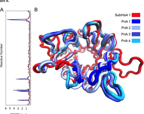
10 β to α loops 1 and 6, as well as in α helix 7, were found to be significantly higher in
PriA than in subHisA (Figure 6A and Movie S1). This observation is in agreement
with the fact that PriA adopts different conformational sub-states related to its
dual-substrate specificity (Due, et al. 2011; Wright, et al. 2008). More importantly, subHisA,
which can only accept ProFAR as a substrate, may have lost conformational diversity
during the process leading to the narrowing of this enzyme’s substrate specificity.
To further investigate the importance of the enzymes accessible conformational
states, we performed a principal component analysis on the molecular dynamics of PriA
and subHisA. This approach allowed us to cluster all conformations adopted by the
enzymes throughout their corresponding dynamics in solution. Indeed, our analyses
revealed the existence of four most populated conformational states in PriA, and only
one in subHisA. Interestingly, two of the four conformational states predicted for PriA
after these analyses were previously reported using co-crystal structures with PRA and
ProFAR analogs (Due, et al. 2011). The latter observation strongly supports the validity
of our findings, which are highlighted in Figure 6B. Given that substrates were not used
for these analyses, moreover, the conformational space explored by PriA thus appears to
be independent of an induced-fit mechanism
Discussion
The most accepted hypothesis regarding enzyme evolution embraces enzyme
substrate ambiguity and the idea that modern enzymes are the result of specialization
processes prompted by gene duplication (Jensen 1976; Piatigorsky 2007). We found,
however, that a generalist enzyme, PriA, is present in approximately 50% of the
organisms belonging to the closely related genera Mycobacterium and Corynebacterium.
11 agreement with early suggestions of a metabolic interlock and common ancestry
between L-Histidine and L-Tryptophan biosynthesis (Jensen 1969; Kane and Jensen
1970; Nester and Montoya 1976). The evidence provided by these reports suggest
cross-regulation, potentially involving the common biosynthetic precursor phosphoribosyl
pyrophosphate, in Bacillus subtilis. Therefore, specialization of these ancient
biosynthetic pathways during the course of evolution in most Actinobacteria must have
been impeded by strong physiological constrains that outweigh the benefits of enzyme
proficiency and pathway specialization.
If enzyme specialization in the sub-clades containing PriA enzymes is
constrained by strong factors, then gene duplication and subsequent divergence can be
expected to occur at low frequency, making it an unlikely event. HGT, as a driving
force for specialization of L-Histidine and L-Tryptophan biosynthesis in the
Corynebacterium lineage receiving the WPTO may have overcome the limitations of
evolution through gene duplication. Indeed, organisms are believed to have evolved
regulation of metabolism in a pathway-specific manner only possible in the absence of
substrate ambiguity (Jensen 1976). Interestingly, in C. glutamicum, where we confirmed
the existence of a subHisA enzyme, feedback gene regulation of L-tryptophan (Brune,
et al. 2007; Ikeda 2006; Xie, et al. 2004; Xie, et al. 2003) and L-histidine (Jung, et al.
2010) biosynthesis, which are specialized pathways, seems to have evolved. The
foregoing physiological regime contrasts with the proficiency of its broad substrate PriA
ancestor, which is encoded within a tightly packed, conserved and constitutively
expressed his-trp gene cluster (Hodgson 2000; Hu, et al. 1999; Parish 2003).
The occurrence of an ancestral-like scenario in modern organisms, i.e. a
generalist enzyme relying in a single active-site that supports committed pathways, not
12 process in enzyme evolution (Depristo 2007; Des Marais and Rausher 2008; Hughes
1994), but raises the interesting question of functional trade-off. Narrowing of enzyme
substrate specificity in subHisA shows that loss of one of the ancestral activities can
occur without compromising the catalytic efficiency of the remaining enzyme function,
even within a highly constrained active site. The conserved substitutions in the branch
where subHisA has evolved suggests that narrowing of substrate specificity in purely
biochemical processes may involve positive selection. In the presence of a PRA
isomerase encoded by a WPTO trpF, the highly conserved mutations leading to
subHisA (e.g. Leu48Ile, Phe51Leu, Ser80Thr, Arg142Asn and shift of Asp127) may
have provided an adaptive mechanism to avoid productive binding of PRA.
This idea is in agreement with the regulatory and physiological regime described
for subHisA-containing Corynebacterium species in previous paragraphs. Therefore,
given the promiscuous-prone active-site of subHisA, as demonstrated by our
site-directed mutagenesis experiment, mutations that would restrain PRA from binding –
without affecting binding of ProFAR – must have been selected for. Solution to such
conundrum speaks out of a complex evolutionary history shaped by the unknown
mechanisms by which HGT operates. Moreover, this may be the reason why subHisA
has not been able to re-specialize to the levels encountered in mono-functional HisA
enzymes, implying a trade-off in terms of evolvability, rather than in absolute enzyme
proficiencies. Investigating the reversibility of subHisA into PriA, to identify mutations
involved in this functional trade-off beyond those that could be pinpointed after our
structural analyses, may shed some light into the raising issues of reversibility (Tokuriki,
et al. 2012).
Our molecular dynamics analyses allowed us to compare the extent of
13 ‘generalist’) and narrow substrate (or ‘specialist’) specificity. Since conformational
diversity has been hypothesized to serve as evolutionary raw material (James and
Tawfik 2003; Tokuriki and Tawfik 2009), our discovery that this conformational
diversity is lost in the narrow substrate, or ‘specialist’ enzyme, is remarkable. PriA has
been previously postulated to accommodate and convert two different substrates
through conformational changes (Due, et al. 2011; Noda-Garcia, et al. 2010; Wright, et
al. 2008). The molecular dynamics results are consistent with these observations. It
should be noted, however, that the conformational states explored by PriA exist
irrespective of the presence of substrates, questioning the likelihood of an induced-fit
mechanism.
In conclusion, during dynamic genome processes, which may include HGT and
differential gene loss, positive selection may be needed to drive both (i) evolution of
narrowing of enzyme substrate specificity from a generalist enzyme; and (ii) efficient
assembly of HGT-acquired biosynthetic pathways within the receiving metabolic
network, as previously postulated (Klassen 2009). Our results also emphasize the need
for an integrated view on the evolution of enzyme substrate specificity, which should
include prokaryotic physiology and genetics. Incorporating HGT into current models of
enzyme evolution, including its formalization within population genetics, seems both a
necessity and an opportunity for evolutionary biology. Finally, our results demonstrate
the importance of multidisciplinary approaches as a powerful conceptual framework to
investigate complex evolutionary mechanisms in biochemical and biophysical processes
(Dean and Thornton 2007).
14
Bioinformatics analysis. The Blast algorithm was used for database searches. The
sequences were aligned with Muscle version 3.6 and edited with Jalview. ProtTest v1.4
(Abascal, et al. 2005) was used to select, out of fifty-six different models, the protein
evolution model that best fit the protein alignments of PriA. According to the statistical
AIC, this model was WAG + I + G + F. The selected protein evolution model and its
parameters were used for the reconstruction of protein phylogenies using the maximum
likelihood methods (Guindon, et al. 2010). The genome context analyses were done
using the Artemis Comparative Tool (Carver, et al. 2005).
Functional characterization of PriA homologs. PriA coding sequence from C. jeikeium
was synthesized by our group (Figure S1) and subHisA coding sequences from C.
amycolatum, C. efficiens, C. matruchotii and C. striatum were synthesized by
GeneART; in both cases codons were optimized for its over expression in E. coli
(Table S3). subHisA from C. diphtheriae and C. glutamicum were cloned from
genomic DNA gently gifted by Androulla Efstratiou (Health Protection Agency, UK)
and from the ATCC collection, respectively. All enzymes were cloned in a pQE-30
derivative (Qiagen) and pET22b (Novagen) using the enzymes NdeI and HindIII. In
vivo E. coli trpF and hisA complementation assays were done as previously reported
(Wright, et al. 2008) other than pQE-30 (Qiagen) derivatives were used, and M9
minimal medium was enriched with a mixture of all the amino acids at 50 μg/ml other
than L-histidine and L-tryptophan. Enzyme purification by Nickel affinity
chromatography was performed as previously reported (Noda-Garcia, et al. 2010). In
vitro Michaelis Menten enzyme kinetic parameters of both PRA and ProFAR isomerase
15 active (positive control) and inactive (negative control) (Henn-Sax, et al. 2002;
Noda-Garcia, et al. 2010).
Construction of the subHisA* mutant. The mutant
subHisA_Cdip_Leu48Ile-Phe50Leu-Thr80Ser was constructed using the site directed mutagenesis kit from
Stratagene. The triple mutant was constructed using the pQEI_subHisA_Cdip_Thr80Ser
as a template (Noda-Garcia, et al. 2010) and the oligonucleotides
Leu48Ile-Phe50Leu_For 5’ggggcatcgtggattcatctggtggatttagat and Leu48Ile-Phe50Leu_Rev
5’atctaaatccaccagatgaatccacgatgcccc. subHisA* was cloned in pET22b (Novagen) using
the enzymes NdeI and HindIII and sequenced before functional analysis.
X-ray crystallography. Overexpressed sub-HisA from Corynebacterium efficiens was
purified as a 6X His-tagged fusion from plasmid pET22-Ceff in E. coli strain BL21star
(DE3) in LB broth. Soluble protein was obtained as reported previously for PriA
(Noda-Garcia, et al. 2010; Wright, et al. 2008). Initial crystallization trials were performed
with screens from Molecular Dimensions Ltd, Hampton Research and Emerald
Bio-structures Inc using the sitting drop vapor diffusion technique. Needle-shaped crystals
were obtained with conditions 20 (0.1M HEPES pH 7.5, 1.4 M Sodium Citrate), 70
(0.1M Bis-Tris pH 5.5, 0.2M MgCl2, 25% w/v PEG 3350) and 71 (0.1 M Bis-Tris pH
6.5, 0.2M MgCl2, 25% w/v PEG 3350) of the Hampton Research screen using 0.2 μl of
protein at 15 mg/ml mixed with an equal volume of mother liquor. After optimization,
crystals grew after 1 or 2 d at 291 K in mother liquor consisting of 0.1 M Bis-Tris pH
7.5, 25% v/w PEG 3350 and 0.2M MgCl2, and mixing 1 μl of protein at 15 mg/ml with
16 Prior to data collection, PriA crystals were cryoprotected by dipping in mother
liquor containing 30% of glycerol and immediately frozen in the N2 cryostream. X-ray
data were collected on the I04 beamline at the Diamond synchrotron (UK) using an
ADSC Q315 CCD detector. All data were indexed, integrated and scaled using the XDS
package. Subsequent data handling was carried out using the CCP4 software package
(1994). Molecular replacement was carried out using the coordinates of S. coelicolor
PriA (PDB: 2vep) as a search model with the PHASER program (McCoy, et al. 2007).
Refinement of the structure was carried out by alternate cycles of REFMAC
(Murshudov, et al. 1997) using non-crystallographic symmetry (NCS) restraints and
manual rebuilding in O (Jones, et al. 1991). Water molecules were automatically added
to the atomic model by Arp/wARP (Perrakis, et al. 1997) and in the last steps of
refinement all the NCS restraints were released. A summary of the data collection and
refinement statistics is given in Table S2.
Molecular dynamics simulations. In order to find the best protocol to perform the
molecular dynamics analysis, an optimization protocol specified in Text S1 & Table S5
was followed. Missing loops from the crystal structure of PriA from M. tuberculosis
(PDB: 2Y89) and a comparative model of subHisA from C. diphtheriae based on the
crystal structure of C. efficiens (PDB: 4AXK, this study) were constructed using Rosetta
3.2.1 (Leaver-Fay, et al. 2011). Addition of missing side-chains and protons was
achieved with the WHATIF tools (Vriend 1990) keeping its predicted protonation state
for His residues and assuming a neutral pH. Topology files, computational cubic box,
solvation, system neutralization by addition of NaCl, system minimization, equilibration
and molecular dynamics simulations were carried out using GROMACS 4.5.3 (Hess, et
17 (MacKerell, et al. 1998) and explicit TIP3P water (Jorgensen, et al. 1983) were used.
Systems were minimized for 5000 conjugate gradient steps and heated up to 300 K
during 600 ps with protein atoms harmonically restrained. This was followed by
equilibration steps done under NvT conditions (300 K) and then under NpT conditions
(1 atm), during 1 ns each, using the V-rescale and isotropic Berendsen barostat methods
without atom restraints.
Long-range electrostatics interactions were included using the Reaction Field
method. A cutoff for the van der Waals interactions was applied with a 1.2 nm radius,
and the LINCS method was used to restrain all bonds involving hydrogen atoms. 300 ns
of molecular dynamics were performed with a time step of 2 fs. Trajectories were
obtained by saving the atomic coordinates of the whole system every 80 ps. Generation
of DCD and PSF files was done with VMD's psfgen plugin (Humphrey, et al. 1996).
Calculation of global RMSDs, radius of gyration and hydrogen bond formation as a
function of time, and average RMSDs per residue were estimated with tools from
GROMACS 4.5.3 (Hess, et al. 2008). Cross-correlation matrix, principal component
analysis (PCA), clustering and average structures were obtained using Carma 1.0
(Glykos 2006). All numerical simulations and corresponding analysis were performed at
the supercomputing center (mazorka) at Langebio. Structure, dynamics and PCA
comparisons amongst subHisA from C. efficiens, subHisA from C. diphtheriae and
PriA from M. tuberculosis are specified in Text S1 & S2.
Acknowledgements. We are indebted with Prof. Therese Markow for useful
discussions and critical reading of this manuscript. We thank Karina Verdel-Aranda,
Helena Wright, Hilda E. Ramos-Aboites, Dean Rea and Ralf Flaig for technical support,
18 a UCMEXUS Conacyt grant to F.B.-G. with Steven E. Brenner and Kimmen Sjölander
as co-applicants, by Conacyt grants to F.B.-G. (No. 50952-Q and No. 83039) and
M.C.-T. (No. 132376), by a joint international Royal Society (UK) grant to V.F. and F.B.-G.,
19
References
1994. The CCP4 suite: programs for protein crystallography. Acta Crystallogr D Biol
Crystallogr 50: 760-763.
Abascal F, Zardoya R, Posada D 2005. ProtTest: selection of best-fit models of protein
evolution. Bioinformatics 21: 2104-2105.
Barona-Gomez F, Hodgson DA 2003. Occurrence of a putative ancient-like isomerase
involved in histidine and tryptophan biosynthesis. EMBO Rep 4: 296-300.
Brune I, Jochmann N, Brinkrolf K, Huser AT, Gerstmeir R, Eikmanns BJ, Kalinowski J,
Puhler A, Tauch A 2007. The IclR-type transcriptional repressor LtbR regulates the
expression of leucine and tryptophan biosynthesis genes in the amino acid producer
Corynebacterium glutamicum. J Bacteriol 189: 2720-2733.
Carver TJ, Rutherford KM, Berriman M, Rajandream MA, Barrell BG, Parkhill J 2005.
ACT: the Artemis Comparison Tool. Bioinformatics 21: 3422-3423.
Dean AM, Thornton JW 2007. Mechanistic approaches to the study of evolution: the
functional synthesis. Nat Rev Genet 8: 675-688.
Depristo MA 2007. The subtle benefits of being promiscuous: adaptive evolution
potentiated by enzyme promiscuity. HFSP J 1: 94-98.
Des Marais DL, Rausher MD 2008. Escape from adaptive conflict after duplication in
an anthocyanin pathway gene. Nature 454: 762-765.
Dittmar K, Liberles D. 2010. Evolution after Gene Duplication. Hoboken, New Jersey:
Wiley-Blackwell.
Due AV, Kuper J, Geerlof A, von Kries JP, Wilmanns M 2011. Bisubstrate specificity
in histidine/tryptophan biosynthesis isomerase from Mycobacterium tuberculosis by
20 Glykos NM 2006. Software news and updates - Carma: A molecular dynamics analysis
program. Journal of Computational Chemistry 27: 1765-1768.
Guindon S, Dufayard JF, Lefort V, Anisimova M, Hordijk W, Gascuel O 2010. New
algorithms and methods to estimate maximum-likelihood phylogenies: assessing the
performance of PhyML 3.0. Syst Biol 59: 307-321.
Henn-Sax M, Thoma R, Schmidt S, Hennig M, Kirschner K, Sterner R 2002. Two
(betaalpha)(8)-barrel enzymes of histidine and tryptophan biosynthesis have similar
reaction mechanisms and common strategies for protecting their labile substrates.
Biochemistry 41: 12032-12042.
Hess B, Kutzner C, van der Spoel D, Lindahl E 2008. GROMACS 4: Algorithms for
highly efficient, load-balanced, and scalable molecular simulation. Journal of Chemical
Theory and Computation 4: 435-447.
Hodgson DA 2000. Primary metabolism and its control in streptomycetes: a most
unusual group of bacteria. Adv Microb Physiol 42: 47-238.
Hu DS, Hood DW, Heidstra R, Hodgson DA 1999. The expression of the trpD, trpC
and trpBA genes of Streptomyces coelicolor A3(2) is regulated by growth rate and
growth phase but not by feedback repression. Mol Microbiol 32: 869-880.
Hughes AL 1994. The evolution of functionally novel proteins after gene duplication.
Proc Biol Sci 256: 119-124.
Humphrey W, Dalke A, Schulten K 1996. VMD: Visual molecular dynamics. Journal of
Molecular Graphics & Modelling 14: 33-38.
Ikeda M 2006. Towards bacterial strains overproducing L-tryptophan and other
aromatics by metabolic engineering. Appl Microbiol Biotechnol 69: 615-626.
James LC, Tawfik DS 2003. Conformational diversity and protein evolution--a
21 Jensen RA 1976. Enzyme recruitment in evolution of new function. Annu Rev
Microbiol 30: 409-425.
Jensen RA 1969. Metabolic interlock. Regulatory interactions exerted between
biochemical pathways. J Biol Chem 244: 2816-2823.
Jones TA, Zou JY, Cowan SW, Kjeldgaard M 1991. Improved Methods for Building
Protein Models in Electron-Density Maps and the Location of Errors in These Models.
Acta Crystallographica Section A 47: 110-119.
Jorgensen WL, Chandrasekhar J, Madura JD, Impey RW, Klein ML 1983. Comparison
of Simple Potential Functions for Simulating Liquid Water. Journal of Chemical
Physics 79: 926-935.
Jung S, Chun JY, Yim SH, Lee SS, Cheon CI, Song E, Lee MS 2010. Transcriptional
regulation of histidine biosynthesis genes in Corynebacterium glutamicum. Can J
Microbiol 56: 178-187.
Kane JF, Jensen RA 1970. Metabolic interlock. The influence of histidine on tryptophan
biosynthesis in Bacillus subtilis. J Biol Chem 245: 2384-2390.
Klassen JL 2009. Pathway evolution by horizontal transfer and positive selection is
accommodated by relaxed negative selection upon upstream pathway genes in purple
bacterial carotenoid biosynthesis. J Bacteriol 191: 7500-7508.
Kuper J, Doenges C, Wilmanns M 2005. Two-fold repeated (betaalpha)4 half-barrels
may provide a molecular tool for dual substrate specificity. EMBO Rep 6: 134-139.
Leaver-Fay A, Tyka M, Lewis SM, Lange OF, Thompson J, Jacak R, Kaufman K,
Renfrew PD, Smith CA, Sheffler W, Davis IW, Cooper S, Treuille A, Mandell DJ,
Richter F, Ban YE, Fleishman SJ, Corn JE, Kim DE, Lyskov S, Berrondo M, Mentzer S,
22 Kuhlman B, Baker D, Bradley P 2011. ROSETTA3: an object-oriented software suite
for the simulation and design of macromolecules. Methods Enzymol 487: 545-574.
Lerat E, Daubin V, Ochman H, Moran NA 2005. Evolutionary origins of genomic
repertoires in bacteria. PLoS Biol 3: e130.
MacKerell AD, Bashford D, Bellott M, Dunbrack RL, Evanseck JD, Field MJ, Fischer
S, Gao J, Guo H, Ha S, Joseph-McCarthy D, Kuchnir L, Kuczera K, Lau FTK, Mattos
C, Michnick S, Ngo T, Nguyen DT, Prodhom B, Reiher WE, Roux B, Schlenkrich M,
Smith JC, Stote R, Straub J, Watanabe M, Wiorkiewicz-Kuczera J, Yin D, Karplus M
1998. All-atom empirical potential for molecular modeling and dynamics studies of
proteins. Journal of Physical Chemistry B 102: 3586-3616.
McCoy AJ, Grosse-Kunstleve RW, Adams PD, Winn MD, Storoni LC, Read RJ 2007.
Phaser crystallographic software. J Appl Crystallogr 40: 658-674.
Murshudov GN, Vagin AA, Dodson EJ 1997. Refinement of macromolecular structures
by the maximum-likelihood method. Acta Crystallogr D Biol Crystallogr 53: 240-255.
Nester EW, Montoya AL 1976. An enzyme common to histidine and aromatic amino
acid biosynthesis in Bacillus subtilis. J Bacteriol 126: 699-705.
Noda-Garcia L, Camacho-Zarco AR, Verdel-Aranda K, Wright H, Soberon X, Fulop V,
Barona-Gomez F 2010. Identification and analysis of residues contained on beta -->
alpha loops of the dual-substrate (beta alpha)8 phosphoribosyl isomerase A specific for
its phosphoribosyl anthranilate isomerase activity. Protein Sci 19: 535-543.
Ohno S. 1970. Evolution by gene duplication: Springer-Verlag.
Pal C, Papp B, Lercher MJ 2005. Adaptive evolution of bacterial metabolic networks by
horizontal gene transfer. Nat Genet 37: 1372-1375.
Parish T 2003. Starvation survival response of Mycobacterium tuberculosis. J Bacteriol
23 Perrakis A, Sixma TK, Wilson KS, Lamzin VS 1997. wARP: Improvement and
extension of crystallographic phases by weighted averaging of multiple-refined dummy
atomic models. Acta Crystallographica Section D-Biological Crystallography 53:
448-455.
Piatigorsky J. 2007. Gene sharing and evolution: the diversity of protein function. .
Cambridge, Massachusetts: Harvard University Press.
Sterner R, Kleemann GR, Szadkowski H, Lustig A, Hennig M, Kirschner K 1996.
Phosphoribosyl anthranilate isomerase from Thermotoga maritima is an extremely
stable and active homodimer. Protein Sci 5: 2000-2008.
Tokuriki N, Jackson CJ, Afriat-Jurnou L, Wyganowski KT, Tang R, Tawfik DS 2012.
Diminishing returns and tradeoffs constrain the laboratory optimization of an enzyme.
Nat Commun 3: 1257.
Tokuriki N, Tawfik DS 2009. Protein dynamism and evolvability. Science 324: 203-207.
Treangen TJ, Rocha EP 2011. Horizontal transfer, not duplication, drives the expansion
of protein families in prokaryotes. PLoS Genet 7: e1001284.
Vriend G 1990. WHAT IF: a molecular modeling and drug design program. J Mol
Graph 8: 52-56, 29.
Wright H, Noda-Garcia L, Ochoa-Leyva A, Hodgson DA, Fulop V, Barona-Gomez F
2008. The structure/function relationship of a dual-substrate (betaalpha)8-isomerase.
Biochem Biophys Res Commun 365: 16-21.
Xie G, Bonner CA, Song J, Keyhani NO, Jensen RA 2004. Inter-genomic displacement
via lateral gene transfer of bacterial trp operons in an overall context of vertical
genealogy. BMC Biol 2: 15.
Xie G, Keyhani NO, Bonner CA, Jensen RA 2003. Ancient origin of the tryptophan
24
Figure 1. L-histidine and L-tryptophan biosynthesis in Actinobacteria. (A)
Convergent pathways, as found in S. coelicolor and M. tuberculosis. (B) Independent
pathways, as found in C. diphtheriae and C. glutamicum. The enzymes for L-histidine
and L-tryptophan biosynthesis are shown in red and blue gradients, respectively.
Phosphoribosyl isomerase A (PriA), at which these pathways converge in (A), is shown
half blue and red. Names and details of the pathways intermediaries and enzymes are
provided as Table S1.
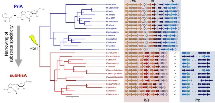
Figure 2. his and trp comparative genomics and priA phylogeny. (A) PriA-based
phylogeny. Numbers at nodes are the approximate likelihood ratio test supporting each
branch. The branches with priA genes, as these sequences co-occur with lack of a trpF
gene, are shown in blue. The branches with subhisA genes, as they co-occur with
HGT-acquired WPTO trpFs, are shown in red. Enzymes selected for further functional
analyses are highlighted with an asterisk. (B) Genomic context analysis of his and trp
genes. The lgt gene, shown in white, was adopted as a genetic marker to define
conservation of gene context. Genes of unknown function, or unrelated to His or
L-Trp biosynthesis, are marked with white triangles (same directionality) or diamonds
(both directionalities). The numbers within these triangles and diamonds indicate how
many predicted genes are in this category. When this is higher than fifteen, disruption of
the his and trp gene cluster is marked with two diagonal black lines. Gene’s
nomenclature and colors are used as in Table S1 & Figure 1.
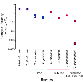
Figure 3. Selected PRA and ProFAR isomerase catalytic efficiencies. ProFAR
isomerase (HisA) and PRA isomerase (TrpF) activities are shown in circles and squares,
25 obtained from (Henn-Sax, et al. 2002) and (Sterner, et al. 1996). PriA are shown in
rblue. Data from S. coelicolor was obtained from (Noda-Garcia, et al. 2010). subHisAs
are shown in red. subHisA* (Leu48Ile, Phe50Leu and Thr80Set) is shown in red/black.
The detailed enzyme kinetic parameters and in vivo characterization is provided as
Table 1.
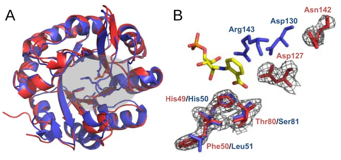
Figure 4. X-ray structural and sequence analysis of subHisA (A) Structure of PriA
(PDB: 2Y85, blue) superimposed on C. efficiens subHisA (chain A, PDB: 4AXK, red).
Key residues in the active site are highlighted. (B) Zoom-in of the superimposed
active-site residues of PriA (blue) and subHisA (red), with rCdRP (yellow), showing at the
bottom of the active site the substrate binding residues His49, Phe50 and Thr80, as well
as variant residues Asp127 and Asn142, which adopt a novel architecture at the top of
the active site.
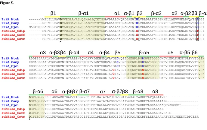
Figure 5. Multiple sequence alignment of PriA (blue) and subHisA (red) sequences.
Catalytic residues, Asp11 and Asp 175, are marked with an asterisk. PRA binding
residues are shown in red/blue. subHisA* gain-of function residues are framed. The
secondary structure is shown at the top of the sequence. Loops are shown in green, α
helixes are shown in red and β sheets are shown in yellow. Sequence corresponding to
loops 1, 5 and 6 is highlighted.
Figure 6. Molecular dynamics of subHisA and PriA. (A) RMSD per residue of PriA
(blue) and subHisA (red) with respect to equilibrated initial structures. (B) Different
average structures, or conformational states, found for PriA (four shades of blue) and
26
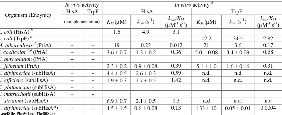
Table 1. Functional characterization of PriA homologs
a
Each data point comes from at least three independent determinations using freshly purified enzyme. n.d., activity not detected, even using active-site saturation conditions. Empty entries reflect our inability to properly express and/or solubilize these
proteins. b Data obtained from (Henn-Sax, et al. 2002) for E. coli HisA and (Sterner, et al. 1996) for E. coli TrpF.c In vivo data
obtained from (Barona-Gomez and Hodgson 2003) d. In vitro data obtained from (Noda-Garcia, et al. 2010) and (Due, et al.
2011) for S. coelicolor and M. tuberculosis, respectively. The discrepancy between the M. tuberculosis data and all otherPriA
enzymes reported here may relate to the fact that sub-optimal conditions were used for determination of the M. tuberculosis
enzyme kinetic parameters, leading to an underestimation of its KM. Standard deviation is not provided for data obtained from
previously published works. Organism (Enzyme)
In vivo activity In vitro activity a
HisA TrpF HisA TrpF
(complementation) KM (M) kcat (s-1)
kcat/KM
(M-1 s-1) KM (M) kcat (s
-1
) kcat/KM
(M-1 s-1)
E. coli (HisA) b 1.6 4.9 3.1
E. coli (TrpF) b 12.2 34.5 2.82
M. tuberculosis d (PriA) + + 19 0.23 0.012 21 3.6 0.17
S. coelicolor c,d (PriA) + + 3.6 0.7 1.3 0.2 0.36 5.0 0.08 3.4 0.09 0.68
C. amycolatum (PriA) + +
C. jeikeium (PriA) + + 2.3 0.2 0.9 0.08 0.39 5.1 1.0 1.6 0.16 0.31
C. diphtheriae (subHisA) + - 4.4 0.5 2.6 0.3 0.59 n.d. n.d n.d.
C. efficiens (subHisA) + - 1.9 0.3 2.7 0.5 1.42 n.d. n.d. n.d.
C. glutamicum (subHisA) + -
C. matruchotii (subHisA) + -
C. striatum (subHisA) + - 6.9 0.7 2.1 0.5 0.3 n.d n.d. n.d
C. diphtheriae (subHisA*)
(Leu48Ile-Phe50Leu-Thr80Ser)
27
28
Figure 2.
0.2
C. jeikeium M. sp. MCS M. ulcerans M. smegmatis M. tuberculosis C. pseudogenitalium C. lipophiloflavum C. striatum C. matruchotii M. vanbaalenii C. resistens C. efficiens C. ammoniagenes C. diphtheriae M. abscessus M. bovis C. accolens C. genitalium M. gilvum C. variabile C. aurimucosum C. urealyticum C. amycolatum C. pseudotuberculosis C. kroppenstedtii C. tuberculostearicum C. glutamicum 0,97 1 1 1 0,95 1 1 1 0,99 0,63 0,95 0,99 0,61 0,91 1 1 0,95 1 0,75 0,95 0,77 1 1 1 0,9
N
a
rr
o
w
in
g
o
f
s
u
b
s
tr
a
te
s
p
e
c
if
ic
it
y
PriA
C R O O
-N N O N CH O NH2 R O H HO OH O
PO3-2
+
subHisA
N N O N CH O NH2 R O H HO OH O
PO3-2
HGT
! " #" $" %" &"
' " ! " (" &" )*+" #" $" ! """""""&" ' " , " - " ! "
#"
$ " !" ! ' " ( " &" %" #" )*+" ( " ! " &"""""""#" $ " , " - " ! " #"
$" ' """""""( " &" %" ' "
$" #" ) " ( "
&" *" ! " ( " +, -" ! "
#"
$ " !" ' " &" %" (" #" )*+" &" ! " %"""""""#" $ " , " - " ! "
#" $ " ! "
%" ! " ' " &" (" )*+" ' " #" "#" &"""""""! " %" , " - " ! "
#"
$ " ! " &" ' " %" (" #" )*+" ' " ! " %"""""""#" $ " , " - " !"
#" $"
%"
&" ' " ( " %" )*+" ( " $" #"""""""%" &" , " - " ! "
#"
$ " ! " &" ' " %" (" #" )*+" , " - " $! #""""""""%" ! " ' " ! "
#" $" %" &"
' " ! " (" &" )*+" #" $" ! """""""&" ' " , " - " !"
#" $"
%"
&" ' " ( " %" )*+" ( " $" #"""""""%" &" , " - " !"
#" $"
%"
&" ' " ( " %" )*+" ( " $" #"""""""%" &" , " - " ! "
#" $" %" &"
' " ! " (" &" )*+" , " - " '! &""""""""! " $" #" ! "
#" $" %" &"
' " ! " (" &" )*+" #" $" ! """""""&" ' " , " - " ! "
#"
$ " %" &" ' """""""(""""""""") " #" ! " &" *+," ! " #" $" %&' " ( " )" *" $" ! " #"
+ " , "
!" #" $"
%"
&" ' " ( " ) " %" $" ( " *+," !"
#" $"
%"
&" ' " ( " ) " %" $" ( " *+," ! "
#" $"
%" &" ' " ," ( " $" #" ' " )*+" ! "
#" $" %" &"
' " ! " )" ( " &" $" #" *+," ! " $" #" &" %" ' " (" !" ! ) " $" #" %" *+," ! " $" #" &" %" ' " (" !" ) " $" #" %" *+," ! " $" #" &" %" ' " (" !" ! ) " $" #" %" *+, " ! " $" #" &" %" ' " (" !" ! ) " $" #" %" *+," ! " $" #" &" %" ' " (" !" ! ) " $" #" %" *+," ! " $" #" &" %" ' " (" !" ! ) " $" #" %" *+," ! " $" #" &" %" ' " (" ) " $" #" %" *+," ! " $" #" &" %" ' " (" !" ! ) " $" #" %" *+,"
his
&
trp
his
trp
29 Figure 3. H is A E . c o li Tr p F E . c o li S . c o e lic o lo r C . je ik e iu m C . e ff ic ie ns C . st ri at um C . d ip h th er ia e C . di p ht he ria e * 0.0001 0.001 0.01 0.1 1 10 subHisA PriA subHisA*
L48I - F50L - T80S
30
Figure 4.
Asn142
Asp127
Thr80
/
Ser81
His49
/
His50
Asp130
Arg143
A
Phe50
/
Leu51
31
Figure 5.
PriA_Mtub ---VMPLILLPAVDVVEGRAVRLVQGKAGSQTEYGSAVDAALGWQRDGAEWIHLVDLDAAFGRGSNHELLAEVVGKLDVQVELSGGI
PriA_Camy ---MSLTLLPAVDVADGQAVRLVQGAAGTETSYGAPLEAAMNWQNAGAEWIHLVDLDAAFGRGSNYDLLADVVGKLDVKVELSGGI
PriA_Cjei MASTDNSRALTLLPAVDVADGQAVRLVQGAAGTETSYGAPIEAALAWQNAGAEWIHLVDLDAAFGRGSNFELLKEVTGQLDVNVELSGGI
subHisA_Cdip ---MTFTLLPAVDVVDGQAVRLDQGEAGTEKSYGSPIAAALKWQEQGASWLHFVDLDAAFNRGSNHELMAEVVKNLDINVELTGGI
subHisA_Ceff ---MTFTILPAVDVVNGQAVRLDQGEAGTEKSYGTPLESALRWQEQGAEWLHFVDLDAAFNRGSNHELMAEITRQLDIKVELTGGI
subHisA_Cstr ---MSFTLLPAVDVVDGQAVRLDKGEAGTEKSYGAPREAAEKWQAQGAEWLHFVDLDAAFNRGSNYELMAEITSSLDIQVELTGGI
PriA_Mtub RDDESLAAALATGCARVNVGTAALENPQWCARVIGEHGDQVAVGLDVQIIDGEHRLRGRGWETDGGDLWDVLERLDSEGCSRFVVTDITK
PriA_Camy RDNASLEAALATGCARVNIGTAALENPEWCREVIANYGDRVAIGLDVLNDEGQWRLRGRGWVSDGGDLWEVLERLDAQGASRFVVTDVSK
PriA_Cjei RDDESLERALSTGCRRVNIGTAALEDPEWCESVISRYGDKVAIGLDTREVDGEWRLRGRGWTSDGGELWEVLERLDSQGVSRLVVTDVSR
subHisA_Cdip RDDASLKRALATGARRVNIGTAALEKPEWIEKVLGEYGDAIAVDIAVRNIDGQWRTRGNGWVSDGGDLWEVLERLDSQGCTRFVVTDVSK
subHisA_Ceff RDDASLERALATGATRVNIGTAALEKPEWIADVIRRHGEKIAVDIAVRLENGEWRTKGNGWVSDGGDLWEVLERLDSQGCSRFVVTDVSK
subHisA_Cstr RDDESLARVLATGARRVNIGTAALENPEWIEKVLAEHGDKIAVDLAVRLEDGEWRTRGNGWVSDGGDLWEVLERLDAAGCTRFVVTDVSK
PriA_Mtub DGTLGGPNLDLLAGVADRTDAPVIASGGVSSLDDLRAIATLTHRGVEGAIVGKALYARRFTLPQALAAVRD---
PriA_Camy DGTLQGPNVELLREVAAATDAPIVASGGVSSLDDIAAIATLVDEGVDSAIVGKALYAGRFTLEEALAIARG---
PriA_Cjei DGMLNGPNIDLLREVAAATDAPVVASGGISSLDDIRALAAVVHEGVDSAIVGKALYAGKFTLEEALEAAQGVARGSDI---
subHisA_Cdip DGTLSGPNIDLLRDVSAATDAKVVASGGISTLEDVLELARYEDEGIDSAIIGKALYEGRFTLKEALAAL---
subHisA_Ceff DGTLTGPNVDLLRDVAAATDAPIVASGGISTLEDVLGLAKYQDEGIDSVIIGKALYEHRFTLAEALEAVEKLG---
subHisA_Cstr DGTLEGPNVQLLREVAAATDAKVTASGGISTLDDLRELALYENQGIDSAIIGKALYEGRFSLEEALAAVAEVEPLPEEDYIDPIEER
β1
β-α1
α1
α-β1
β2
β-α2
α2
α-β2
β3
β-α3
α3
α-β3
β4
β-α4
α4
α-β4
β5
β-α5
α5
α-β5
β6
β7
β8
α6
α7
α8
β-α6
α-β6
β-α7
α-β7
β-α8
*
32