Original citation:
Turner, Matthew S. , Platts, James A. and Deeth, Robert J.. (2016) Modeling of platinum-aryl interaction with amyloid-β peptide. Journal of Chemical Theory and Computation, 12 (3). pp. 1385-1392.
Permanent WRAP URL:
http://wrap.warwick.ac.uk/78916
Copyright and reuse:
The Warwick Research Archive Portal (WRAP) makes this work by researchers of the University of Warwick available open access under the following conditions. Copyright © and all moral rights to the version of the paper presented here belong to the individual author(s) and/or other copyright owners. To the extent reasonable and practicable the material made available in WRAP has been checked for eligibility before being made available.
Copies of full items can be used for personal research or study, educational, or not-for profit purposes without prior permission or charge. Provided that the authors, title and full bibliographic details are credited, a hyperlink and/or URL is given for the original metadata page and the content is not changed in any way.
Publisher’s statement:
This document is the Accepted Manuscript version of a Published Work that appeared in final form in Journal of Chemical Theory and Computation, copyright © American Chemical Society after peer review and technical editing by the publisher.
To access the final edited and published work see :
http://dx.doi.org/10.1021/acs.jctc.5b01045
A note on versions:
The version presented here may differ from the published version or, version of record, if you wish to cite this item you are advised to consult the publisher’s version. Please see the ‘permanent WRAP url’ above for details on accessing the published version and note that access may require a subscription.
Modeling of Platinum-Aryl Interaction with Amyloid- Peptide
Matthew Turner1 James A. Platts1* and Robert J. Deeth2†
1 School of Chemistry, Cardiff University, Park Place, Cardiff CF10 3AT, UK; 2 Department of
Chemistry, University of Warwick, Gibbet Hill, Coventry, CV4 7AL
Abstract
Ligand field molecular mechanics (LFMM), density functional theory (DFT) and semi-empirical
PM7 methods are used to study the binding of two Pt(II)-L systems to an N-terminal fragment of
the amyloid-β peptide, where L = 2,2-bipyridyl or 1,10-phenanthroline. Molecular dynamics
simulations are used to explore the conformational freedom of the peptide using LFMM combined
with AMBER molecular mechanics parameters. We establish a modelling protocol, allowing for
identification and analysis of favorable platinum-binding modes and peptide conformations.
Preferred binding modes are identified for each ligand investigated; metal coordination occurs via
Nε in His residues for both ligands - His6ε-His13ε and His6ε-His14ε for the bipyridyl and
phenanthroline ligands, respectively. The observed change in binding mode for the different
ligands suggests that the binding mode of these platinum-based structures can be controlled by the
choice of ligand. In the bipy systems, Boltzmann population at 310K is dominated by a single
to the ensemble. The relative stability of these conformations is due to the inherent stability of
binding platinum via Nε in addition to subtle H-bonding effects.
Introduction
Alzheimer’s disease (AD) is a widespread neurodegenerative condition associated with
progressive cognitive decline in patients. AD is the most common cause of dementia in adults and
the fourth most common cause of death in Western countries.1 The pathogenesis of AD is
complex, involving the interplay of a range of molecular, cellular and physiological processes2;
the causes and development of AD are not well understood, with links to both lifestyle3,4 and
genealogy.5 Amyloid-β (Aβ) peptide was first recognized as a factor in AD in 19856 and has been
the subject of the much research in the decades since. The well-publicized amyloid-cascade
hypothesis remains popular, if controversial, and suggests that individual Aβ peptides - between
39-43 residues in length - agglomerate, leading to plaque formation.7,8,9,10 These plaques are
thought to be the cause of local inflammation and neuronal cell death. Subsequent effects seen in
AD may be caused by this imbalance between Aβ generation and clearance. As such, the early
stages of Aβ aggregation form a realistic druggable target for the development of anti-AD
therapeutics.2
The N-terminal region of Aβ contains histidine-rich metal binding sites that are responsible for
physiological coordination of transition metals. These metal ions are believed to play an important
role in the development of amyloid plaques; analysis of fibrils in Alzheimer’s brains proves that
they show increased concentrations of transition metal ions such as Cu, Zn and Fe.11,12 However,
the exact metal binding sites are the subject of debate, with authors showing varied metal
development of compounds that selectively occupy Cu/Zn binding sites on Aβ and so prevent
aggregation and oligomerisation.
Pioneering work by Barnham et al.17 showed that PtII(phenanthroline) complexes are able to
inhibit Aβ aggregation, and inhibit its neurotoxicity in vitro. Additionally, Pt(II) compounds are
stable (i.e. kinetically and redox inert) in biological systems, meaning that once the Pt(II) complex
is bound to a target, it is very difficult to displace. However, there has been a great deal of debate
over exactly how these platinum-ligand systems interact with the Aβ peptide. Of particular
relevance to the current study, Streltsov et al combined DFT and EXAFS data for a
Pt(phen)(imidazole)2 model complex to arrive at experimentally derived predictions of binding
modes.18
Spectroscopic evidence19 suggests that several platinum adducts are formed in the coordination
of PtII(phen) complexes to Aβ, but that His-6 and His-14 are preferentially bound. Recent work
has also shown that aromatic ligands such an phenanthroline are vital for activity - it is believed
that π-π stacking interactions between the ligands and aromatic residues Phe, Tyr and His are
important in the association of PtII(phen) to Aβ.17,20,21
Computational modelling of molecules of this type present significant challenges; quantum
mechanical (QM) methods such as DFT are not routinely applicable to large protein systems,
especially flexible ones such as Aβ. Classical modelling techniques using molecular mechanics
(MM) forcefields typically fail to model the effects of transition metal d-electrons on the structure
and properties of coordination complexes. This problem may be circumvented by combining the
quantum and classical techniques, using QM for the metal center and MM for the remainder of the
similar to those investigated here22,23,24. However, there is still a significant cost associated with
the QM calculation compared to pure MM.
An alternative is ligand field molecular mechanics (LFMM)25,26, designed to provide ‘the
flexibility and generality of quantum mechanics with the speed of molecular mechanics.’26 This
technique has been applied to the study of a variety of complex metal-based systems, such as d9
Cu(II)27, high- and low-spin d8 Ni(II)28 and d8 Pt(II)29,30. The aim of this work is to establish a
protocol for generating and subsequently analyzing conformations of a series of platinum(II)
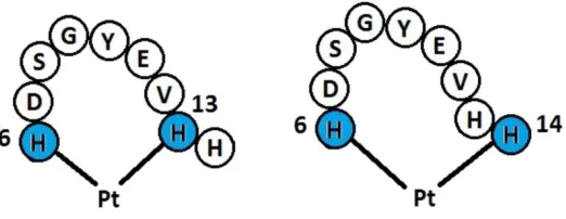
species bound to a fragment of Aβ peptide (residues 6-14 – see Figure 1), with the aim of
identifying favorable metal-binding modes and peptide conformations. This model is too small to
truly represent the biology of Aβ, but this subset of residues is large enough to contain those
important for platinum binding, while remaining such a size that we can benchmark computational
results against DFT. This preliminary work presents findings for two platinum-ligand systems:
2,2-bipyridyl (bipy) and 1,10-phenanthroline (phen), the choice of which stem from recent
literature. Ultimately, this protocol could be extended to study other ligands, different transition
metals such as those of physiological importance (e.g. copper and zinc)31 or other potential
[image:5.612.175.436.529.630.2]therapeutics of current research interest (e.g. ruthenium)32.
Computational Methods
The peptide sequence His6-Asp-Ser-Gly-Tyr-Glu-Val-His-His14, taken from the N-terminal
domain of Aβ was built in an extended conformation in MOE33. Pt was added in eight distinct
coordination modes (i.e. to His6 and either His13 or His14 through N or N), and the bipyridine
or phenanthroline ligand manually constructed. For conformational searching, the resulting
complexes were described using molecular mechanics using a combination of ligand field
molecular mechanics (LFMM) for Pt26,29,30 and AMBER9434 parameters for all other atoms, as
implemented in the DommiMOE35 extension to MOE. Partial charges for Pt and coordinated
groups were calculated for model Pt(imidazole)2(ligand) systems using the Merz-Kollman scheme
from HF/6-31G(d)/SDD electrostatic potential in Gaussian0936, with Pt given a van der Waals
radius of 2.0 Å. The remaining peptide atoms, i.e. those beyond coordinated imidazole, were given
AMBER94 charges as calculated by MOE. Solvation effects were modelled using the
reaction-field model with default parameters.
Conformational freedom was explored through the LowMode MD37 method in MOE. LowMode
MD searches were configured to terminate after 100 successive failures to generate a new
molecular conformation, up to a maximum of 10,000 iterations. An arbitrarily large energy cut-off
(10,000 kcalmol-1) was used alongside an RMSD cut-off of 0.25 Å for retention of all new
conformations. LowMode MD is able to analyze complex molecular systems such as protein loops
and macrocycles, taking into account complex non-bonded interactions, and is able to locate
low-energy conformations for a variety of structures in a computationally efficient manner37,38,39.
During initial conformational searches, it was found that often a large portion of the kinetic energy
conformations since their final position is determined by the LFSE contribution to LFMM. This
lead to the LowMode MD search becoming ‘stuck’ and unable to escape a certain conformation,
resulting in the premature termination of the conformational search. We resolved this by fixing the
platinum centre, making the mass of this atom effectively infinite.
DFT calculations were performed using Gaussian09 with the BHandH40 functional with a
6-31G(d) basis set on all atoms in conjunction with a Stuttgart-Dresden (SDD)41,42 effective core
potential for platinum(II), within a polarizable continuum model (PCM) model of aqueous
solvation43,44. The choice of functional was determined by modelling relevant Pt(II) crystal
structures, while BHandH has been widely used in description of -stacking and other
dispersion-based interactions.45 In addition, two more modern functionals, B97D and M06-2X,46,47
specifically designed to account for dispersion were tested for a subset of complexes.
Semi-empirical calculations were performed using MOPAC48 with the PM7 method49 and the COSMO
model of aqueous solvation50; full relaxation was achieved through use of keywords LET and
DDMIN=0. Overlay plots were obtained using Chimera imaging software51. RMSD values were
calculated using a python script obtained via GitHub (https://github.com/charnley/rmsd)52,53.
Results and Discussion
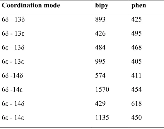
Using the conformational searching protocol described above, searches were performed on the
eight platinum binding modes of interest for each ligand system. This produced between 400 and
1600 conformers for each platinum binding mode (
Table 1). There is appreciable variation between sets which may be at least partially due to the
stochastic nature of the LowMode MD method as well as the inherent flexibility of the different
Pt-coordination results in 9,962 conformations from 10,000 starting attempts. It is therefore apparent
from
Table 1 that Pt binding to two His residues allows the peptide chain significant flexibility, no
matter which coordination mode is considered, but that this conformational freedom is
[image:8.612.167.447.305.524.2]significantly reduced compared to the free peptide.
Table 1. Number of conformers found using LowMode MD for the ligands studied.
Coordination mode bipy phen
6δ - 13δ 893 425
6δ - 13ε 426 495
6ε - 13δ 484 468
6ε - 13ε 995 405
6δ -14δ 574 411
6δ -14ε 1570 454
6ε - 14δ 429 618
6ε - 14ε 1135 450
While the MM protocol used to generate data in
Table 1 has been successfully applied to many similar problems, it is important to validate the
results it provides against other theoretical approaches. In particular, we note that LFMM has
long-standing use for metal-peptide binding, but that Pt(II) is not yet among the metals used. Pt(II)
parameters are available, and have been used to model Pt-DNA complexes29,30. In the current case,
LFMM has typically been used. DFT was therefore applied to a subset of 158 unique low-energy
conformers, encompassing several different platinum-binding modes, in order to test further
computational methods. We first consider the relative energies of LFMM-generated structures,
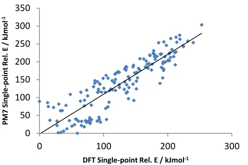
calculated without further geometry relaxation. We find little correlation between LFMM and DFT
relative energies at LFMM geometry (R2 = 0.14), suggesting that the combined LFMM/AMBER
approach does not accurately predict the relative energies of different conformers.
In order to check the reliability of DFT methods used as a benchmark, a set of non-platinated
conformations was constructed by removing PtL from conformations located as discussed above,
and their energies calculated using DFT and AMBER. Three different DFT methods (BHandH,
B97D and M06-2X) show excellent agreement with one another (R2 BHandH vs. B97D = 0.94,
BHandH vs. M06-2X 0.93, B97D vs. M06-2X 0.95), supporting our conclusion that BHandH is a
suitable method for these structures. Furthermore, these DFT methods show little agreement with
the AMBER energies for these non-platinated systems (R2 BHandH vs. AMBER = -0.02). This
suggests that the poor agreement between DFT and LFMM/AMBER energies described above
stems from the use of AMBER94 for the peptide, rather than from use of LFMM for the metal
centre.
Due to the high computational expense of DFT calculations, a faster computational method was
required to identify conformers of interest. PM7 and DFT energies of platinated conformations at
MM geometries are in good agreement with one another (R2 = 0.78); PM7 therefore reproduces
DFT energies at a much lower computational cost. This suggests that PM7 is a suitable theoretical
method for structures of this type. This assertion is reinforced by the modelling of the
non-platinated conformations: BHandH and PM7 calculations show strong agreement (R2 = 0.77),
Figure 2. Plot of DFT vs. PM7 relative energies at LFMM geometry.
Table 2. Correlation between relative energies of 158 conformers.
LFMM PM7 DFT
LFMM 1.00 0.38 0.14
PM7 - 1.00 0.78
DFT - - 1.00
The other key consideration of the LFMM results is whether the geometries produced are
reliable. We therefore carried out DFT and PM7 geometry optimization of all 158 platinated
conformers, and evaluated the all-atom RMSD of the resulting structures from those obtained from
LFMM energy minimization.
0 50 100 150 200 250 300 350
0 100 200 300
P M 7 Si ng le -poi nt R e l. E / kJ m ol -1
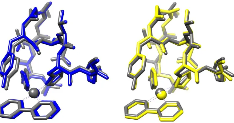
[image:10.612.73.534.436.533.2]Table 3 reports the results of such comparison, and demonstrates that LFMM and DFT are in
good general agreement, i.e. DFT optimization of a selected structure does not significantly alter
[image:11.612.142.472.233.335.2]the overall geometry, indicating that LFMM produces robust geometries.
Table 3. Summary of geometry comparison between LFMM, PM7 and DFT.
RMSD VALUES LFMM vs. DFT PM7 vs. DFT
Mean 0.670 0.817
SD 0.191 0.307
Min. 0.336 0.339
Max. 1.293 1.982
Figure 3. Left: LFMM (blue) vs DFT (grey) geometry overlay. Right: PM7 (yellow) vs DFT
geometry (grey) overlay.
[image:11.612.118.498.396.597.2]approach might be suitable for this purpose. Table 3 shows that, on average, PM7-optimised
structures are slightly further from DFT ones than LFMM structures (mean RMSD 0.82 vs. 0.67
Å). These values include data from 9 conformers for which PM7 predicts large (> 1.3 Å) RMSD
from DFT-optimized geometry. For the remaining 149 structures, the mean RMSD is 0.77 Å.
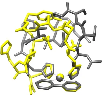
Interestingly, further DFT optimization for the 9 complexes with large RMSD, starting from the
PM7 endpoint produced significant reduction in energy, indicating that in some cases PM7
geometry optimization in MOPAC is able to escape from the local minimum located by LFMM
conformational search. One such case is illustrated in Figure 4 below, in which DFT optimization
following initial PM7 refinement leads to a structure that is 62.3 kJ mol-1 more stable than that
[image:12.612.205.404.358.542.2]reached by optimization directly from the LFMM structure.
Figure 4. DFT optimised structure (grey) overlaid with PM7-DFT optimised structure (yellow).
We therefore surmise that PM7/COSMO optimization following initial LFMM conformational
search is a useful step in obtaining reliable energy and geometry data. Although the extra step
minutes per structure on a single compute node), the evidence above indicates that it provides a
more balanced set of data than either LFMM alone or PM7 at the LFMM geometry. We therefore
performed this step for all conformations located for both ligands, a total of 6506 for bipy and
3726 for phen. The resulting relative energies were collated and used in Boltzmann weighting to
determine the binding mode(s) and conformation(s) that contribute significantly to the overall
[image:13.612.163.453.439.702.2]ensemble at 310 K, the results of which are reported in Table 4.
Table 4. Relative energy and Boltzmann factors for low energy conformations.
Ligand Coordination Rel. E / kJ mol-1
Boltzmann
fraction
Bipy 6ε - 13ε 0.00 0.79
“ 6.27 0.07
“ 7.07 0.05
“ 7.51 0.04
Phen 6ε - 14ε 0.00 0.38
“ 0.77 0.28
“ 1.96 0.18
“ 4.14 0.08
On the basis of these results, we predict that Pt(II) binds to histidine residues via Nε rather than
Nδ. The first conformations that feature binding via Nδ of any His residue are found over 22 and
15 kJ mol-1 for bipy and phen ligands, respectively, and so contribute almost nothing to the
Boltzmann-weighted ensemble of structures. This is an inherent property of the histidyl residue:
calculations on a model system Pt(bipy)(Cl)His gives a preference for Nε over Nδ of 5 kJ mol-1
using DFT, and 3 kJ mol-1 using PM7. The data in Table 4 indicate that, out of the several thousand
conformations considered, just a small number contribute significantly to the Boltzmann-weighted
ensemble. A single conformation accounts for almost 80% of the ensemble of bipy complexes,
and four conformations for over 95%. Three low energy conformations account for over 80% of
the ensemble of phen complexes, and five account for 99%. This shows that coordination of Pt
massively reduces the conformational freedom of the peptide by “pinning” two histidines to the
metal center.
There is also a clear preference in coordination mode for each ligand; metal binding occurs via
Nε of His-6 and His-13 for the bipy systems and via Nε of His-6 and His-14 for the phenanthroline
systems. This result is particularly promising for the phenanthroline case, since the preferred
metal-binding mode identified here (6ε - 14ε) was also determined to be the binding mode of this
ligand system in experimental work carried out by Ma et al.19 The fact that a relatively small
change in ligand from bipy to phen alters the preferred coordination mode is somewhat surprising,
and suggests that binding mode can be controlled through suitable choice of ligand.
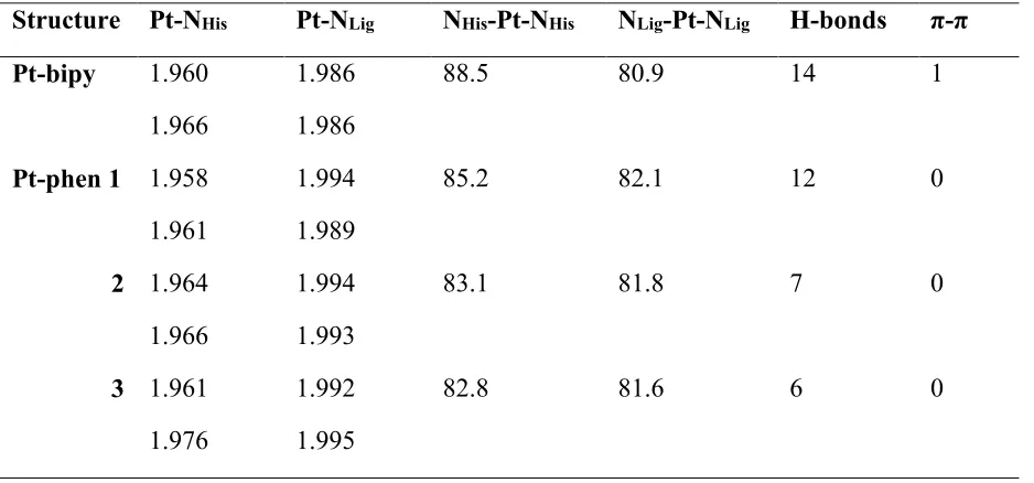
Table 5 reports selected details of the DFT-optimized structures of the low energy
conformation(s) for each ligand system. These data show that there is almost no variation in Pt—
N bond lengths following change in conformation and/or coordinated ligand. In the bipy complex,
the bonds to the N-donor ligands are slightly longer than those to the histidine residues of Aβ
(mean values 1.963±0.004 vs 1.986±0.000 Å). Moreover, Pt—NHis distances are slightly shorter
in the low energy bipy conformation than the average over all conformations located for the same
binding mode (mean = 1.966 Å ±0.005), whereas Pt—NLig distances are slightly larger (mean =
1.981 Å ± 0.004). However, differences are typically within 1 or 2 standard deviations, and so not
statistically significant. One apparently significant difference lies in the NHis-Pt-NHis angle that is
on average 84.5 (±1.75) for the collection of 6ε - 13ε conformations but 88.5 in the single
low-energy conformation. It is notable that these Pt—N distances are in good agreement with those
reported by Streltsov et al,18 Pt–N(imidazole) and Pt–N(phen) bond distances of 1.99(1) Å and
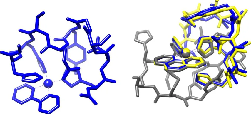
Figure 5. Left: Low energy bipy conformation. Right: Lowest (grey), second- and third-lowest
Table 5. Selected geometrical details of single low energy conformations (Pt-bipy) and three low
energy conformations (Pt-Phen)
Structure Pt-NHis Pt-NLig NHis-Pt-NHis NLig-Pt-NLig H-bonds π-π
Pt-bipy 1.960
1.966
1.986
1.986
88.5 80.9 14 1
Pt-phen 1 1.958
1.961
1.994
1.989
85.2 82.1 12 0
2 1.964
1.966
1.994
1.993
83.1 81.8 7 0
3 1.961
1.976
1.992
1.995
82.8 81.6 6 0
As expected for biomolecular systems, a large number of intramolecular hydrogen bonds are
present in the low energy conformations, including backbone-backbone, backbone-side chain and
side chain-side chain interactions. The low energy Pt-bipy complex contains 14 H-bonds, as judged
by geometrical criteria. Of these, five are concentrated around Glu-11 and a further three around
Asp7. Four of these H-bonds are relatively long at over 2 Å, but a number of strong interactions
were also identified, including those between backbone and residue (Asp7 – backbone N-H at
1.659 Å and Glu11 – N-terminal N-H at 1.794 Å) and residue-residue (Glu11 – His14 at 1.691 Å
and Asp7 – His13 at 1.775 Å) which may be responsible for the stability of this conformation. In
addition, four backbone-backbone H-bonds were identified in residues Gly9-His14. The low
side chain of His14. The two ring systems lie in an approximately parallel orientation (angle
between mean planes = 29.9) with an inter-centroid distance of approximately 4 Å.
The Pt-phen complexes identified exhibit markedly fewer H-bonds than the bipy complex, and
also no evidence for π-π stacking interactions is found in these low energy conformations (see
Figure 5). As before, the H-bonds are generally found around residues Glu11 and Asp7, though
strong interactions (average length 1.526 Å over the three phen conformations) were identified
between the hydroxyl group of Tyr10 and Asp7.
Conclusions
LFMM has previously been demonstrated to be a powerful tool in predicting geometries and
conformations of biomolecules bound to TM species. In the present work, we aim to apply this
methodology to the study of platinum-complexes of the type studied by Barnham17,21 bound to
model amyloid-β fragments in order to determine favourable metal-binding modes and peptide
conformations. Thorough exploration of the conformational space of these biomolecules was
achieved using LowMode MD in conjunction with AMBER molecular mechanics parameters.
Comparison of calculated relative energies of a subset of these conformations indicates that LFMM
fails to reproduce DFT results (R2 = 0.14). However, QM evaluation of structures of this size is
computationally expensive, so the less expensive semi-empirical PM7 method was utilized, which
agrees well with QM results (R2 = 0.78); PM7 therefore reproduces DFT energies at a much lower
computational cost. In addition, PM7 optimization generally retains the DFT structure, with
average all-atom RMSD of 0.817 over 158 conformers. This illustrates that PM7 accurately
reproduces DFT geometries as well as energies. The present results indicate that PM7 is an
appropriate and computationally manageable approach to modelling these metal-biomolecule
This semi-empirical approach was subsequently extended to all conformations identified by
LFMM. Statistical thermodynamics was applied to the generated databases in order to generate
relative populations of each conformer at 310K. It was found that platinum coordination occurs
via the Nε atom in His residues for both of the ligands investigated here. Favorable
platinum-coordination modes were identified for each ligand: 6ε-13ε and 6ε-14ε for bipy and phen,
respectively. Furthermore, this change in binding mode for different ligands suggests that the
binding mode can be controlled by the choice of ligand.
In the bipy systems, the Boltzmann population is dominated by a single conformer that accounts
for almost 80% of the weighted ensemble containing a large number of intramolecular H-bonds
and a possible π-stacking interaction between His 14 and the bipy ligand. In the phenanthroline
case, three conformations make significant contributions to the ensemble. Each of these
conformations contains fewer intramolecular H-bonds than the dominant bipy conformation and
no evidence of π-π stacking interactions was found.
As this work continues, research will focus on two areas. Firstly, the peptide fragment will be
extended to at least the Aβ1-16 unit studied in experimental work19,54. This would allow
confirmation of platinum binding modes on a more realistic model of biological Aβ. Additionally,
this modelling protocol may be used to study other ligands, such as those investigated by Barnham
et al.,21 Collin et al.55 and Yellol et al.56 and Furthermore, the flexibility of LFMM allows the study
of different transition metals such as those of physiological importance (e.g. copper and zinc)31 or
other potential therapeutics of current research interest (e.g. ruthenium32 or mixed-metal
complexes57).
This work was carried out using resources of the Advanced Research Computing @ Cardiff
(ARCCA) and HPC Wales facilities. MT is grateful to Cardiff University for a studentship. RJD
acknowledges the University of Edinburgh for granting an Honorary Fellowship. Molecular
graphics were obtained using the UCSF Chimera package. Chimera is developed by the Resource
for Biocomputing, Visualization, and Informatics at the University of California, San Francisco
(supported by NIGMS P41-GM103311). RJD acknowledges the support of Chemical Computing
Group.
References
(1) Brookmeyer, R.; Johnson, E.; Ziegler-Graham, K.; Arrighi, H. M. Forecasting the Global Burden of Alzheimer’s Disease. Alzheimers Dement. J. Alzheimers Assoc. 2007, 3, 186– 191.
(2) Valensin, D.; Gabbiani, C.; Messori, L. Metal Compounds as Inhibitors of Beta-Amyloid Aggregation. Perspectives for an Innovative Metallotherapeutics on Alzheimer’s Disease.
Coord. Chem. Rev.2012, 256, 2357–2366.
(3) Elwood, P.; Galante, J.; Pickering, J.; Palmer, S.; Bayer, A.; Ben-Shlomo, Y.; Longley, M.; Gallacher, J. Healthy Lifestyles Reduce the Incidence of Chronic Diseases and Dementia: Evidence from the Caerphilly Cohort Study. PLoS ONE2013, 8, e81877.
(4) Cai, W.; Uribarri, J.; Zhu, L.; Chen, X.; Swamy, S.; Zhao, Z.; Grosjean, F.; Simonaro, C.; Kuchel, G. A.; Schnaider-Beeri, M.; Woodward, M.; Striker, G. E.; Vlassara, H. Oral Glycotoxins Are a Modifiable Cause of Dementia and the Metabolic Syndrome in Mice and Humans. Proc. Natl. Acad. Sci.2014, 201316013.
(5) Amouyel, P. et al. Meta-Analysis of 74,046 Individuals Identifies 11 New Susceptibility Loci for Alzheimer’s Disease. Nat. Genet.2013, 45, 1452–U206.
(6) Masters, C.; Simms, G.; Weinman, N.; Multhaup, G.; Mcdonald, B.; Beyreuther, K. Amyloid Plaque Core Protein in Alzheimer-Disease and down Syndrome. Proc. Natl. Acad. Sci. U. S. A.1985, 82, 4245–4249.
(7) Selkoe, D. The Molecular Pathology of Alzheimers-Disease. Neuron1991, 6, 487–498. (8) Walsh, D. M.; Klyubin, I.; Fadeeva, J. V.; Cullen, W. K.; Anwyl, R.; Wolfe, M. S.; Rowan,
M. J.; Selkoe, D. J. Naturally Secreted Oligomers of Amyloid Beta Protein Potently Inhibit Hippocampal Long-Term Potentiation in Vivo. Nature2002, 416, 535–539.
(9) Haass, C.; De Strooper, B. Review: Neurobiology - The Presenilins in Alzheimer’s Disease - Proteolysis Holds the Key. Science1999, 286, 916–919.
(10) Cleary, J. P.; Walsh, D. M.; Hofmeister, J. J.; Shankar, G. M.; Kuskowski, M. A.; Selkoe, D. J.; Ashe, K. H. Natural Oligomers of the Amyloid-Protein Specifically Disrupt Cognitive Function. Nat. Neurosci.2005, 8, 79–84.
(12) Barnham, K. J.; Masters, C. L.; Bush, A. I. Neurodegenerative Diseases and Oxidative Stress. Nat. Rev. Drug Discov.2004, 3, 205–214.
(13) Bolognin, S.; Messori, L.; Drago, D.; Gabbiani, C.; Cendron, L.; Zatta, P. Aluminum, Copper, Iron and Zinc Differentially Alter Amyloid-A beta(1-42) Aggregation and Toxicity.
Int. J. Biochem. Cell Biol.2011, 43, 877–885.
(14) Kenche, V. B.; Barnham, K. J. Alzheimer’s Disease & Metals: Therapeutic Opportunities.
Br. J. Pharmacol.2011, 163, 211–219.
(15) Zirah, S.; Kozin, S. A.; Mazur, A. K.; Blond, A.; Cheminant, M.; Segalas-Milazzo, I.; Debey, P.; Rebuffat, S. Structural Changes of Region 1-16 of the Alzheimer Disease Amyloid Beta-Peptide upon Zinc Binding and in Vitro Aging. J. Biol. Chem. 2006, 281, 2151–2161.
(16) Giannozzi, P.; Jansen, K.; La Penna, G.; Minicozzi, V.; Morante, S.; Rossi, G.; Stellato, F. Zn Induced Structural Aggregation Patterns of Beta-Amyloid Peptides by First-Principle Simulations and XAS Measurements. Metallomics2012, 4, 156–165.
(17) Barnham, K. J.; Kenche, V. B.; Ciccotosto, G. D.; Smith, D. P.; Tew, D. J.; Liu, X.; Perez, K.; Cranston, G. A.; Johanssen, T. J.; Volitakis, I.; Bush, A. I.; Masters, C. L.; White, A. R.; Smith, J. P.; Cherny, R. A.; Cappai, R. Platinum-Based Inhibitors of Amyloid-Beta as Therapeutic Agents for Alzheimer’s Disease. Proc. Natl. Acad. Sci. U. S. A. 2008, 105, 6813–6818.
(18) Streltsov, V. A.; Chandana Epa, V.; James, S. A.; Churches, Q. I.; Caine, J. M.; Kenche, V. B.; Barnham, K. J. Structural Insights into the Interaction of Platinum-Based Inhibitors with the Alzheimer’s Disease Amyloid-Β Peptide. Chem. Commun.2013, 49, 11364.
(19) Ma, G.; Huang, F.; Pu, X.; Jia, L.; Jiang, T.; Li, L.; Liu, Y. Identification of [PtCl2(phen)] Binding Modes in Amyloid-Beta Peptide and the Mechanism of Aggregation Inhibition.
Chem.- Eur. J.2011, 17, 11657–11666.
(20) Yao, S. G.; Cherny, R. A.; Bush, A. I.; Masters, C. L.; Barnham, K. J. Characterizing Bathocuproine Self-Association and Subsequent Binding to Alzheimer’s Disease Amyloid Beta-Peptide by NMR. J. Pept. Sci.2004, 10, 210–217.
(21) Barnham, K. J.; Kenche, V. B.; Hung, L. W.; Perez, K.; Volitakes, I.; Ciccotosto, G.; Kwok, J.; Critch, N.; Sherratt, N.; Cortes, M.; Lal, V.; Masters, C. L.; Murakami, K.; Cappai, R.; Adlard, P. A. Development of a Platinum Complex as an Anti-Amyloid Agent for the Therapy of Alzheimer’s Disease. Angew. Chem.-Int. Ed.2013, 52, 3374–3378.
(22) Robertazzi, A.; Platts, J. A. A QM/MM Study of Cisplatin-DNA Oligonucleotides: From Simple Models to Realistic Systems. Chem.- Eur. J.2006, 12, 5747–5756.
(23) Gkionis, K.; Platts, J. A. QM/MM Investigation into Binding of Square-Planar Platinum Complexes to DNA Fragments. JBIC J. Biol. Inorg. Chem.2009, 14, 1165–1174.
(24) Gkionis, K.; Mutter, S. T.; Platts, J. A. QM/MM Description of Platinum-DNA Interactions: Comparison of Binding and DNA Distortion of Five Drugs. Rsc Adv.2013, 3, 4066–4073. (25) Burton, V.; Deeth, R.; Kemp, C.; Gilbert, P. Molecular Mechanics for Coordination-Complexes - the Impact of Adding D-Electron Stabilization Energies. J. Am. Chem. Soc. 1995, 117, 8407–8415.
(27) Deeth, R. J.; Hearnshaw, L. J. A. Molecular Modelling of Jahn-Teller Distortions in Cu(II)N-6 Complexes: Elongations, Compressions and the Pathways in between. Dalton Trans.2006, 1092–1100.
(28) Deeth, R. J.; Randell, K. Ligand Field Stabilization and Activation Energies Revisited: Molecular Modeling of the Thermodynamic and Kinetic Properties of Divalent, First-Row Aqua Complexes. Inorg. Chem.2008, 47, 7377–7388.
(29) Anastasi, A. E.; Deeth, R. J. Capturing the Trans Influence in Low-Spin d(8) Square-Planar Platinum(II) Systems Using Molecular Mechanics. J. Chem. Theory Comput. 2009, 5, 2339–2352.
(30) Tai, H.-C.; Brodbeck, R.; Kasparkova, J.; Farrer, N. J.; Brabec, V.; Sadler, P. J.; Deeth, R. J. Combined Theoretical and Computational Study of Interstrand DNA Guanine-Guanine Cross-Linking by Trans-[Pt(pyridine)(2)] Derived from the Photoactivated Prodrug Trans,trans,trans-[Pt(N-3)(2)(OH)(2)(pyridine)(2)]. Inorg. Chem.2012, 51, 6830–6841. (31) Maynard, C. J.; Bush, A. I.; Masters, C. L.; Cappai, R.; Li, Q. X. Metals and Amyloid-Beta
in Alzheimer’s Disease. Int. J. Exp. Pathol.2005, 86, 147–159.
(32) Valensin, D.; Anzini, P.; Gaggelli, E.; Gaggelli, N.; Tamasi, G.; Cini, R.; Gabbiani, C.; Michelucci, E.; Messori, L.; Kozlowski, H.; Valensin, G. Fac-{Ru(Co)(3)}(2+) Selectively Targets the Histidine Residues of the Beta-Amyloid Peptide 1-28. Implications for New Alzheimer’s Disease Treatments Based on Ruthenium Complexes. Inorg. Chem.2010, 49, 4720–4722.
(33) Molecular Operating Environment (MOE),(2013.08); Chemical Computing Group Inc.: 1010 Sherbooke St. West, Suite #910, Montreal, QC, Canada, H3A 2R7, 2013.
(34) Cornell, W.; Cieplak, P.; Bayly, C.; Gould, I.; Merz, K.; Ferguson, D.; Spellmeyer, D.; Fox, T.; Caldwell, J.; Kollman, P. A 2nd Generation Force-Field for the Simulation of Proteins, Nucleic-Acids, and Organic-Molecules. J. Am. Chem. Soc.1995, 117, 5179–5197.
(35) Deeth, R. J.; Fey, N.; Williams–Hubbard, B. DommiMOE: An Implementation of Ligand Field Molecular Mechanics in the Molecular Operating Environment. J. Comput. Chem. 2005, 26, 123–130.
(36) Frisch, M. J.; Trucks et al., G. W. Gaussian09; Gaussian Inc.: Wallingford, CT, 2009. (37) Labute, P. LowModeMD—Implicit Low-Mode Velocity Filtering Applied to
Conformational Search of Macrocycles and Protein Loops. J. Chem. Inf. Model.2010, 50, 792–800.
(38) Chen, I.-J.; Foloppe, N. Tackling the Conformational Sampling of Larger Flexible Compounds and Macrocycles in Pharmacology and Drug Discovery. Bioorg. Med. Chem. 2013, 21, 7898–7920.
(39) Kobayashi, M.; Kinjo, T.; Koseki, Y.; Bourne, C. R.; Barrow, W. W.; Aoki, S. Identification of Novel Potential Antibiotics against Staphylococcus Using Structure-Based Drug Screening Targeting Dihydrofolate Reductase. J. Chem. Inf. Model.2014, 54, 1242–1253. (40) Becke, A. D. Density‐functional Thermochemistry. III. The Role of Exact Exchange. J.
Chem. Phys.1993, 98, 5648–5652.
(41) Fuentealba, P.; Preuss, H.; Stoll, H.; Vonszentpaly, L. A Proper Account of Core-Polarization with Pseudopotentials - Single Valence-Electron Alkali Compounds. Chem. Phys. Lett.1982, 89, 418–422.
(42) Andrae, D.; Haussermann, U.; Dolg, M.; Stoll, H.; Preuss, H. Energy-Adjusted Abinitio Pseudopotentials for the 2nd and 3rd Row Transition-Elements. Theor. Chim. Acta 1990,
(43) Miertus, S.; Scrocco, E.; Tomasi, J. Electrostatic Interaction of a Solute with a Continuum - a Direct Utilization of Abinitio Molecular Potentials for the Prevision of Solvent Effects.
Chem. Phys.1981, 55, 117–129.
(44) Cammi, R.; Tomasi, J. Remarks on the Use of the Apparent Surface-Charges (asc) Methods in Solvation Problems - Iterative Versus Matrix-Inversion Procedures and the Renormalization of the Apparent Charges. J. Comput. Chem.1995, 16, 1449–1458.
(45) Waller, M. P.; Robertazzi, A.; Platts, J. A.; Hibbs, D. E.; Williams, P. A. Hybrid Density Functional Theory for Π-Stacking Interactions: Application to Benzenes, Pyridines, and DNA Bases. J. Comput. Chem.2006, 27, 491–504.
(46) Grimme, S.; Ehrlich, S.; Goerigk, L. Effect of the Damping Function in Dispersion Corrected Density Functional Theory. J. Comput. Chem.2011, 32, 1456–1465.
(47) Zhao, Y.; Truhlar, D. G. The M06 Suite of Density Functionals for Main Group Thermochemistry, Thermochemical Kinetics, Noncovalent Interactions, Excited States, and Transition Elements: Two New Functionals and Systematic Testing of Four M06-Class Functionals and 12 Other Functionals. Theor. Chem. Acc.2007, 120, 215–241.
(48) Stewart, J. J. P. Http://openmopac.net/index.html. Accessed 1st July 2015.
(49) Stewart, J. J. P. Optimization of Parameters for Semiempirical Methods VI: More Modifications to the NDDO Approximations and Re-Optimization of Parameters. J. Mol. Model.2013, 19, 1–32.
(50) Klamt, A.; Schüürmann, G. COSMO: A New Approach to Dielectric Screening in Solvents with Explicit Expressions for the Screening Energy and Its Gradient. J. Chem. Soc. Perkin Trans. 21993, 799–805.
(51) Pettersen, E. F.; Goddard, T. D.; Huang, C. C.; Couch, G. S.; Greenblatt, D. M.; Meng, E. C.; Ferrin, T. E. UCSF Chimera--a Visualization System for Exploratory Research and Analysis. J. Comput. Chem.2004, 25, 1605–1612.
(52) Kabsch, W. A Solution for the Best Rotation to Relate Two Sets of Vectors. Acta Crystallogr. Sect. A1976, 32, 922–923.
(53) Charnley, J. GitHub: Calculate RMSD for Two XYZ Structures. Accessed 1st September
2015
(54) Ma, G.; Wang, E.; Wei, H.; Wei, K.; Zhu, P.; Liu, Y. PtCl2(phen) Disrupts the Metal Ions Binding to Amyloid-Β Peptide. Metallomics2013, 5, 879.
(55) Collin, F.; Sasaki, I.; Eury, H.; Faller, P.; Hureau, C. Pt(II) Compounds Interplay with Cu(II) and Zn(II) Coordination to the Amyloid-Beta Peptide Has Metal Specific Consequences on Deleterious Processes Associated to Alzheimer’s Disease. Chem. Commun.2013, 49, 2130– 2132.
(56) Yellol, G. S.; Yellol, J. G.; Kenche, V. B.; Liu, X. M.; Barnham, K. J.; Donaire, A.; Janiak, C.; Ruiz, J. Synthesis of 2-Pyridyl-Benzimidazole Iridium(III), Ruthenium(II), and Platinum(II) Complexes. Study of the Activity as Inhibitors of Amyloid-Β Aggregation and Neurotoxicity Evaluation. Inorg. Chem.2015, 54, 470–475.
(57) Rangachari, V.; Kumar, A.; Moody, L.; Olaivar, J. F.; Lewis, N. A.; Khade, R. L.; Holder, A. A.; Zhang, Y. Inhibition of A Beta 42 Peptide Aggregation by a Binuclear Ruthenium(II)-Platinum(II) Complex: Potential for Multimetal Organometallics as Anti-Amyloid Agents.
ASSOCIATED CONTENT
Supporting Information. Comparison of the performance of different DFT methods against
X-ray crystallographic geometry for a series of model compounds, and Cartesian coordinates for
structures reported in Table 5. This material is available free of charge via the Internet at
http://pubs.acs.org.
AUTHOR INFORMATION
Corresponding Author
* Dr. James A. Platts, School of Chemistry, Cardiff University, Park Place, Cardiff CF10 3AT,
UK. Email: [email protected] ; Phone: +44-2920-874950
Present Addresses
Table of Contents Graphic
A combination of molecular mechanics, semi-empirical and DFT methods are used to predict the
binding of two different platinum-aryl complexes to a fragment of the N-terminal region of