Autumn 2011, Volume 3, Number 1 Basic and Clinical
1. Introduction
t is well established that systemic administra-tion of morphine produces antinocicepadministra-tion in part through the activation of supraspinal sys-tems that inhibit spinal nociresponsive neu-rons through descending projections (Yeung & Rudy, 1980). The antinociceptive effects of mor-phine and related compounds on formalin-induced pain
Contribution of the Nucleus Cuneiformis to the Antinociceptive
Effects of Systemic Morphine on Inflammatory Pain in Rats
Abdolaziz Ronaghi, Mohammad Ebrahimzadeh, Abbas Haghparast*
Neuroscience Research Center, Shahid Beheshti University of Medical Sciences, Tehran, Iran.
I
* Corresponding Author:
Abbas Haghparast, PhD.
Neuroscience Research Center, Shahid Beheshti University of Medical Sciences, P.O. Box 19615-1178, Tehran, Iran. Tel./fax: +98- 21 -22431624
Introduction: The role of midbrain reticular formation, which includes the nucleus cuneiformis (NCF), as a crucial antinociceptive region in descending pain modulation has long been investigated. In this study, we tried to highlight the role of NCF in morphine-induced antinociception in formalin-induced pain model in rats.
Methods: A total of 201 male Wistar rats weighing 260-310 g were used in this study. The effective dose of morphine in systemic administration (intraperitoneal; i.p.) was determined after a dose- and time-response protocol. In consequent groups, bilateral electrolytic lesion (500 μA, 30 sec) or reversible inactivation (lidocaine 2%) were used in the NCF before systemic administration of morphine, and then, the nociceptive test was immediately carried out.
Results: The results showed that administration of 6 mg/kg morphine, 30 min before the formalin test, is the best dose- and time-response set in these experiments. The obtained data also indicated that bilateral electrical destruction or reversible inactivation of the NCF significantly decreased antinociceptive responses of systemic morphine (6 mg/kg; i.p.) during the second phase of formalin test (P<0.05).
Discussion: Therefore, it seems that opioid receptors located in the NCF may be involved in modulation of central sensitization which occurred in inflammatory pain in rats.
A B S T R A C T
Article info:
Received: 10 August 2011 First Revision: 20 August 2011 Accepted: 8 September 2011
behaviors have already been demonstrated. Systemic morphine inhibited both the early and late phases of the formalin-induced licking responses, and this action was naloxone-sensitive (Dubuisson & Dennis, 1977; Oluyo-mi, Hart, & Smith, 1992) as well.
It has been shown that wide variety of brain regions including the frontal lobe, anterior cingulate cortex, insula, amygdala, hypothalamus, periaqueductal gray Key Words:
Nucleus Cuneiformis, Electrolytic Lesion, Reversible Inactivation, Morphine,
bilateral lesions of the dorsolateral funiculus markedly decreased the analgesic effect of systemic morphine in the tail-flick (TF) test, but not in the formalin test (Ab -bott & Melzack, 1982; Ab-bott, Melzack, & Leber, 1982; Ryan, Watkins, Mayer & Maier, 1985). Previous study showed that the antinociceptive response of morphine microinjected into the NCF was attenuated by lesion of the nucleus raphe magnus (NRM) in the TF test (A. Haghparast, Ordikhani-Seyedlar, & Ziaei, 2008). It pro-vided strong support that descending pathways to the spinal cord play an important role in the analgesic effect of morphine (Abbott, Hong, & Franklin, 1996). In a pre-vious study, which morphine analgesia was abolished in rats transected rostral to the pons, it was suggested that forebrain areas also participate in the analgesic effect of morphine in the formalin test (Matthies & Frank-lin, 1992). This could be plausible whereas higher brain centers interact in this test. In addition, formalin test was extensively utilized as a model of persistent pain such as postoperative hyperalgesia (Franklin, et al., 1990) in human. Despite the above evidence, very few evidences have shown the role of NCF in the morphine analgesic effect in the inflammatory pain.
The NCF is a nucleus with the nociceptive control-ling action in which its role in other aspects such as human migraine has been recently disclosed (Moulton, et al., 2008). Evidences also implicated that the NCF is a mediator of morphine analgesia (A. Haghparast & Ahmad-Molaei, 2009; A. Haghparast, Gheitasi, & Lashgari, 2007; Rezvanipour, Haghparast, & Millan, 2006). Recent studies in our laboratory demonstrated that morphine application into the NCF depresses the rat TF reflex (A. Haghparast & Ahmad-Molaei, 2009; A. Haghparast, et al., 2008; A. Haghparast, Soltani-Hek-mat, Khani, & Komaki, 2007). An electrophysiologi-cal study undertaken in this laboratory also revealed that subcutaneous injection of formalin into the plan-tar surface of one hind paw significantly increases the spontaneous activity of NCF neurons in rat (A. Hagh-parast & Ahmad-Molaei, 2009). Although it has been hypothesized that lack of function in some descending neurotransmitter systems are responsible for the cen-tral sensitization and pain chronification (Vanegas & Schaible, 2004); previous reports also demonstrated that some other structures such as NCF and PAG are possibly involved in the same processes in both animal and human (Ossipov, Lai, Malan, & Porreca, 2000; Zambreanu, Wise, Brooks, Iannetti, & Tracey, 2005).
nucleus to systemic morphine-induced antinociception in formalin test as a model of persistent inflammatory pain in rats.
2. Methods
2.1. Animals
A total of 201 male Wistar rats weighing 260-310 g were used in this study. Animals were kept under stan-dard laboratory conditions, with tap water and regular rat chow ad libitum. They were housed in a temperature controlled vivarium on a 12-h light-dark (7:00 h–19:00 h) cycle. All experiments executed with the Guide for the Care and Use of Laboratory Animals (National In-stitute of Health Publication No. 80-23, revised 1996) and were approved by the Research and Ethics Commit-tee of Shahid Beheshti University of Medical Sciences.
2.2. Formalin Test
Autumn 2011, Volume 3, Number 1 Basic and Clinical
nociceptive score, ranging from 0 to 3 was calculated by multiplying the time spent in each category by the category weight, summing these products and dividing by the total time (300 sec) for each 5-min block of time.
Nociceptive score = (t0 × 0) + (t1 × 1) + (t2 × 2) + (t3 × 3)/t0 + t1 + t2 + t3
By utilizing this method, an ordinal scale (Coderre, et al., 1993) of nociceptive scores was generated with a range of 0-3.
2.3. Stereotaxic Surgery
Rats were anesthetized with a cocktail of 100 mg/kg ketamine HCl and 10 mg/kg xylazine prior to surgery. Bilateral stainless steel guide cannulae (23-gauge nee-dle, 9 mm in length and 0.6 mm outer diameter) were implanted directly overlying the NCF, using standard stereotaxic technique. Jeweler’s screws were anchored to the skull and attached to the cannulae with dental acrylic. Stainless steel guide cannulae were bilaterally directed, in accordance with stereotaxic coordinates in atlas of Paxinos and Watson (Paxinos & Watson, 2005), to the NCF as AP= 8.2-8.5 mm caudal to bregma, Lat = ±1.9 mm lateral to midline, DV= 6.2-6.4 mm ventral from the skull surface (guide cannulae were aimed 1 mm above the appropriate injection place). They were sealed with occluding stylette in recovery period (7 days). During the recovery period, rats were handled daily to decrease stress associated with handling. This procedure habituated the animals to the microinjection procedure and reduced effects resulting from mechani-cal damage to neurons on the test day. Microinjections were made using 30-gauge injection cannulae inserted through and extending 1 mm beyond the tip of the guide cannulae. In the reversible inactivated animals, the lidocaine (0.3 µl/side) were injected into the NCF over 45 sec while the rat was awake and gently re-strained. The injection cannulae remained in place for 60 additional seconds and then stylette were replaced to minimize backflow of the drug. In the non-revers -ible inactivated rats, electrolytic lesions (500 μA DC, 30 sec) were made by anodal microelectrode, at the same coordinates. The chemicals used were: morphine (Temad Co., Iran) and lidocaine HCl (Sigma-Aldrich, Germany). All agents were freshly dissolved in saline at the day of examination.
2.4. Experimental Design
This study was constructed in two sections; in the first section, the 50% effective dose (ED50) of intraperito-neal (i.p.) administration of morphine was determined after a dose- and time-response protocol and then, in the second part, the selected systemic dose of morphine was administered after bilateral destruction (electrolytic lesion) or reversible inactivation (lidocaine 2%) of the NCF.
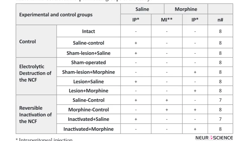
In the first stage of this experiment, a total of 15 groups (n = 7-8 rats in each group) containing one control (sa-line) group (1 ml/kg) and four experimental groups, who were treated with systemic doses of morphine (1, 3, 6 and 12 mg/kg/ml, i.p.) in 3 different times (15, 30 and 60 min) before the formalin test, were considered. At the end of this set of experiment, the ED50 value for antinociceptive effect of systemic morphine and the optimal time of injection before the formalin test were determined. In the second stage, the ED50 dose of mor-phine was used in experimental groups (see Table 1) in order to find out the effect of destruction or reversible inactivation of the NCF on systemic morphine-induced analgesia.
2.5. Statistical Analysis
The obtained results are expressed as mean ±SEM (standard error of mean). An average of the scores ob-tained in the first 5 min was considered as phase 1, and the area under curve (AUC) of pain scores obtained us-ing the trapezoidal rule durus-ing 15-60 min after formalin injection was considered as phase 2. Data were ana-lyzed by GraphPad Prism® (Version 5.0) software. The calculated and normalized AUC values in all groups were subjected to one- or two-way ANOVA and were respectively followed by protected Tukey’s or Bonfer-roni’s test for multiple comparisons, as needed. P-values less than 0.05 were considered to be statistically signifi -cant.
2.6. Histological Verification
logically verified.
3. Results
In the present study, the saline control group consid-ered as a group without any antinociceptive treatment, and the AUC of weighted pain scores in early and late phases were normalized by AUC values of respective saline control groups in all experimental groups. There-fore, the baseline values are equal to zero according to normalization of AUC values in experimental groups. On the other hand, the percentage of decrease in AUC was considered as a drug-induced antinociception dur-ing two phases of formalin test. Additionally, there were no significant differences between saline microinjected groups versus morphine misplacement control.
3.1. Dose- and time-response effects of systemic administration of morphine on time-course of formalin-induced pain behaviors
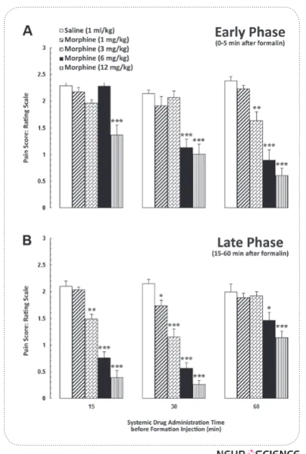
Fig. 1 shows the dose- and time-response effects of different doses of morphine (1, 3, 6 and 12 mg/kg; i.p.) in three injection times on time-course of formalin-in-duced pain behaviors in early and late phases. Based on the data of these curves, the ED50 value was calculated using linear regression. Mean pain score values in Fig.
group. One-way ANOVA followed by Tukey’s post-hoc test showed that different doses of systemic morphine, 15 min before the formalin test, could not induce anti-nociception except for the dose of 12 mg/kg. While ad-ministration of other doses of morphine, 30 and 60 min before the formalin test, could produce significant anti -nociceptive effects in early phase (Fig. 2A). Additional-ly, the antinociceptive effect of morphine appeared well in late phase of formalin test in all set injection times (Fig. 2B). However, one-way ANOVA revealed that systemic administration of different doses of morphine, 60 min before the formalin test induce antinociception only at two high doses (6 and 12 mg/kg) in late phase. Therefore, for evaluating the contribution of the NCF to morphine-induced antinociception, we chose the dose of 6 mg/kg for systemic administration 30 min before formalin test. It seems that this dose is near to ED50 of morphine and 30-min injection time is the best time for induction of antinociception in both early and late phases of formalin test.
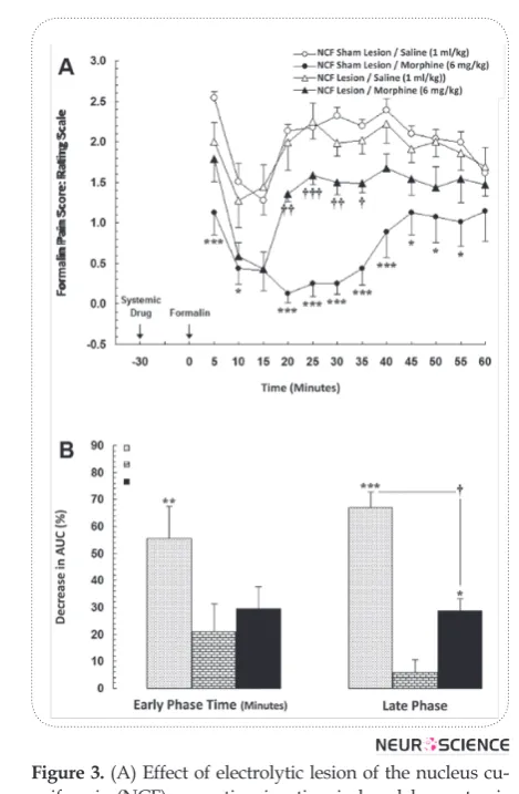
3.2. Effects of bilateral electrolytic lesion of the NCF on analgesic response of systemic morphine
In the present study, the control (NCF sham lesion+Saline) group was considered as a group without any antinociceptive treatment and the AUC of weighted
Table 1. The Control and experimental groups of the study.
Experimental and control groups Saline Morphine
IP* MI** IP* n#
Control
Intact - - - 8
Saline-control + - - 8
Sham-lesion+Saline + - - 8
Electrolytic Destruction of the NCF
Sham-operated - - - 8
Sham-lesion+Morphine - - + 8
Lesion+Saline + - - 8
Lesion+Morphine - - + 8
Reversible Inactivation of the NCF
Saline-Control + + - 7
Morphine-Control - + + 8
Inactivated+Saline + - - 7
Inactivated+Morphine - - + 8
* Intraperitoneal injection
Autumn 2011, Volume 3, Number 1 Basic and Clinical
pain scores, in both early and late phases in all experi-mental groups, were normalized by AUC values in con-trol group. In this set of experiments, animals received saline (1 ml/kg; i.p.) or morphine (6 mg/kg; i.p.), 30 min before the formalin test. Two-way ANOVA for repeated measures over time, followed by Bonferroni’s test for obtained pain score values shown in Fig. 3A, revealed a significant difference in time-course of formalin-induced pain behaviors between NCF sham lesion+Morphine and NCF lesion+Morphine groups [treatment main effect: F(3,336)=84.21, P<0.0001; time main effect
F(11,336)=6.298, P<0.0001; treatment×time interaction effect: F(33,336)=1.359, P=0.0957]. On the other hand, the Tukey’s multiple comparison test for normalized AUC values in Fig. 3B showed that administration of morphine in sham lesion group significantly increased the antinociception by the percentage of decrease in normalized AUC values in both early [F(3,31)=5.204, P<0.01] and late phases [F(3,31)=41.24, P<0.0001]. Nevertheless, this figure showed that normalized de -crease percentages of AUCs, as analgesic index in bi-lateral electrolytic lesion of the NCF that received sys-temic saline (NCF lesion +Saline), are not significantly different from the baseline in both early and late phases of formalin test. Furthermore, data obtained in this ex-periment indicated that the bilateral electrolytic lesion in the NCF could significantly decrease the morphine-induced antinociception in the late (P<0.05; Fig. 3B), but not early phase in NCF lesion +Morphine group as compared to the control group.
Figure 1. Time-course of formalin-induced pain behaviors after intraperitoneal administration of saline (1 ml/kg) or different doses of morphine (1, 3, 6 and 12 mg/kg) when in-jected (A) 15 min (B) 30 min and (C) 60 min before the forma-lin test. The dose of 6 mg/kg of morphine at 30 min before the formalin test was determined as the 50% effective dose in this protocol. Each point is the mean ± SEM for 7-8 rats.
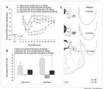
3.3. Effects of bilateral reversible inactivation of the NCF on analgesic response of systemic ad-ministration of morphine
In this section, animals received saline (0.3 µl/side) or lidocaine 2% (0.3 μl/side) into the NCF after adminis -tration of morphine (6 mg/kg; i.p.) or saline (1 ml/kg; i.p.), just before formalin injection, and formalin test was carried out immediately. Two-way ANOVA for re-peated measures over time, followed by Bonferroni’s test for obtained pain score values [treatment main
Saline+Morphine and Lidocaine+Morphine at the late phase. However, the increase of formalin-induced pain behaviors in Lidocaine +Morphine group was not sig-nificant in both phases compared to the baseline saline control (Saline+Saline) group (Fig. 4A). Nevertheless, as shown in Fig. 4B, the normalized decrease percent-ages of AUCs as analgesic index in NCF-inactivated an-imals that received systemic saline (Lidocaine+Saline) showed the hyperalgesic responses in both early and late phases of formalin test; however, it is not signifi -cantly different from the baseline. Furthermore, data obtained in this experiment indicated that the bilateral reversible inactivation of NCF could significantly de -crease the morphine-induced antinociception compared to Saline+Morphine group only in the late phase of the formalin test (P<0.05; Fig 4B).
4. Discussion
The findings of the present study were: (1) systemic administration of morphine, dose- and time-depend-ently, attenuated the formalin-induced pain behaviors in early and late phases and (2) neither electrolytic nor reversible inactivation of NCF had no effect on baseline formalin-induced pain sensitivity. However, (3) both electrolytic lesion and reversible inactivation of the NCF significantly decreased antinociceptive responses of systemic administration of morphine.
There is some evidence that the specific areas involved in chronic nociception in animals are the midbrain PAG and adjacent NCF, the parabrachial nucleus in the rostral pons, and the RVM (Suzuki, Morcuende, Webber, Hunt, & Dickenson, 2002; Urban & Gebhart, 1999; Williams & Beitz, 1993). One of the neighboring areas of the NCF, which has long been known to be a major site of descending pain modulation, is the PAG and its sub-re-gions involving in pain modulatory effects through the opioid receptors (Dostrovsky & Deakin, 1977; Manning & Franklin, 1998; Smith, Monroe, & Hawranko, 1994; Wiedenmayer & Barr, 2000). It has been demonstrat-ed that action by morphine at the CNS region/regions may be followed by release of endogenous opiates in the PAG (da Costa Gomez & Behbehani, 1995), which is somehow causally linked to the development of an-algesia. Moreover, direct application of morphine into the PAG elicits analgesia in the formalin test (Manning & Franklin, 1998), suggesting that the PAG is the site of action of morphine in the modulation of persistent
Figure 3. (A) Effect of electrolytic lesion of the nucleus cu-neiformis (NCF) on antinociception induced by systemic administration of morphine (ED50; 6 mg/kg, i.p.) during formalin test. Lesions were bilaterally made into the NCF and morphine was administered 30 min before formalin test. Each point is the mean ± SEM for 8 rats. (B) The percent-age of reduction (analgesic effect) in area under the curves (AUC) of weighted pain scores using the time-response curves shown in A during the early (0-5 min) and late (15-60 min) phases of formalin test. Normalized data are repre-sented as mean ± SEM.
* P<0.05; ** P<0.01; *** P<0.001 compared to NCF sham lesion+Saline group
Autumn 2011, Volume 3, Number 1 Basic and Clinical
inflammatory pain. It has been reported that intra-PAG injection of the µ-opioid antagonist, blocked an opioid receptor-mediated antinociception in the rat in hot-plate test. In addition, injection of naloxone into the PAG blocked the action of local opioid, resulting in attenua-tion of analgesia (Manning & Franklin, 1998). Several studies have also shown that PAG lesions reduce mor-phine analgesia in rats (Bouhassira, Villanueva, & Le Bars, 1992; Dostrovsky & Deakin, 1977; McGaraughty, Farr, & Heinricher, 2004). With respect to the similari-ties between the NCF and PAG areas in ultrastructural (Gioia & Bianchi, 1987b) and functional (Gioia & Bi-anchi, 1987a) characteristics and their anatomical pro-jections to the same regions such as NRM (A. Hagh-parast, et al., 2008; A. HaghHagh-parast, Soltani-Hekmat, et
Figure 4. (A) Effect of reversible inactivation (lidocaine 2%) of the nucleus cuneiformis (NCF) on antinociception induced by systemic administration of morphine (ED50; 6 mg/kg, i.p.) dur-ing formalin test. Morphine was systemically administered 30 min before formalin test and lidocaine was bilaterally injected into the NCF, 28-29 min after the morphine, just before the for-malin test. Each point is the mean ± SEM for 7-8 rats. (B) The percentage of decrease (analgesic effect) in area under the curves (AUC) of weighted pain scores using the time-response curves shown in A during the early (0-5 min) and late (15-60 min) phases of formalin test. Normalized data are represented as mean ± SEM. (C) Coronal schematic sections showing the locations of
microinjections of lidocaine (●) and saline (○) in the NCF during the formalin test. 4n, trochlear
nerve; 4v, 4th ventricle; Aq, aqueduct; LPAG, lateral periaqueductal gray; NCF, nucleus cunei-formis; vlPAG, ventrolateral periaqueductal gray.
* P<0.05; ** P<0.01; *** P<0.001 compared to Saline+Saline (saline control) group
† P<0.05; †† P<0.01; ††† P<0.001 compared to Saline+Morphine (morphine control) group
tion, sole lesion of NCF was not effective on baseline pain threshold. The relative failure of effectiveness of NCF lesions, but not concurrently with systemic admin-istration of morphine, on preventing the antinocicep-tion suggests that the NCF may not initially participate in triggering of the antinociception and/or it might be compensated by other pathways. We suppose that it may be involved as a functional unit in the mediation of morphine-induced analgesia in a complexity of neu-ral connections. On the other hand, our findings in the present study indicate that the NCF participates in the morphine-induced analgesia, whereas this analgesic ef-fect is significantly reduced following the electrolytic and reversible inactivation of the NCF in the formalin test. Therefore, this function of NCF would be mostly through mechanisms involving the opioid receptors lo-cated in this region. These results confirm some reports that opioidergic system directly acts in NCF descend-ing pain modulatory system (A. Haghparast & Ahmad-Molaei, 2009; A. Haghparast, Gheitasi, et al., 2007; A. Haghparast, et al., 2008) but it still need more investiga-tions.
Acknowledgements
This work was supported by the grant (No. 86-349-A) from Neuroscience Research Center, Shahid Beheshti University of Medical Sciences, Tehran, Iran.
References
Abbott, F. V., Hong, Y., & Franklin, K. B. (1996). The effect of le-sions of the dorsolateral funiculus on formalin pain and mor-phine analgesia: a dose-response analysis. Pain, 65(1), 17-23.
Abbott, F. V., & Melzack, R. (1982). Brainstem lesions dissociate neural mechanisms of morphine analgesia in different kinds of pain. Brain Research, 251(1), 149-55.
Abbott, F. V., Melzack, R., & Leber, B. F. (1982). Morphine
an-algesia and tolerance in the tail-flick and formalin tests:
dose-response relationships. Pharmacology Biochemistry and Be-havior, 17(6), 1213-9.
Bouhassira, D., Villanueva, L., & Le Bars, D. (1992). Effects of systematic morphine on diffuse noxious inhibitory controls: Role of the periaqueductal grey. European Journal of Phar-macology, 216(2), 149-56.
da Costa Gomez, T. M., & Behbehani, M. M. (1995). An elec-trophysiological characterization of the projection from the central nucleus of the amygdala to the periaqueductal gray of the rat: the role of opioid receptors. Brain Research, 689(1), 21-31.
Dostrovsky, J. O., & Deakin, J. F. W. (1977). Periaqueductal grey lesions reduce morphine analgesia in the rat. Neuroscience Letters, 4(2), 99-103.
Dubuisson, D., & Dennis, S. G. (1977). The formalin test: a quantitative study of the analgesic effects of morphine, meperidine, and brain stem stimulation in rats and cats. Pain, 4(2), 161-74.
Fields, H. L., Basbaum, A. L., & Heinricher, M. M. (2006). Cen-tral nervous system mechanisms of pain modulation. In K. M. McMahon (Ed.), Wall and Melzack’s textbook of pain. New York: Churchill Livingstone Press.
Franklin, K. B., Abbott, F. V., English, M. J., Jeans, M. E., Tasker, R. A., & Young, S. N. (1990). Tryptophan-morphine interac-tions and postoperative pain. Pharmacology Biochemistry and Behavior, 35(1), 157-63.
Gioia, M., & Bianchi, R. (1987a). The cytoarchitecture of the nucleus cuneiformis. A Nissl and Golgi study. Journal of anatomy, 155, 165-76.
Gioia, M., & Bianchi, R. (1987b). Ultrastructural study of the nucleus Cuneiformis in the cat. Journal für Hirnforschung, 28(4), 375-83.
Haghparast, A., & Ahmad-Molaei, L. (2009). Effects of electro-lytic lesion of dorsolateral periaqueductal gray on analgesic response of morphine microinjected into the nucleus cunei-formis in rat. Neuroscience Letters, 451(2), 165-9.
Haghparast, A., Gheitasi, I. P., & Lashgari, R. (2007). Involve-ment of glutamatergic receptors in the nucleus cuneiformis in modulating morphine-induced antinociception in rats. Eurpean Journal of Pain, 11(8), 855-62.
Haghparast, A., Naderi, N., Khani, A., Lashgari, R., Motamedi, F. (2010). Formalin-Induced Differential Activation of Nu-cleus Cuneiformis Neurons in the Rat. The Journal of Pain, 11, 32-43.
Haghparast, A., Ordikhani-Seyedlar, M., & Ziaei, M. (2008). Electrolytic lesion of the nucleus raphe magnus reduced the antinociceptive effects of bilateral morphine microinjected into the nucleus cuneiformis in rats. Neuroscience Letters, 438(3), 351-5.
Autumn 2011, Volume 3, Number 1 Basic and Clinical
Hasanein, P., Parviz, M., Keshavarz, M., & Javanmardi, K. (2007). CB1 receptor activation in the basolateral amygdala produces antinociception in animal models of acute and tonic nociception. Clinical Experiment of Pharmacology and Physiology, 34(5-6), 439-49.
Haws, C. M., Williamson, A. M., & Fields, H. L. (1989). Puta-tive nocicepPuta-tive modulatory neurons in the dorsolateral pon-tomesencephalic reticular formation. Brain Reseaech, 483(2), 272-82.
Hudson, P. M., & Lumb, B. M. (1996). Neurones in the mid-brain periaqueductal grey send collateral projections to nu-cleus raphe magnus and the rostral ventrolateral medulla in the rat. Brain Research, 733(1), 138-41.
Jiang, M., & Behbehani, M. M. (2001). Physiological characteris-tics of the projection pathway from the medial preoptic to the nucleus raphe magnus of the rat and its modulation by the periaqueductal gray. Pain, 94(2), 139-47.
Manning, B. H., & Franklin, K. B. (1998). Morphine analgesia in the formalin test: reversal by microinjection of quaternary naloxone into the posterior hypothalamic area or periaque-ductal gray. Behavoral Brain Research, 92(1), 97-102.
Matthies, B. K., & Franklin, K. B. (1992). Formalin pain is ex-pressed in decerebrate rats but not attenuated by morphine. Pain, 51(2), 199-206.
McGaraughty, S., Farr, D. A., & Heinricher, M. M. (2004). Le-sions of the periaqueductal gray disrupt input to the rostral ventromedial medulla following microinjections of mor-phine into the medial or basolateral nuclei of the amygdala. Brain Research, 1009(1-2), 223-7.
Moulton, E. A., Burstein, R., Tully, S., Hargreaves, R., Becerra, L., & Borsook, D. (2008). Interictal dysfunction of a brainstem descending modulatory center in migraine patients. PLoS ONE, 3(11), e3799.
Oluyomi, A. O., Hart, S. L., & Smith, T. W. (1992). Differential antinociceptive effects of morphine and methylmorphine in the formalin test. Pain, 49(3), 415-8.
Ossipov, M. H., Lai, J., Malan, T. P., Jr., & Porreca, F. (2000). Spinal and supraspinal mechanisms of neuropathic pain. Annals of the New York Academy of Sciences, 909, 12-24.
Paxinos, G., & Watson, C. R. (2005). The Rat Brain in Stereotaxic Coordinates. (5th ed.). San Diego: Elsevier Academic Press.
Rezvanipour, M., Haghparast, A., & Millan, H. (2006). The role of GABAA receptor inhibitor on morphine antinociception action in cuneiformis nucleus. International Journal of Phar-macology, 2, 400-5.
Ryan, S. M., Watkins, L. R., Mayer, D. J., & Maier, S. F. (1985). Spinal pain suppression mechanisms may differ for phasic and tonic pain. Brain Research, 334(1), 172-5.
Smith, D. J., Monroe, P. J., & Hawranko, A. A. (1994). Multi-ple opioid receptors mediate descending pain modulation from the periaqueductal gray (PAG). Regulatory Peptides, 53(Supplement 1), S99-S100.
Suzuki, R., Morcuende, S., Webber, M., Hunt, S. P., &
Dick-enson, A. H. (2002). Superficial NK1-expressing neurons
control spinal excitability through activation of descending pathways. Nature Neuroscience, 5(12), 1319-26.
Tracey, I., & Mantyh, P. W. (2007). The cerebral signature for pain perception and its modulation. Neuron, 55(3), 377-91.
Urban, M. O., & Gebhart, G. F. (1999). Supraspinal contribu-tions to hyperalgesia. Proceedings of the National Academy of Sciences U S A, 96(14), 7687-92.
Vanegas, H., & Schaible, H. G. (2004). Descending control of persistent pain: inhibitory or facilitatory? Brain Research Re-view, 46(3), 295-309.
Wiedenmayer, C. P., & Barr, G. A. (2000). [mu] Opioid Recep-tors in the Ventrolateral Periaqueductal Gray Mediate Stress-Induced Analgesia but Not Immobility in Rat Pups. Behavio-ral Neuroscience, 114(1), 125-36.
Williams, F. G., & Beitz, A. J. (1993). Chronic pain increases brainstem proneurotensin/neuromedin-N mRNA expres-sion: a hybridization-histochemical and immunohistochemi-cal study using three different rat models for chronic nocic-eption. Brain Research, 611(1), 87-102.
Yeung, J. C., & Rudy, T. A. (1980). Multiplicative interaction be-tween narcotic agonisms expressed at spinal and supraspinal sites of antinociceptive action as revealed by concurrent in-trathecal and intracerebroventricular injections of morphine. Journal of Pharmacology and Experimental Therapeutics, 215(3), 633-42.