Design and Evaluation of Floating Microspheres of Pantoprazole Sodium
Behin Sundara Raj
1*, Jigar Pancholi
2, Punitha Isaac Samraj
31School of Pharmacy, Curtin University, Bentley, Perth, Western Australia – 6102
2Department of Pharmaceutics, Shree Devi college of Pharmacy, Airport Road, Kenjar, Mangal
ore, Karnataka, India – 574 142
3Department of Pharmacognosy, Shree Devi college of Pharmacy, Airport Road, Kenjar, Mangal
ore, Karnataka, India – 574 142
Article Information Received 17 June 2015
Received in revised form 21 Sep 2015 Accepted 23 Sep 2015
Abstract
In the present study, an attempt was made to prepare floating microspheres of Pantoprazole
sodium by a non-aqueous solvent evaporation method. The half-life of Pantoprazole sodium is
1-1.5 hours and rapidly eliminated from the body. It is in a perfect world suited to be conveyed
through floating multiunit measurements structure. Biocompatible polymers, Eudragit S100 and
HPMC K 100M were utilized alongside the medication as a part of diverse extents. The prepared
six formulations (F1-F6) were characterized for their drug polymer compatibility (IR study),
micromeritic properties, particle size, percentage yield, scanning electron microscopy, buoyancy
studies, drug encapsulation efficiency and in vitro drug release studies. The formulated
microspheres were free flowing. The optical microscopic studies revealed that the particles were
of the size range of 193.29-517.16 μm. SEM studies indicated that the microspheres were porous
and almost spherical in shape. The prepared floating microspheres were found to produce the
percentage yield of 84.43-91.93%, drug encapsulation efficiency was 65.83-90.03% and
buoyancy percentage was 61.7-78.46%. In-vitro drug release studies showed cumulative
percentage drug release between 69.27-79.06%. The information acquired in this study
recommends that a micro particulate floating dose type of Pantoprazole sodium can be effectively
intended to give delayed arrival of medication and thus enhanced bioavailability. Keywords:
Gastro retentive system, Pantoprazole sodium, Floating microspheres, Non- aqueous solvent evaporation Corresponding Author: E-mail: [email protected] Mob.: +61-0406307140
1 Introduction
Late experimental and patent writing shows expanded enthusiasm
for scholastics and mechanical exploration gatherings with respect to
the novel measurement shapes that can be held in the stomach for a
delayed and unsurprising timeframe. A standout amongst the most
attainable methodologies for accomplishing a drawn out and
unsurprising medication conveyance profile in the gastro intestinal
(GI) tract is to control the gastric residence time (GRT) , utilizing
gastro retentive drug delivery systems (GRDDS) that will give us new
and imperative remedial alternatives.1.
GRDDS can remain in the gastric region for several hours and
significantly prolong the gastric residence time of drugs. Drawn out
gastric maintenance enhances bioavailability, lessens drug wastage,
and enhances solvency of medications that are less dissolvable in a
high pH environment. It has applications likewise for nearby
medication conveyance to the stomach and proximal small insides.
Gastro maintenance serves to give better accessibility of new items
with new helpful potential outcomes and considerable advantages for
patients2.
A number of frameworks have been used to increase the GRT of
dosage forms by employing a variety of concepts. These frameworks
have been characterized by essential standards of gastric
maintenance as floating medication frameworks, bioadhesive
frameworks, swelling and growing frameworks and high-thickness
frameworks3.
Floating microspheres are gastro retentive drug delivery systems
based on the non-effervescent approach. Empty microspheres are in
a strict sense, circular vacant particles without a center. These
microspheres are distinctively free streaming powders comprising of
proteins or engineered polymers, in a perfect world having a size
under 200 micrometers. Strong biodegradable microspheres fusing a
UK Journal of Pharmaceutical and Biosciences
Available at www.ukjpb.com
UK J Pharm & Biosci, 2015: 3(6); 10 medication scattered or broke down all through molecule lattice have
the potential for controlled arrival of medications4,5.
Floating microspheres can be prepared by solvent diffusion and
evaporation, solvent evaporation and spray drying methods. In the
present work, we prepared floating microspheres of Pantoprazole
sodium by non-aqueous solvent evaporation method. We worked on
prolonging its gastric residence time, with the aim of improving the
oral bioavailability of the drug.
2 Material and Methods 2.1 Materials
Pantoprazole sodium and HPMC K100M were gift samples and
provided by Cadila Pharmaceuticals, Ahmedabad, Gujarat, India.
Eudragit S100 was purchased from Yarrow Chemicals, Mumbai,
India. Ethanol was procured from Poly Pharma Laboratories,
Gujarat. Dichloromethane was purchased from Finar Chemicals,
Ahmedabad. All other chemicals and reagents used were of
laboratory or analytical grade.
2.2 Methods
2.2.1 Compatibility studies of Pantoprazole sodium and polymers
An FTIR spectrum helps to confirm the identity of the drug and to
detect the interaction of the drug with the carriers6,7,8. IR spectroscopy of pure drug and the physical mixture of the drug with
polymers were carried out to check the compatibility of drug and
polymers. The IR spectra of the drug with polymers was compared
with the standard IR spectrum of the pure drug. Infrared spectra of
Pantoprazole Sodium, HPMC K100M, Eudragit S-100 and
formulations F1, F4 were carried out by using KBr pellet technique
and recorded on an FTIR 4100 type A Jasco International co ltd,
Japan.
2.2.2 Preparation of floating microspheres of Pantoprazole sodium
The floating microspheres of Pantoprazole sodium were prepared by
nonaqueous solvent evaporation method6,9,-11 using different polymers as follows:
Microspheres containing Pantoprazole sodium as a core material
were prepared by Non-aqueous Solvent Evaporation method. Drug
and HPMC or Drug and Eudragit S-100 were mixed in Ethanol:
Dichloromethane at various ratios. The slurry was slowly introduced
into 50 ml of liquid paraffin containing 1% tween 80 as an emulsifying
agent while being stirred at 1400 rpm by a mechanical stirrer
equipped with a three bladed propeller at room temperature. The
solution was stirred for 4h to allow the solvent to evaporate
completely, and the microspheres were collected by filtration. The
microspheres were repeatedly washed with n-hexane until free from
oil. The collected microspheres were dried for 1h at room
temperature and subsequently stored in a desiccator.
2.3 Evaluation of Pantoprazole sodium floating microspheres
2.3.1 Micromeritic properties
The microspheres were characterized by their micromeritic
properties such as particle size, bulk density, tapped density,
compressibility index, Hausners ratio and angle of repose12,13.
2.3.2 Particle size
The particle size was measured by microscopic technique7,8. The suspension of floating microspheres was prepared using castor oil. A
drop of suspension was mounted on a slide and observed under the
optical microscope. About 100 particles were measured with the help
of the eye piece micrometer. All the microspheres in a field were
counted.
2.3.3 Bulk density
In this method, floating microspheres were transferred to a
measuring cylinder and is tapped manually till a constant volume is
obtained7,12. This volume is bulk volume, and it includes the true volume of the powder and the void space between the
microspheres.
2.3.4 Tapped density
In this method, floating microspheres were transferred to a
measuring cylinder and tapped for 100 times8,12,14,15. After tapping the volume of microspheres was visually examined. The ratio of the
mass of microspheres to the volume of microspheres after tapping
gives tapped density of the floating microspheres.
2.3.5 Carr’s compressibility index
UK J Pharm & Biosci, 2015: 3(6); 11 Lower compressibility values indicate better flow.
2.3.6 Hausners ratio
Hausners ratio of microspheres was determined by comparing
tapped density to bulk density8,16 using the equation.
Values less than 1.25 indicates good flow (= 20% Carr), where as
greater than 1.25 indicates poor flow (= 33% Carr).
2.3.7 Angle of repose
The angle of repose (θ) of the microspheres, measures the
resistance to particle flow, was determined by a fixed funnel
method18. The height of the funnel was adjusted in such a way that the tip of the funnel just touches the heap of the blends. Accurately
weighed microspheres were allowed to pass through the funnel
freely on to the surface. The height and radius of the powder cone
were measured, and angle of repose was calculated using the
following equation.
θ = tan-1 h / r
Where,
θ - Angle of repose
h - Height of granules above the flat surface
r - Radius of the circle formed by the granule heap.
2.4 Percentage yield of floating microspheres
The prepared floating microspheres with a size range of 102–420 µm
was collected and weighed7,16,18. The measured weight was divided by the total amount of all non-volatile components which were used
for the preparation of microspheres.
2.5 In-vitro buoyancy
Floating microspheres (equivalent to 150 mg) were dispersed in 900
ml of 0.1 N hydrochloric acid solution (pH 1.2) containing tween 80
(0.01 W/V %) / tween 20 (0.02 W/V %) to simulate gastric fluid at
37°C. The mixture was stirred with a paddle at 100 rpm and after 12
h; the layer of buoyant microspheres (Wf) was pipetted and
separated by filtration. Simultaneously sinking microspheres (Ws)
were also separated. Both microspheres were dried at 40°C
overnight. Each weight was measured, and buoyancy was
determined by the weight ratio of the floating microspheres to the
sum of floating and sinking microspheres7,16,19.
Wf and Ws are the weights of the floating and settled microspheres.
All the determinations were made in triplicate.
2.6 Estimation of drug loading/encapsulation efficiency
Microspheres weighing 25 mg were taken for evaluation. The
amount of drugs entrapped was estimated by crushing the
microspheres and extracting the drug using simulated gastric fluid
(SGF) (pH 7.4) (10 ml). The extract was transferred to 100 ml
volumetric flask and volume was made up using SGF (pH 7.4). The
solution was filtered, and from the filtrate 10 ml was taken and further
diluted to 100 ml, and the absorbance was measured at 230 nm
against SGF (pH 7.4) as blank7, 16, 18, 20.
2.7 Scanning electron microscopy
The morphological study was carried out by Scanning Electron
Microscope6,18. SEM studies were carried out by using JEOL JSM- 6380 LA scanning electron microscope (Japan). The samples of
SEM were prepared by lightly sprinkling the microspheres powder on
a double adhesive tape, which was stuck on an aluminium stub. The stubs were then coated with gold to the thickness of about 200Ǻ
using a sputter coater. The photomicrographs were taken with the
help of SEM analyzer.
2.8 In-vitro drug release studies
A USP XXIII basket type dissolution apparatus was used to study the
in-vitro drug release from microspheres6,18. A weighed amount of floating microspheres equivalent to 40 mg drug was filled into the
capsule and placed in the basket. Dissolution medium used was
SGF (pH 1.2) 900 ml, containing 0.02% tween 20 maintained at
37±0.1ºC and stirred at 100 rpm for 1 h. 10 ml of sample was
withdrawn at predetermined time interval, diluted and was analyzed
for drug content spectrophotometrically at 289 nm against suitable
blank. The volume was replenished with the same amount of the
fresh dissolution medium each time to maintain sink condition and
10 h in phosphate buffer pH 6.8 with tween 80 (0.01 W/V%) / tween
UK J Pharm & Biosci, 2015: 3(6); 12 withdrawn at different time intervals and replaced with fresh
phosphate buffer. The amount of drug released was analyzed at 289
nm using UV-visible spectrophotometer (UV-1800, shimadzu, japan).
2.9 Drug release analysis
To analyze the mechanism for the release and release rate kinetics
of the formulated dosage form, the data obtained from conducted
studies was fitted into Zero order, First order, Higuchi's matrix,
Peppas and Hixson Crowell model. By comparing the r-values
obtained, the best-fit model was selected6,21.
3 Results and Discussions
3.1 Compatibility studies of Pantoprazole sodium and polymers
The FTIR spectra of Pantoprazole and polymers are shown in figure
1A-1C. Our experimental results were assessed on the basis of
physical data obtained for drug and polymers as well as
formulations. The medication Pantoprazole displayed CH retentions
from 2941.3 cm-1 demonstrates the vicinity of aromatic and
additionally aliphatic CH vibrations. The strong S=O absorption peak
was noticed at 1041.37cm-1. The peak at 1775.5cm-1 shows N-H stretching indicating the presence of amine group. The spectrum
also shows a peak at 1590.02cm-1. The peak in this range is due to
C-N stretching. The peak at 1304.61 cm-1 is the presence of C-F stretching.
Another polymer Eudragit S 100 showed broad peak at 3500 cm-1
corresponding to carboxylic acid moiety present in the polymer. The
aliphatic C-H absorption peaks of this molecule were observed
around 2953cm-1. These data are in conformity with the structure of polymer taken for the formulation.
The floating microspheres were obtained with drug Pantoprazole and
polymer eudragit S 100 (Formulation F4- F6) as per the procedure
described earlier. The strong S=O absorption peak was noticed at
1041.37cm-1. The peak at 1729.83 cm-1 indicates N-H stretching confirming the presence of amine group. The peak at1590.02 cm-1 is due to C-N stretching. The spectrum showed the peak at 1304.61
cm-1 was due to C-F stretching.
The IR spectra of the medication and physical blend, showed peaks
in the same area, which affirmed there was no association in the
middle of medication and polymer and there was no incompatibility of
medication in the vicinity of the used polymers.
3.2 Preparation of floating microspheres of Pantoprazole sodium
Calibration curve for the estimation of Pantoprazole sodium was
constructed in 7.4 pH buffer at 289 nm. The method obeyed Beer’s
Lambert law in the range of 10 to 50 mcg/ml. Floating microspheres
of Pantoprazole sodium using different grades of HPMC polymer
was prepared by emulsion solvent evaporation method as shown in
table 1. The emulsion was stabilized by tween-80 and the volatile
solvent evaporate leaving a solidified thin film at the interface
between the aqueous phase and the organic phase, where
Pantoprazole sodium is encapsulated in the core-coat of polymers.
UK J Pharm & Biosci, 2015: 3(6); 13 Table 1: Formulation of floating microspheres of Pantoprazole
sodium
Ingredients (gm)
Formulation code
F1 F2 F3 F4 F5 F6
HPMC K100M 0.50 1.00 1.50 - - -
Eudragit S 100 - - - 0.50 1.00 1.50
Lansoprazole 0.50 0.50 0.50 0.50 0.50 0.50
Dichloromethane 30 30 30 30 30 30
Ethanol 30 30 30 30 30 30
3.3 Evaluation of floating microspheres of Pantoprazole sodium
3.3.1 Micromeritic properties
The mean particle size of the microspheres formulation F1 to F3
containing the different polymer ratio of HPMC K 100 M and
Eudragit S-100 were in the range of 193.29±13.8 µm to 517.16±3.25
µm respectively. The mean molecule size of the microspheres
arranged by utilizing HPMC K 100 M was observed to be bigger
contrasted with the microspheres arranged by utilizing Eudragit
S-100. The medium's thickness is expanded at a higher HPMC K 100M
fixation bringing about improved interfacial pressure. Shearing
proficiency additionally lessened at higher viscosities. This brought
about the development of bigger particles.
The bulk density, tapped density, hausners ratio of formulation F1 to
F6 containing different polymer concentration of HPMC K 100M and
Eudragit S-100 formulation was in the range of 0.3522±0.007 to
0.4220±0.010 gm/cm3 (Table 2).
The carr’s index of formulation F1 to F6 containing different polymer
concentration of HPMC K 100M and Eudragit S-100 was in the range
of 13.92±0.26 to 19.03±0.33 %. The angle of repose of formulation
F1 to F6 containing the distinctive polymer grouping of HPMC K
100M and Eudragit S-100 details was in the reach 28.84±1.80 ° to
36.96±1.93 ° (Table 2). The estimations of carr's file and angle of
repose show great stream property.
3.4 Percentage yield of floating microspheres
The percentage yield of floating microsphere formulation F1 to F6
containing the different polymer concentration of HPMC K 100M and
Eudragit S-100 formulation was in the range of 84.43±0.53 to
91.93±0.43 (Table 3). To observe the effect of polymer concentration
on the percentage yield of the floating microspheres, formulations
were prepared at varying concentration of HPMC K100M and
Eudragit S-100 (Table 3). The yield of the floating microspheres
increased with enhancing the polymer concentration. At low
concentration of polymers part of the polymer solution aggregated in
a fibrous structure, as it solidified prior to forming droplets, or the
transient droplets were broken before solidification. It was completely
due to poor mechanical strength resulting in low yield.
3.5 In-vitro buoyancy
The motivation behind get ready floating microspheres was to
amplify the gastric living arrangement time of a medication. The
lightness test was done to examine the floatability of the arranged
microspheres. The microspheres were spread over the surface of a
reenacted gastric liquid and the division of microspheres light and
settled down as a component of time was quantitated. The in-vitro
buoyancy of formulation F1 to F6 containing the different
concentration of HPMC K100M and Eudragit S-100 formulations was
in the range from 61.71±1.52 h to 78.46±1.07 h (table 3). Among all
formulations, F5 had the best in-vitro buoyancy time 78.46±1.07.
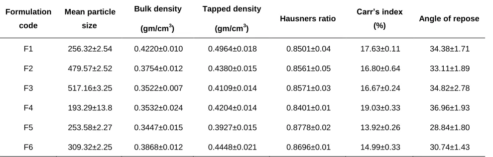
Table 2: Micromeritic properties of floating microspheres of Pantoprazole sodium
Formulation code
Mean particle size
Bulk density (gm/cm3)
Tapped density (gm/cm3)
Hausners ratio Carr’s index
(%) Angle of repose
F1 256.32±2.54 0.4220±0.010 0.4964±0.018 0.8501±0.04 17.63±0.11 34.38±1.71
F2 479.57±2.52 0.3754±0.012 0.4380±0.015 0.8561±0.05 16.80±0.64 33.11±1.89
F3 517.16±3.25 0.3522±0.007 0.4109±0.014 0.8571±0.03 16.67±0.24 34.82±2.78
F4 193.29±13.8 0.3532±0.024 0.4204±0.014 0.8401±0.01 19.03±0.33 36.96±1.93
F5 253.58±2.27 0.3447±0.015 0.3927±0.015 0.8778±0.02 13.92±0.26 28.84±1.80
UK J Pharm & Biosci, 2015: 3(6); 14 Table 3: Percentage yield, in-vitro buoyancy and incorporation efficiency of floating microspheres of Pantoprazole sodium
Formulation
code Percentage yield (%) In vitro buoyancy (h) Drug loading (%)
Drug entrapment efficiency (%)
F1 84.43±0.53 61.71±1.52 44.91±1.69 69.80±2.72
F2 87.63±0.69 64.23±2.07 26.64±1.76 70.02±2.84
F3 86.36±0.51 62.96±1.04 19.07±1.53 65.83±2.17
F4 88.30±0.66 71.34±2.02 43.83±1.19 77.41±1.01
F5 91.93±0.43 78.46±1.07 32.64±1.24 90.03±1.59
F6 87.05±0.72 74.59±2.06 24.21±1.03 84.31±2.46
3.6 Drug entrapment efficiency
The drug entrapment efficiency of formulations F1 to F6 containing
different concentrations of HPMC K100M and Eudragit S-100
formulations was in the range of 65.83±2.17% to 90.03±1.59%
(Table 3). Among all the prepared formulations, F5 (90.03±1.59%)
results demonstrated that the increase in the concentration of
polymer increased the entrapment of the drug. The drug entrapment
efficiency was found to be good in all the formulations.
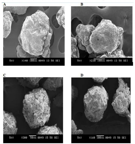
3.7 Scanning electron microscopy (SEM)
Morphology of microspheres was examined by scanning electron
microscopy. The view of the microspheres showed a spherical
structure with a smooth surface morphology (Figure 2A-2D). Some of
the microspheres showed a dented surface structure, but they
showed good floating ability on the surface of the medium, indicating
intact surface. The outer surface of the microspheres was smooth
and dense while the internal surface was porous. The shell of the
microspheres also showed some porous structure. It may be caused
by the evaporation of solvent entrapped within the shell of
microspheres after forming a smooth and dense skin layer.
3.8 In-vitro drug release
In-vitro drug release studies of pantoprazole sodium from floating
microspheres were performed at pH 1.2 and pH 7.4 for 10 h in the
dissolution test apparatus. Formulations F1- F6 showed the
percentage drug release in the range of 69.27±1.10 to 79.06±0.49%
at the end of 10 h. Amongst the formulation F3 was found to be the
best formulation as it released pantoprazole 79.06±0.49% in a
sustained manner with constant fashion over an extended period of
time (after 10 h).
It was seen as the grouping of polymers was expanded rate arrival of
pantoprazole sodium diminished. The increment in polymer focus
prompts the expanded thickness of polymer grid in the microspheres
which brought about an expanded diffusional way length. This may
diminish the general medication discharge from the polymer grid.
Besides, littler microspheres were framed at lower polymer fixation
and has bigger surface territory presented to the disintegration
medium. The r values of Zero order of the above six formulations
were in the range of 0.9665 to 0.9988. Similarly, the r-values of the
first order were in between 0.9195 to 0.9761 (Table 4). The results
suggest that the drug was released by mixed order kinetics. It
suggests that the Higuchi diffusion plots of all the formulations were
fairly linear, and we can conclude that the drug was released by Higuchi’s diffusion mechanism. The formulations were also treated to Peppa’s plot by taking log percent versus log time. The plots were
fairly linear, and the regression values (n value) of all the
formulations ranged from the lowest 0.9374 to highest 1.1730 (Table 4), which is in the range of ˃ 0. 89. This suggested that the drug was
released by super case-II transport with swelling.
4 Conclusions
Gliding microspheres of Pantoprazole sodium can be effectively
arranged utilizing HPMC and Eudragit S-100 as polymers by
emulsion solvent evaporation. The rate yield of all floating
microspheres was more than 80% recommending that the system
UK J Pharm & Biosci, 2015: 3(6); 15 expanding the measure of polymer in definition it upgrades the rate
of gliding microspheres. The entanglement proficiency was more
than 80%. This recommended that improved parameters were
utilized as a part of the strategy for readiness.
Figure 2: A - Scanning Electron Microscope Photographs of Formulation F1, B - Formulation F3, C - Formulation F5 and D - Formulation F6
The in-vitro buoyancy was more than 65% after 10 h indicates the
satisfactory performance of proposed formulations. The percentage
buoyancy increased significantly as the amount of polymer was
increased in each preparation method. The mean particle size of
microspheres was in the range of 193.29-517.16 µm depending upon
the type of polymer used. The particle size increased significantly as
the amount of polymer increased.
The flow properties of all the prepared microspheres were good as
indicated by the low angle of repose (Ɵ<40º) and low carr’s index
(I<20). The good flow properties suggested that the microspheres
produced were non-aggregated.
The in-vitro release of floating microspheres of Pantoprazole sodium
was found to be in following the order F3>F1>F2>F4>F6>F5. In vitro
UK J Pharm & Biosci, 2015: 3(6); 16 of formulations F1-F3 containing HPMC K 100M and formulations
F4-F6 containing only Eudragit S-100. In-vitro release data fitted into
various kinetic models suggests that the release obeyed mixed order
kinetic, Higuchi’s diffusion mechanism, and super case-II transport.
Finally, it was concluded that the prepared floating microspheres of
Pantoprazole sodium may prove to be a potential candidate for safe
and effective sustained drug delivery over an extended period of time
which can reduce dosing frequency.
Table 4: Kinetics data obtained from in-vitro release profile for floating microspheres of Pantoprazole sodium
Formulation code
Zero-order kinetic data
First-order kinetic data
Higuchi matrix
data Peppas kinetic data
Regression Coefficient (r)
Regression Coefficient (r)
Regression Coefficient (r)
Regression Coefficient (r)
n-value
F1 0.9877 0.9249 0.9566 0.9900 0.9374
F2 0.9988 0.9761 0.9836 0.9991 1.0124
F3 0.9981 0.9457 0.9739 0.9990 1.0183
F4 0.9959 0.9507 0.9579 0.9982 1.0567
F5 0.9866 0.9411 0.9531 0.9986 1.1020
F6 0.9665 0.9195 0.9315 0.9969 1.1730
5 Acknowledgements
The authors would like to thank Shree Devi Education Trust,
Mangalore for providing the necessary facilities to carry out the
research work. We also thank Cadila Pharmaceuticals, Ahmedabad,
Gujarat for supplying free samples of Pantoprazole sodium and
HPMC.
6 Conflicts of interests
The authors declare that they have no competing interests.
7 Authors contributions
BS and PIS carried out literature review and preparation of the
manuscript. JP participated in the collection of data. All authors read
and approved the final manuscript.
8 References
1. Yeole PG, Khan S, Patel VF. Floating drug delivery systems:
Need and Development. Indian J Pharm Sci 2005; 67(3):
265-72.
2. Arora S, Ali J, Ahuja A, Khar RK, Baboota S. Floating drug
delivery system. A review. AAPS Pharm Sci Tech 2005; 6(3):
372-90.
3. Chawla G, Gupta P, Koradia V, Bansal AK. Gastro retention
A means to address regional variability in intestinal drug
absorption. Pharmaceutical Technology 2003:50-68.
4. Vyas SP, Khar RK. Gastro-retentive system In Controlled
Drug Delivery System: Concept & Advances. 1st ed. New Delhi:Vallabh Prakashan; 2002.
5. Shivkumar HG, Vishakanta GD, PramodKumar TM.
Formulation and evaluation of muco adhesive drug delivery
system for some anti-asthmatic drugs. IJPE 2004 Oct-Dec.;
38(4): 22-26.
6. Bhardwaj P, Chaurasia D, Singh R, Swarup A. Development
and characterization of novel site specific hollow floating
microspheres bearing 5-Fu for stomach targeting. The
Scientific World Journal. 2014; 705259.
7. Suresh F, Sajal KJ, Manvendra SK, Rohit G, Ankit V.
Formulation and Evaluation of Floating Microspheres of
Boswellic acid. Int.J. PharmTech. Res. 2011; 3(1): 76-81.
8. Najmuddin M, Aejaz A, Sachin S, Patel V, Khan T. Floating
microspheres of ketoprofen: formulation and evaluation. Int.
J. Pharm. Pharm. Sci. 2010; 2(2): 164-168.
9. Biswal I, Dinda A, Das D, Chowdary KA. Encapsulation
protocol for highly hydrophilic drug using nonbiodegradable
polymer. Int. J. Pharm. Pharm. Sci. 2011; 3(2): 256-259.
10. Deepa MK, Karthikeyan M. Cefpodoxime proxetil floating
microspheres: formulation and In vitro evaluation. IJPS.
UK J Pharm & Biosci, 2015: 3(6); 17 11. Yogesh SG, Durgacharan AB, Akhil PM. Formulation and
evaluation of intra gastric floating drug delivery system of
diltiazem hydrochloride. Asian Journal of Pharmaceutics.
Oct-Dec 2008: 228-231.
12. Aulton ME. Pharmaceutics: The Science of Dosage Form
Design. 2nd ed. Livingstone C. Elsevier science Ltd; 2002.
13. Jianhu LIU, Rong Yang, Lulu Jing. Synthesis and
characterization of novel buoyant photo catalyst; Nano
science 2006; 126-130.
14. Sarode SM, Mittal M, Magar RM, Shelke AD, Shrivastava B,
Vidyasagar G. Formulation and evaluation of floating
microspheres of glipizide. J. Chem. Pharm. Res. 2011; 3(3):
775-783.
15. Ibrahim EG. Development and in vitro evaluation of novel
floating chitosan microcapsules for oral use: comparison with
non-floating chitosan microspheres. Int. J. Pharm. 2002; 249:
7-21.
16. Mallikarjuna KR, Gnanaprakash K, ChandraKBS,
Madhusudhana CC. Formulation and in-vitro characterization
of floating microspheres of Amoxycillin trihydrate against
H.pylori. Journal of Pharmacy Research. 2011; 4(3):836-840.
17. Sunil KJ, Govind PA, Narendra KJ. Evaluation of porous
carrier-based floating orlistat microspheres for gastric
delivery. AAPS PharmSciTech. 2006; 7 (4): E1-E9.
18. Anand G, Chirag N, Krunal P, Panchaxari D, Vinayak M.
Formulation and evaluation of floating microspheres of
captopril for prolonged gastric residence time. Indian Journal
of Novel Drug Delivery. Jan-Mar 2011; 3(1): 17-23.
19. Nila MV, Sudhir MR, Cinu TA, Aleykutty NA, Jose S. Floating
microspheres of carvedilol as gastro retentive drug delivery
system: 3(2) full factorial design and in vitro evaluation. Drug
Deliv.2014; 21(2): 110-117.
20. Himansu BS, Suddhasattya D, Dhiraj K, Sandeep Dk,
Sreenivas SA, Rahul V. Formulation, characterization and
in-vitro evaluation of floating microspheres of nateglinide.
International Journal of Pharma and Bio Sciences. Jan- Mar
2011; 2(1): 147- 156.
21. Harris SM, Jaweria T, Hamid AM, Rabia IY. Evaluation of
drug release kinetics from ibuprofen matrix tablets using
HPMC. Pak. J. Pharm. Sci. 2006, Vol.19 (2), 119-124.
22. Shah MB, Israni SA, Kapadia NS. A simple method of
isolation of plumbagin from P. rosea. Pharmaceutical