E
WAS
AWICKA1, A
NNAD
ŁUGOSZ1, K
RYSTYNAP
ORĘBA2, A
GATAC
HOWANIEC1Dissimilar Effects of Cisplatin and the Pyridopyrazolo−
pyrimidine Derivative KP972 on Free Radicals
Odmienny wpływ na procesy wolnorodnikowe cisplatyny i preparatu
KP972 – pochodnej pirydopirazolopirymidyny
1 Department of Toxicology, Wroclaw Medical University, Poland
2 Department of Technology of Drugs, Wroclaw Medical University, Poland Adv Clin Exp Med 2009, 18, 6, 551–557
ISSN 1230−025X
ORIGINAL PAPERS
© Copyright by Wroclaw Medical University
Abstract
Background.The toxicity of cytostatic medications and their side effects pose a great problem and there is a con− stant need for new and effective anticancer drugs of potentially lower toxicity. The pyridopyrazolopyrimidine de− rivative KP972 showed antiproliferative activity on the HL−60 cell line similar to that of cisplatin.
Objectives. Assessment of the participation of free−radical processes in the activity of KP972 compared with ci− splatin.
Material and Methods. The study was performed on in vitro mitochondria isolated from human placenta and ery− throcytes. The degree of lipid peroxidation in the erythrocytes was measured as the TBARS level according to Stocke’s method. The hydroxyl radical (·OH) concentration in mitochondria was determined by deoxyribose de− gradation. KP972 and cisplatin in concentrations of 1–70 µg/ml were used.
Results. KP972 in concentrations of 50–70 µg/ml caused a statistically significant decrease in TBARS level in ery− throcytes compared with the control. A statistically significant negative correlation between KP972 concentration and TBARS level (r= –0.5962, p< 0.0005) was observed. This shows that KP972 is able to decrease lipid pero− xidation in erythrocytes. Cisplatin showed quite the opposite effect. All concentrations of cisplatin (1–70 µg/ml) increased the TBARS level compared with the control. The statistically significant positive linear correlation be− tween cisplatin and TBARS level (r= 0.5014, p< 0.0005) also differed from the KP972 relationship. The influen− ce on ·OH generation also differed. KP972 at all concentrations caused a decrease in ·OH radical in mitochondria compared with the control while cisplatin at concentrations of 1–50 µg/ml caused an increase (p < 0.01).
Conclusions. The results show that KP972 and cisplatin acted in an opposite manner; however, the final antipro− liferative effects were comparable. It appears that the mechanisms of these compounds’ action are completely dif− ferent, at least in their influence on free radicals. It seems that the proradical effect of cisplatin is connected with its toxicity (increased MDA levels in the urine of patients was noted), but the antiradical effect of KP972 could ha− ve considerable significance in its antiproliferative activity (Adv Clin Exp Med 2009, 18, 6, 551–557).
Key words:cisplatin, KP972, hydroxyl radical, lipid peroxidation, antiproliferative effect.
Streszczenie
Wprowadzenie. Toksyczność leków cytostatycznych oraz pojawiające się działania niepożądane, zagrażające na− wet życiu pacjenta, rodzą potrzebę poszukiwania nowych, skutecznych leków przeciwnowotworowych o mniej− szej toksyczności. Preparat KP972, będący przedmiotem badań, jest pochodną pirydopirazolopirymidyny. W eks− perymentach in vitro na linii komórek białaczki HL−60 wykazał aktywność antyproliferacyjną, porównywalną z aktywnością cisplatyny i stał się przedmiotem patentu P379.
Cel pracy. Badanie udziału reakcji wolnorodnikowych w aktywności przeciwnowotworowej preparatu KP972 w porównaniu z cisplatyną.
Free radicals are factors able to cause perma− nent and irreversible structural changes in DNA, such as base−pair mutations in purine and pyrimi− dine bases, deletions (loss of one or several pairs of nucleotides in DNA), insertions (adding one or several nucleotides), and rearrangements. They can also damage and modulate the activity of stress proteins and stress genes that regulate effec− tor genes related to growth, differentiation, and cell proliferation. Oxygen radicals have all these properties [1, 2]. A very reactive one is hydroxyl radical (OH·). The high redox potential of OH· (+2.31 V) is seen in its very strong oxidative pro− perties. Some investigations showed that the level of reactive oxygen species (ROS) in cancer cells was much higher than in healthy cells. Although the participation of free radicals in carcinogenesis is undeniable, it is still not fully explained [3]. The mechanism responsible for increasing ROS level in cancer cells probably depends on the Warburg effect, which is that cancerous cells produce ener− gy by glycolysis, i.e. from anaerobic respiration, rather than by oxidation, as normal cells do. Gly− colysis, the anaerobic enzymatic transformation of glucose into lactic acid, provides cells with far mo− re energy (even 18 times more) than oxidation. It was suggested that cancers differ in some way in metabolizing glucose. Studies of whether and how glycolytic activity affects ROS level in cancer cells are of interest [4, 5].
The compound KP972, the subject of the pre− sent study, is a derivative of pyridopyrazolopyri− midine (i.e. 4−phenyl−2−(4’−trifluoromethyl−β− −styryl) pyrido (2’,3’:3,4) pyrazolo (1.5−a) pyri− midine). In in vitro experiments on the HL−60 leukemia cell line, the antiproliferative activity of KP972 was similar to that of cisplatin (patent P 379). The purpose of this study was to examine the participation of free radicals in the antiproli−
ferative action of KP972 in comparison with ci− splatin.
Material and Methods
The study was performed on two in vitro mo− dels. Human erythrocytes were obtained from pa− tients of a surgical clinic and human placenta mi− tochondria from natural deliveries in the Depart− ment of Reproduction and Obstetrics, Wroclaw Medical University.
The concentration of TBARS (thiobarbituric active reagent species), an index of lipid peroxida− tion in human erythrocytes, was measured using thiobarbituric acid according to Stocke’s method [6]. The hydroxyl radical concentration in mito− chondria was assessed using the method of deoxy− ribose degradation according to Rice−Evans [7]. Mitochondrial proteins in mitochondria were mea− sured by the Lowry method [8]. Mitochondria we− re isolated using the methods of Lass et al. [9] and Radi et al. [10] by homogenization of placenta in 5 mM Tris−HCl buffer, pH 7.4, containing 0.23 M mannitol, 0.07 M sucrose, 1 mM EDTA, and 0.2% bovine serum albumin (BSA). The homoge− nate was centrifuged at 1000 ×g (4°C) and then centrifuged again under the same conditions at 12,000 × g. The mitochondrial suspension was stored at –80°C for no longer than 3 months until use [9, 10]. The method was adapted for hydro− carbons and antioxidants by Długosz and Pio− trowska [11].
The fresh blood received from the surgical cli− nic, drawn onto the sodium citrate, was centrifuged and the blood plasma was discarded. The erythro− cytes were washed with physiological salt solution (0.9% NaCl) and a 10% suspension of blood corpu− scles in PBS (phosphate−buffered saline, buffered
Wyniki. Preparat KP972 w stężeniach 50–70 µg/ml powodował obniżenie stężenia TBARS w stosunku do grupy kontrolnej istotnie statystycznie. Zaobserwowano istotną statystycznie ujemną korelację między dawką KP972 a ilością produktów peroksydacji lipidów, ze współczynnikiem Pearsona r = – 0,5962 (p = 0,000). Cisplatyna na− tomiast w całym stosowanym zakresie stężeń (1–70 µg/ml) powodowała istotne zwiększenie stężenia TBARS. Za− leżność liniowa między dawką cisplatyny a stężeniem TBARS również była inna niż zależność między dawką KP972 a stężeniem TBARS w krwince. Zanotowano istotną statystycznie, dodatnią korelację między dawką cis− platyny a stężeniem TBARS w krwince czerwonej (p = 0,000) ze współczynnikiem Pearsona r = 0,5014. Preparat KP972 we wszystkich stosowanych stężeniach (1–70 µg/ml) powodował zmniejszenie stężenia rodnika ·OH w mi− tochondriach. Cisplatyna, odwrotnie, w większości badanych stężeń, tzn. 1–50 µg/ml, powodowała zwiększenie generacji rodnika ·OH.
Wnioski. Przedstawione wyniki wskazują, że chociaż działanie antyproliferacyjne obu preparatów, tj. KP972 i ci− splatyny, jest zbliżone, mechanizmy leżące u podstaw tego działania mogą być całkowicie odmienne, gdyż odmienny jest wpływ tych związków na badane przemiany wolnorodnikowe. Ponieważ toksyczność cisplatyny jest związana z jej prorodnikowym działaniem (zwiększenie MDA w moczu leczonych), można przypuszczać, że an− tyrodnikowe działanie KP972 przyczyni się do zmniejszenia toksyczności preparatu (Adv Clin Exp Med 2009, 18, 6, 551–557).
physiological salt solution without calcium and magnesium ions), pH = 7.4, was prepared. The level of hemoglobin (Hb) in the blood corpuscle suspension was determined by Drabkin’s method using a commercial kit. The TBARS concentration was expressed in nmol/g of hemoglobin [12].
The General Method
of Measuring Lipid
Peroxidation After Exposure
to KP972 or Cisplatin
To 2 ml of blood erythrocyte suspension pre− pared as described above was added 25 µl of a 66 mmol/l solution of cumene peroxide in etha− nol and physiological salt solution (1 : 3). The samples were incubated for 1 hour in 25°C and then 20 µl of a solution of KP972 dissolved in DMSO (dimethylsulfoxide) at concentrations of 1, 3.5, 10, 30, 40, 50, 60, and 70 µg/ml or cisplatin dissolved in 0.9% NaCl at the same concentrations was added and incubated again for 0.5 h in 37°C. Then the TBARS concentration was measured ac-cording to Stocke’s. The concentration of the lipid peroxidation products was read from a standard curve and expressed in nmol/g Hb. For each dose of KP972 and cisplatin, 10–12 samples were ana-lyzed. The results were compared with the control K1 (for cisplatin) or K2 (for KP972) prepared without the examined drugs.
The General Method
of Measuring Hydroxyl Radical
After Exposure to KP972
Cisplatin
A mitochondrial suspension (0.5 ml) was in− cubated at 37°C for 15 min with 0.5 ml of
20 mmol/l deoxyribose, 15 µl of 1% tert−butyl hydroperoxide (t−BOOH), and 50 µl of KP972 or cisplatin at the doses as above (1, 3.5, 10, 30, 40, 50, 60, and 70 µg/ml) and then centrifuged. To 0.8 ml of supernatant, 0.5 ml of 20% TCA, 1.5 ml of 0.67% TBA, and 30 µl of 1% BHT were added and the samples were incubated at 85°C for 15 min. The hydroxyl radical level was measured by spec− trophotometry at the same conditions as above [11–13]. The results were compared with the con− trol K3 (for cisplatin) or K4 (for KP972) without the examined drugs with the same procedure.
Statistical Analysis
Results were evaluated statistically with the program Statistica PL 7.1. The variability of the distribution was checked with Lillefor’s test. Data with a normal distribution were estimated using one−factor analysis of variance (ANOVA) and the post hoc RIR Tukey test. Pearson’s correlation co− efficient (r) was used to test relationships. Diffe− rences with p< 0.05 were regarded as statistically significant.
Results
The different effects of cisplatin and KP972 on free radical processes were observed. It was noted that KP972 in concentrations of 50–70 µg/ /ml caused statistically significant decreases in TBARS level in erythrocytes compared with the control. The strongest lowering effect on lipid pe− roxidation was demonstrated at the dose of 60 µg/ml KP972 (119.75 ± 23.47 vs. 199.14 ± ± 11.47 nmol/g Hb in the control, p< 0.001) (Fig. 1). A statistically significant negative correlation be− tween the dose of KP972 and the level of lipid pe− roxidation expressed by TBARS was observed (r =
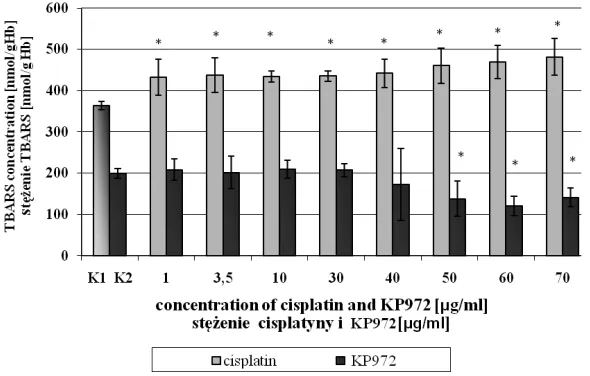
Fig. 1.Comparison of the effects of cisplatin and KP972 on TBARS con− centration in erythrocytes vs. con− trols K1 and K2; *significantly diffe− rent from controls (p< 0.05)
–0.5962, p < 0.0005) (Fig. 2). This means that KP972 is able to decrease lipid peroxidation in erythrocytes. Cisplatin showed quite an opposite effect. All cisplatin concentrations (1–70 µg/ml) raised the TBARS level from appropriately 432.52 ± 43.16 to 481.58 ± 44.47 nmol/g Hb compared with the control (363.57 ± 10.32 nmol/g Hb) (Fig. 1). Also, the linear correlation between ci− splatin dose and TBARS differed from that of the KP 972 relationship. A statistically significant posi− tive correlation between cisplatin dose and TBARS level in erythrocytes was noted (r = 0.5014, p< < 0.0005) (Fig. 4).
In the next experiment, the mechanism of the influence of KP972 and cisplatin on free radical processes was determined by measuring hydroxyl radical generation in mitochondria. Also in this case there was a different influence of the compo−
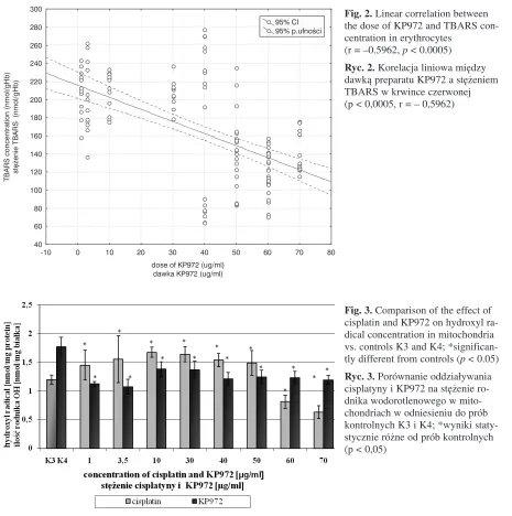
unds on OH· radical formation. KP972 at all con− centrations (1–70 µg/ml) caused a decrease in ·OH radical in the mitochondria compared with the control. The strongest effect was noted at the dose of 3.5 µg/ml (1.07 ± 0.13 vs. control: 1.77 ± 0.16 nmol/mg protein, p< 0.001) (Fig. 3). Cisplatin, in contrast, caused an increase in hydroxyl radical generation compared with the control at most of the concentrations used, i.e. 1–50 µg/ml (1.20 ± 0.08 nmol/mg protein, p< 0.01). The most inten− sive formation of OH· appeared at the 10 µg/ml concentration of cisplatin (1.68 ± 0.13 nmol/mg protein). Two doses of cisplatin (60 and 70 µg/ml) decreased ·OH generation.
The results show that the two compounds ac− ted in opposite ways; however, the final antiproli− ferative effects were comparable. It appears that the mechanisms of the compounds’ action can be
-10 0 10 20 30 40 50 60 70 80
dose of KP972 (ug/ml) dawka KP972 (ug/ml) 40
60 80 100 120 140 160 180 200 220 240 260 280 300
95% Cl 95% p.ufnoœci
TBARS concentration (nmol/gHb)
stê¿enie TBARS
(nmol/gHb)
Fig. 2.Linear correlation between the dose of KP972 and TBARS con− centration in erythrocytes
(r = –0.5962, p< 0.0005)
Ryc. 2.Korelacja liniowa między dawką preparatu KP972 a stężeniem TBARS w krwince czerwonej (p < 0,0005, r = – 0,5962)
Fig. 3. Comparison of the effect of cisplatin and KP972 on hydroxyl ra− dical concentration in mitochondria vs. controls K3 and K4; *significan− tly different from controls (p< 0.05)
completely different, at least regarding their influ− ence on free radicals. It seems that the antiradical effect of KP972 could have considerable signifi− cance in its antiproliferative activity, but the prora− dical action of cisplatin results in its harmful side effects, probably connected with the presence of metal in its structure.
Discussion
The toxicity of cytostatic medications presents a huge problem. Moreover, their side effects crea− te a constant need to search for new and effective anticancer compounds with potentially lower toxi− city. The examined pyridopyrazolopyrimidine de− rivative KP972 appeared in the Bulletin of the Pa− tent Office in 2006. The investigations conducted at the Institute of Immunology and Experimental Therapy, Polish Academy of Sciences, in Wrocław demonstrated a cytotoxic activity of KP972 in vi− troat a dose of 3.59 ± 0.56 µg/ml exceeding that of cisplatin. Whether free radical mechanisms par− ticipate in the anticancer activity of KP972 seemed interesting. The degrees of lipid peroxidation and hydroxyl radical generation were measured. The study was performed in an in vitro mitochondrial model from human placenta and isolated erythro− cytes from human blood. Cisplatin was used as the standard.
The results showed that the roles of the exami− ned compounds in free radical processes differ. Ci−
splatin induces oxidiative stress, which was re− vealed in the increase in the TBARS level in ery− throcytes at all concentrations with a statistically significant dose−response correlation. The literatu− re shows that the toxic effects of cisplatin, mainly ototoxicity, nephrotoxicity, and hepatotoxicity, could be connected with ROS generation. Ototoxi− city is explained by the fact that oxygen free radi− cals influence the antioxidant system of the middle ear, lowering the level of glutathione and antioxi− dant enzyme activity and causing an increase in the lipid peroxidation product MDA (malonyl dial− dehyde) [13–15].
In vivoexaminations in rats pointed out cispla− tin’s ability to disturb the GSH/GSSG (redu− ced/oxidized glutathione) balance, an indicator of oxidative stress. Among the side effects of cispla− tin is also protein p53 activation, connected with apoptosis. Supplementation with antioxidants such as dimethylthiourea (DMTU) or N−acetylocystein (NAC) suppressed the PUMA gene responsible for the regulation of protein p53 and helped block the protein’s activation when cisplatin is used [16–18]. To discover the type of mechanism responsible for the nephrotoxic effect of cisplatine, MDA and 8−hy− droxy−2’−deoxyguanosine (8−OHdG) concentrations in the urine of patients treated with cisplatin were measured [19]. Increased concentrations of mar− kers of DNA damage, such as 8−oxo−7,8−dihydro− guanosine (8−oxoGua) and 8−oxo−7,8−dihydro− −2’−deoxyguanosine (8−oxodG), were already ob− served 24 hours after cisplatin use. This indicates
-10 0 10 20 30 40 50 60 70 80
dose of cisplatin (ug/ml) dawka cisplatyny (ug/ml) 360
380 400 420 440 460 480 500 520
95% CI 95% p.ufnoœci
TBARS concentration (nmol/gHb)
stê¿enie TBARS
(nmol/gHb)
Fig. 4.Linear correlation between the dose of cisplatin and TBARS concentration in erythrocytes (r= 0.5014, p < 0.0005)
the participation of free radical mechanisms in the toxicity of this anticancer drug [20]. The above products are proposed as useful markers of acute renal failure after cisplatin treatment [19]. The in− fluence of cisplatin−induced ROS on liver micro− somal enzymes, in particular on CYP2E1, was de− monstrated. Hepatotoxicity stimulated by cisplatin accompanies changes in MDA level as well as GSH and the activity of the antioxidant enzymes glutathione peroxidase (GSH−Px) and catalase (CAT) [21].
An investigation by Chirino et al. [15] conduc− ted on rats showed a significant increase in hydro− xyl radical concentration after cisplatin treatment. The raised level of this radical induces nephrotoxi− city. The present study with the in vitromitochon− drial model of human placenta also showed that ci− splatin at doses of 1–50 µg/ml generated hydroxyl radical. KP972, in contrast, decreased the ·OH level in comparison with the control at all the tested concentrations. It appears that KP972 acted pro− tectively on mitochondria by inhibiting oxidative stress caused by ·OH radical. It could be an impor− tant factor in the anticancer activity of KP972. At
doses 40–70 µg/ml, this compound reduced the concentration of MDA, while cisplatin induced this aldehyde at all the applied concentrations (1–70 µg/ml) with a statistically significant linear correlation between dose and the level of MDA (r= 0.5014, p< 0.0005).
Based on the rich scientific literature concer− ning cisplatin and the present experiments, it can be concluded that the participation of free radicals in the toxicity of cisplatin is very significant. Thus it can be supposed that the pyridopyrazolopyrimi− dine KP972 might be less toxic. The free radical generation of cisplatin is connected with its side effects, but the antiradical activity of KP972 could be connected with its antiproliferative activity. Li− terature data confirm the antiproliferative action of certain pyridopyrazolopyrimidines [2]. They sup− press protein p21 formation, which is an inhibitor of cyclin and cyclin−dependent kinases. This influ− ences Ras, oncogenes which regulate the cell cyc− le and cause perturbations in growth and division of neoplastic cells [23]. These properties of pyry− dopyrazolopyrimidine encourage the synthesis of new active derivatives.
References
[1] Cerutti PA: Oxy−radicals and cancer. Lancet 1994, 24, 344, 862–863.
[2] Guyton K, Hensler T:Oxidative mechanisms in carcinogenesis. Br Med Bull 1993, 523–544.
[3] Singh KK: Oxidative stress, disease and cancer. Eds.: Roswell Park Cancer Institute, Imperial College Press, New York, USA 2006, 705–731, 1013–1022.
[4] Sun Y:Free radicals, antioxidants enzymes, and carcinogenesis. Free Radic Biol Med 1990, 8, 583–599.
[5] Jung−Whan K, Chi VD:Cancer’s Molecular Sweet Tooth and the Warburg Effect. Cancer Res 2006, 66, 15, 8927–8930.
[6] Stocks J, Offerman EL, Model CB, Dormandy TL: The susceptibility to autooxidation of human red cell lipids in health and disease. Br J Haematol 1972, 23, 713–724.
[7] Rice−Evans C, Burdon R:Free radical−lipid interactions and their pathological consequences. Prog Lipid Res 1993, 32, 1, 71–110.
[8] Lowry OH, Rosenbrough JN, Farr AL, Randell R:Protein measurement with the Folin Phenol reagent. J Biol Chem 1951, 193, 265–275.
[9] Lass A, Sohal R: Electron transport−linked ubiquinone−dependent recycling of α−tocoferol inhibits autooxidation of mitochondrial membranes. Arch Biochem Biophys 1998, 352(2), 229–236.
[10] Radi R, Sims S, Cassina A, Turrens JR:Role of catalase and cytochrome c in hydroxyperoxide−dependent lipid peroxidation and chemiluminescence in rat heart and kidney mitochondria. Free Radic Biol Med 1993, 15, 653–659.
[11] Długosz A, Piotrowska D: Lipid peroxidation stimulated by Solvesso, Bawanol and methanol, and its counterac− tion by antioxidants in human placental mitochondria. Toxicol in Vitro 2002, 16, 649–659.
[12] Gawlik M: Zastosowanie modelu krwinki czerwonej człowieka do badania właściwości antyoksydacyjnych związków zawartych w wodnych ekstraktach produktów spożywczych na przykładzie herbaty. Bromat Chem To− ksykol XXXVII, 2004, 4, 311–316.
[13] Rybak LP, Whitworth C, Somani S:Application of antioxidants and other agents to present cisplatin ototoxici− ty. Laryngoscope 1999, 109, 1740–1744.
[14] Rybak LP, Husain K, Morris C, Whitworth C, Somani S: Effect of protective agents against cisplatin ototoxi− city. Am J Otol 2000, 21(4), 513–520.
[15] Chirino YI, Sanchez−Gonzalez DJ, Matrinez−Martinez CM, Cruz C, Pedraza−Chaverri J:Protective effects of apocynin against cisplatin−induced oxidative stress and nephrotoxicity. Toxicology 2008, 245 (1–2), 18–23.
[16] Youngke L, Cederbaum AI:Cisplatin−induced hepatotoxicity is enhanced by elevated expression of cytochrome P450 2E1. Toxicol Sci 2005, 89 (2), 515–523.
[18] Santos NAG, Bezerra CSC, Martins NM, Curti C, Bianchi MLP, Santos AC: Hydroxyl radical scavenger ameliorates cisplatin−induced nephrotoxicity by preventing oxidative stress, redox state unbalance, impairment of energetic metabolism and apoptosis in rat kidney mitochondria. Cancer Chemother Pharmacol 2008, 6, 145–155.
[19] Zhou H, Kato A, Miyaji T, Yasuda H, Fujigaki Y, Yamamoto T, Yonemura K, Takebayashi S, Mineta H, Hishida A: Urinary marker of oxidative stress in kidneys in cisplatin−induced acute renal failure in rats. Nephrol Dial Transplant 2006, 21, 616–623.
[20] Siomek A, Oliński R, Tujakowski J:Severe oxidatively damaged DNA after cisplatin treatment of cancer pa− tients. Int J Cancer 2006, 119, 2228–2230.
[21] Yuce A, Atessahin A, Ceribas AO, Akasakal M:Ellagic acid prevents cisplatin−induced oxidative stress in li− ver and heart tissue of rats. Basic Clin Pharmacol Toxicol 2007, 101, 345–349.
[22] Poręba K, Opolski A, Wietrzyk J, Kowalska M:Synthesis and antiproliferative activity in vitroof new deriva− tives of 3−aminopyrazolo[3,4−b]pyridine. Arch Pharm Pharm Med Chem 2001, 334, 219–223.
[23] Wolin RM, Afonio A, Kelly JM, Njorge FG: Pat. USA 5595998, 1997; C.A. 126, 199793j, 1997.
Address for correspondence:
Ewa Sawicka
Department of Toxicology Wroclaw Medical University Traugutta 57/59
50−417 Wrocław Poland
Tel./fax: +48 71 344 43 75 E−mail: [email protected]
Conflict of interest: None declared