The source of HLA molecules on platelets: Does platelets adsorb
soluble HLA molecules from their environment?
Tahereh Dargahi MSC1, Fatemeh Yari PhD1,*, Negar Rezaei MD PhD1
1. Blood Transfusion Research Center, High Institute for Research and Education in Transfusion Medicine, Tehran, Iran, E-mail: [email protected]
*Corresponding author: Dr Fatemeh Yari, Associate Professor of Blood Transfusion Research Center, High Institute for Research and Education in Transfusion Medicine, Tehran, Iran, E-mail: [email protected]. ORCID ID: 0000-0003-1036-3801
Received: 06 April 2019 Accepted: 02 August 2019
Abstract
Background: The origin and function of human leukocyte antigen (HLA) class I molecules on platelets are still highly arguable. Given the differences in the results of the previous studies in this regard, the lack of research in recent years, and the clinical importance of HLA class I molecules, the absorption capacity of platelets for soluble HLA class I molecules was studied in this investigation.
Materials and Methods: In this experimental study, HLA-A2 antigen was purified from a B cell precursor leukemia cell line (Nalm-6) by cell membrane protein solubilization and usage of HLA-A2 affinity column. Platelet concentrates (PCs) were received from Tehran Blood Transfusion Center. Eighteen bags of HLA-A2-negative PCs were prepared randomly and treated with various concentrations of the purified HLA antigen (100, 500, and 1000 ng/ml) for 48 to 72 hours. Subsequently, the HLA-A2 levels were evaluated on platelets by flow cytometery technique. Data were evaluated using repeated measure ANOVA.P-values less than 0.05 were considered significant.
Results: The results of this study showed that the purified protein was an HLA molecule (HLA-A2). After the treatment of platelets and HLA molecules, platelets inability was shown for the attracting of HLA molecules. This finding was true in both media of RPMI and plasma. The differences between the case (HLA-treated platelets) and control (untreated platelets) were not significant (p-values> 0.05).
Conclusion: Platelets were unable to significantly adsorb exogenous HLA antigens from their environment. Further studies are needed to unravel the nature and origin of HLA molecules on platelets.
Key words: Absorption, HLA, Platelets
Introduction
Platelets are small, non-nucleated cells. Platelets have many physiological roles, including involvement in homeostasis, thrombosis, clot formation, vascular repair, inflammation, immune responses, and play an important role in intercellular communication (1-3). Platelets express human leukocyte antigen (HLA) class I, ABO antigens, several platelet-specific, and carbohydrate antigens on their membrane (4). Among HLA molecules, there are no HLA class II molecules on the platelets, and HLA-C is also expressed in low amounts. HLA molecules can potentially act as an immune stimulant in the recipient of platelet transfusion (5,6). Platelets express HLA class I molecules
less than mononuclear cells, but due to the high platelet count in the circulation, two-thirds of the HLA class I molecules are related to platelets (7). The function of HLA molecules is well described in white blood cells, while in other types of cells, such as platelets, it is not clear yet (8). In addition to HLA class I antigen expression, platelets express T cell co-stimulatory molecules. Platelets process and present antigen and directly activate naïve T cells in a HLA class I -dependent manner in vivo (9). Today, platelet transfusion is used to prevent or treat bleeding in patients with thrombocytopenia, and patients with platelet function impairment. However, after injection of platelet concentrate, a percentage of patients develop antibodies
against HLA antigens, or less likely against platelet antigens. These antibodies can induce platelet refractoriness upon subsequent platelet injections (10, 11). Alloimmunization against the HLA class I antigens is the most common cause of the immunologic loss of the platelets (5, 6, 12). Different studies have listed several ways to prevent platelet refractoriness, but none of the strategies is fully effective. Reducing the number of HLA class I molecules was suggested by some researchers (13-16). If HLA antigens are absorbed from the environment, the isolation of HLA class I antigens from platelets will be feasible without platelet degradation.Theoretically, if conditions for the isolation of HLA-A and HLA-B antigens are not provided, HLA matching, crossmatching, and antibody specificity prediction will be appropriate for the management of the platelet refractory patient (17). Some studies have shown that HLA-A and HLA-B antigens on the platelet surface are the main components of the membrane. Other studies based on chloroquine treatment or incubating platelets with allogeneic plasma show that HLA molecules are absorbed from the plasma to the platelet surface (18). The origin of HLA class I molecules on platelets is still to some extent unclear, so it is argued that these HLA antigens are either integral or absorbed from the environment on the platelet surface. This study was attempted to find out the ability of platelets for absorbing the HLA molecules. The HLA-A2 molecules were added to the storage media of A2-negative platelets and the extent of HLA-A2 adsorption of platelets was determined.
Materials and Methods
Preparation of platelet concentrate
In this experimental study, conducted in 2015 and 2016, eighteen bags of PC units were prepared randomly from blood donors referring to Tehran Transfusion Center. No choice was made regarding the blood group, volume, and appearance of
the platelet bag. Due to the time needed to perform viral screening tests including hepatitis B virus (HBV), hepatitis C virus (HCV), and human immunodeficiency viruses (HIV)) on blood products, platelets concentrate were analysed within 24 hours. The day of entering the PC into the lab was considered as the first day. Eighteen HLA-A2-negative PC bags were chosen using flow cytometry technique.
NALM-6 cell line cultivation, cell lysis, and membrane protein solubilization
In this experimental study, NALM-6 (a human B-cell precursor leukemia cell line) was cultured in RPMI medium containing 10% FBS. This cell line was selected for this study because of the high level of HLA-A2 expression. After the cultivation of the cells, they were collected, centrifuged (at 900 g for 5 minutes), and lysed by lysis buffer including 25 mM Tris-HCL, 150 mM NaCL, 10 mM EDTA, 1.0% NP-40, pH 7.5, and 2 mM phenylmethylsulfonyl fluoride. In order to facilitate the release of membrane proteins such as HLA, two methods of homogenization and sonication were used. The cells were sonicated on ice for 10 minutes or mixed on a stirrer for 30-40 minutes. The cells were sonicated for 10 min with the Hielscher-Ultrasound Technology Sonicator (Hielscher, Teltow, Germany) at amplitude 80% and cycle 0.5. Then, for both methods, the tubes were centrifuged for 10 minutes at 1000 g, the supernatants were collected, and dialysed to replace the lysis buffer with phosphate buffer (PBS). Determination of the total protein concentration of the purified protein was performed using Bradford method. For this purpose, 10 μl of each of the standard samples (BSA) or purified protein were added into 200 μl of Bradford reagent in the microplate wells. Their absorbance was then read at 595 nm with a NanoDrop spectrophotometer (WPA, Biowave UK).
Preparation of an affinity
chromatography resin in a column
Preparation of carbonate bicarbonate (conjugate) buffer was done by mixing 4 ml of 0.2 M Na2CO3 solution with 46 ml
of 0.2 M NaHCO3 solution and bringing the final volume to 100 ml. Then, 0.4 g of cyanogen bromide-activated sepharose 4B was placed in 5 ml conjugate buffer overnight at 4˚ C to swell up. On the next day, 60 μg of mouse anti-human HLA-A2 antibody (BioRad, UK) was added and placed on the rotator for 3 hours. The gel containing the antibody was then added gently to the glass column and washed with PBS until the OD of the output buffer became zero. The column was then washed with 0.2 M glycine buffer (pH=2.8). Ethanolamine 3.4ml/ lit was prepared in 0.05M carbonate bicarbonate buffer and passed through the column to block the empty active sites of the gel. Finally, the column was washed 2 times with PBS.
Purification of the HLA-A2 protein using the affinity chromatographic separation
After passing the dialyzed sample through the column, the column was washed with PBS. Washing was continued until the optical density (OD) of the outlet fluid reached zero at 280 nm. Then, the glycine buffer was added to the column to separate the proteins attached to the antibody. The output of the column was collected. The purified protein was condensed using a Spin-X® UF 20 (5K MWCO) concentrator tube (Corning, UK).Finally, the protein concentration was measured using Bradford method (19).
Analysis of the purified protein by ELISA method
ELISA method was used to analyze the specificity of the purified HLA-A2 antigen. At first, 50 μl of 5 µg/ml antigen was added to the ELISA plate in PBS buffer. In addition, 50 μl of human chorionic gonadotropin (hCG) (Sigma, USA) protein and PBS solution were added as negative control in other wells of the plate. The plate was covered and incubated overnight at 4° C. The wells
were drained and 100 μl of 2% BSA solution (blocking solution) was added to each of the wells. The plate was incubated for three hours at room temperature. In the next step, anti-HLA-A2 specific antibody was added to the antigen-coated plate, and incubated for 1 hour at 37°C. After washing steps, an anti-mouse Immunoglobulin (IgG) conjugate attached to the HRP enzyme (Sigma, USA) was used. After washing, TMB substrate was added forming a colored product. In the final step, sulfuric acid was added as a stopper for enzymatic reaction.
Studying the specificity of the purified protein using dot blot test
In this test, the specificity of the protein was proven by direct loading of the purified sample onto membrane paper and the use of a specific antibody. First, the polyvinylidene difluoride (PVDF) membrane was cut in appropriate dimensions and the locations of proteins were marked. The membrane was placed in methanol for a short time. After drying the proteins, the membrane was blocked with 5% skim milk (2.5 gr in 50 ml of PBS) for 2 hours. Then, the membrane was immersed overnight in anti-HLA-A2 specific antibody. On the next day, the membrane was washed 3 times using a PBST washing solution. Then, the secondary HRP-conjugated anti-mouse IgG was placed on the membrane on a shaker for about 1 hour. After washing step, a luminescence substrate was added for 30 seconds in dark. Antibody binding was measured by a chemiluminometer (Biorad chemidoc XRS system, USA).
Exposure of platelets with the purified HLA-A2 antigen and the capacity of platelets to absorb soluble HLA-A2 protein
Pure and concentrated HLA-A2 antigen was sterile-filtered. Platelets were treated with the purified HLA-A2 antigen in both the plasma and RPMI media. For obtaining plasma which lacked microparticles (MPs), the plasma of a HLA-A2-negative sample was centrifuged at 15,000 g for 12
minutes. In a 24-well culture plate, 5×10⁷ platelets were treated with HLA-A2 protein at the concentrations of 100, 500, and 1000 ng/mL in plasma or RPMI media for 48 and 72 hours. The wells includingthe platelets + plasma or platelet + RPMI medium without HLA-A2 protein were considered as control. After the incubation time, HLA-A2-treated platelets were collected and washed. Then, the levels of HLA-A2 expression were evaluated by flow cytometry technique in a two step method. At first, platelets were treated with anti HLA-A2 monoclonal antibody for 30 minute and in the second step, washed with PBS and incubated with FITC-conjugated anti mouse IgG for 30 minuets at 4ºC.
Statistical analysis
Data were analyzed using SPSS (version 22). Repeated measure ANOVA was run for the evaluation of HLA-A2 levels on platelets. P-value less than 0.05 was considered significant.
Results
The antigenic property of the purified antigen
The specificity of the purified protein was studied by ELISA technique. The mean optical density (OD450 nm) was determined
as 0.424± 0.002 for the purified samples, while this value was 0.17±0.03 for the negative control. In ELISA method, the reaction of the purified protein with anti-HLA-A2 antibody indicated the specificity of the antigen. Additionally, the specificity of the purified protein was also evaluated by dot blot assay in which the reaction of anti-HLA-A2 antibody with the purified protein indicated that the nature of the antigen coincided with the HLA-A2 ,while the reaction was completely negative for the irrelevant protein (HCG) (Figure 1).
Evaluation of HLA-A2 levels on
platelets after exposure to HLA-A2 antigen in plasma or RPMI media
Absorption of platelets for HLA molecules was not significant in both plasma and RPMI media. The difference in HLA-A2
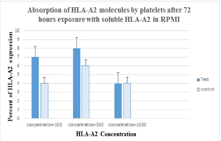
protein levels on platelets was not significant before and after exposure with soluble HLA-A2 at the concentrations of 100, 500 and 1000 ng/ml. The mean HLA-A2 expression on platelets was measured after treatment with HLA-A2 and compared with the control (0 ng/ml of HLA-A2). The difference between the case and control was not significant after 48 hours of treatment in plasma (p-values were 0.528, 0.152, and 0.075 in HLA-A2 at concentrations of 100, 500, and 1000 ng/ml, respectively) and RPMI medium (p-values were 0.221, 0.133 and 0.878 in HLA-A2 at concentrations of 100, 500, and 1000 ng/ml, respectively). Similar results were obtained after 72 hours of treatment in RPMI (with the p-values; 0.285, 0.621 and 0.197) and plasma medium (with the p-values; 0.141, 0.600 and 0.068) in HLA-A2 with concentrations of 100, 500, and 1000 ng/ml (Figur 2 and Figure 3).
Figure 1. Analysis of the specificity of the purified protein by dot-blot method.
Figure 2. Expression of the HLA-A2 molecule on the platelet surface after 72 hours of treatment with HLA-A2 antigen in the plasma indicated a limited absorption of HLA molecules on the platelet surface. This rate of absorption did not significantly affect the expression of HLA molecules on the platelet surface (p>0.05).
Figure 3. The expression of the HLA-A2 molecule on the platelet surface after 72 hours of treatment with the HLA-A2 antigen in RPMI medium indicated a limited absorption of the HLA molecule on the surface of the platelets. This rate of absorption did not significantly change the expression level of HLA molecules on platelets (p>0.05).
Discussion
In this study, platelets were treated with different concentrations of HLA-A2 protein for 48 to 72 hours in both plasma and RPMI media. Then, the mean HLA-A2 expression was measured on platelets by flow cytometry after treatment and compared with the control (0 ng/ml of HLA-A2). The difference between the case and control was not significant after 48 and 72 hours of treatment in RPMI and plasma media.
When HLA antigens are absorbed from the environment, removing them will be feasible without platelet degradation (13, 18). What distinguishes this study from other studies is the use of purified soluble HLA molecules rather than HLA containg plasma to detect the absorption potential of HLA molecules by platelets. We found that the exposure of platelets with HLA-A2 in RPMI or plasma environment did not significantly change the HLA-A2 levels on platelets after 48 or 72 hours of exposure.
The result of this study was consistent with finding reported by Zeileret al. They incubated HLA-A2 negative blood platelets in the plasma of HLA-A2 positive donors. Then, the expression levels of HLA-A2 were measured on platelets after 4 days of storage using flow cytometry technique. They stated that the absorption of HLA-A2 molecules on HLA-A2-negative platelets was unlikely in the routine conditions. In general, they found no significant uptake and absorption of HLA class I antigens on platelets during storage (20).
On the contrary, the results of our study were not correlated with the results of a study conducted by Lalezari and Driscol in 1982, in which the HLA-A1 and HLA-A2-negative platelets were treated with the plasma obtained from HLA-A2 positive patients. Plasma-treated platelets received HLA from plasma, which was demonstrated by lymphocytotoxicity test (21). It seems that the method of their evaluation might influence the results.
Additionally, from the other point of view, some studies implied the transmembrane feature of HLA molecules on the platelet surface because HLA‐stripping using acid treatment of platelets showed losing of beta-2 microglobulin and peptides and keeping denatured heavy chain on the membrane (15). These kinds of studies can be consistent with the integral and transmembrane nature of HLA molecules on platelets, and lack of adsorption of HLA molecules from the environment can be consistent with the result of our study. In contrast, some studies have shown loose attachment of HLA molecules to platelet surface. For instance, Valsami et al., reported that the majorities of HLA antigens on the surface of platelets mostly comprised of heavy chains with only a minimal amount of beta-2 microglobulin and have the ability to disconnect from their surface mainly during platelet storage (22). Further studies are needed to unravel the nature and origin of HLA molecules on platelets.
Conclusion
Platelets were unable to significantly adsorb exogenous HLA antigens from their environment. Further studies are needed to unravel the nature and origin of HLA molecules on platelets.
Acknowledgments
This study was the result of a thesis (the ethics code ; IR.TMI.REC.1394.21), which was financially supported by Blood Transfusion Research center, High Institute for Research and Education in Transfusion Medicine, Iranian Blood Transfusion Organization, Tehran, Iran .
Conflict of interest
None of the authors have any conflicts of interest to declare.
References
1. Vogel S, Thein SL. Platelets at the crossroads of thrombosis, inflammation
and haemolysis. Br J Haematol 2018; 180: 761-767.
2. Austin SK. Haemostasis. Medicine 2009; 37:133-136.
3. Shaiegan M. Platelet Immunology. Sci J Iran Blood Transfus Organ 2012; 9:72-93.
4. Baghdadi V, Yari F, Rezaei N, Rafiee MH. The surface markers and survival rate of platelets during storage at 4°C: The influence of sodium octanoate. J Ped Hematol Oncol 2019; 9(2): 105-116. 5. Waterman HR, Kapp LM, Munday A, Odem‐Davis K, Zimring JC. Transfusion‐induced alloimmunization and platelet refractoriness in a mouse model: mechanisms and interventions. Transfusion 2016; 56: 91-100.
6. Pavenski K, Freedman J, Semple J. HLA alloimmunization against platelet transfusions: pathophysiology, significance, prevention and management. Tissue antigens 2012; 79 : 237-245.
7. Yari F, Ahmadzadeh N, Azadpour S, Vaeli S. HLA antigens shed from the surface of synthetic or naturally occurred platelet-derived microparticles during storage of platelet concentrate. Indian J Hematol Blood Transfus 2012; 28(3):152-156.
8. Valsami S, Dimitroulis D, Gialeraki A, Chimonidou M, Politou M. Current trends in platelet transfusions practice: The role of ABO-RhD and human leukocyte antigen incompatibility. Asian J Transfus Sci 2015; 9:117-123.
9. Chapman LM, Aggrey AA, Field DJ, Srivastava K, Ture S, Yui K, et al. Platelets present antigen in the context of MHC class I. J Immunol 2012; 189:916-923.
10. Hod E, Schwartz J. Platelet transfusion refractoriness. Br J Haematol 2008; 142(3):348-360.
11. Stanworth SJ, Navarrete C, Estcourt L, Marsh J. Platelet refractoriness– practical approaches and ongoing dilemmas in patient management. Br J Haematol 2015; 171:297-305.
12. Jeremiah ZA, Atiegoba A, Mgbere O. Alloantibodies to human platelet glycoprotein antigens (HPA) and HLA class 1 in a cross section of Nigerian antenatal women. Hum Antibodies 2011; 20 (3):71-75.
13. Wiegmann B, Figueiredo C, Gras C, Pflaum M, Schmeckebier S, Korossis S, et al. Prevention of rejection of allogeneic endothelial cells in a biohybrid lung by silencing HLA-class I expression. Biomaterials 2014; 35(28):8123-8133.
14. Figueiredo C, Goudeva L, Horn PA, Eiz-Vesper B, Blasczyk R, Seltsam A. Generation of HLA-deficient platelets from hematopoietic progenitor cells. Transfusion 2010; 50 (8):1690-1701. 15. Meinke S, Sandgren P, Mörtberg A, Karlström C, Kadri N, Wikman A, et al. Platelets made HLA deficient by acid treatment aggregate normally and escape destruction by complement and phagocytes in the presence of HLA antibodies. Transfusion 2016; 56: 370-382.
16. Ofem OE. Comparative effect of two anti-malaria drugs (chloroquine and coartem) on haematological parameters in albino Wistar rats. Afr J Biomed Res 2013; 16:39-46.
17. Forest SK, Hod EA. Management of the Platelet Refractory Patient. Hematol Oncol Clin North Am 2016; 30(3):665-677.
18. Kapur R, Zufferey A, Boilard E, Semple JW. Nouvelle cuisine: platelets served with inflammation. J Immunol 2015; 194(12):5579-5587.
19. Cheng Y, Wei H, Sun R, Tian Z, Zheng X. Rapid method for protein quantitation by Bradford assay after elimination of the interference of polysorbate 80. Anal Biochem 2016; 494:37-39.
20. Zeiler T, Heim M, Dempfle A, Kretschmer V. Platelets do not adsorb HLA class I molecules during storage of pooled platelet concentrates. Transfus Med 2006; 16(3):176-183.
21. Lalezari P, Driscoll AM. Ability of thrombocytes to acquire HLA specificity from plasma. Blood 1982; 59:167-170. 22. Valsami S, Dimitroulis D, Gialeraki A, Chimonidou M, Politou M. Current trends in platelet transfusions practice: The role of ABO-RhD and human leukocyte antigen incompatibility. Asian J Transfus Sci 2015; 9(2):117-123.