M E E T I N G A B S T R A C T S
Open Access
Sepsis 2019
Rio de Janerio, Brazil. 09 - 10 May 2019
Published: 27 June 2019
Sepsis 2019 Abstracts
P1
Withdrawn
P2
Epigenetic changes are reported in animal model of sepsis Monique Michels1, Mariane Rocha Abatti1, Andriele da Silva Vieira1, Heloisa Borges1, Amanda Indalécio Goulart1, Roger Varella2, Samira Valvassori2, Felipe Dal-Pizzol1
1
Laboratory of Experimental Pathophysiology, Extreme University South of Santa Catarina, Criciúma, Brazil;2Laboratório de Neurociências, Programa de Pós-Graduação em Ciências da Saúde (PPGCS), Universidade do Extremo Sul Catarinense (UNESC), Criciúma, SC, Brazil Correspondence:Monique Michels ([email protected]) Intensive Care Medicine Experimental2019,7(Suppl 2):P2
Background
The presence of oxidative stress and inflammatory mediators in sep-sis may lead to epigenetic changes [1,2]. Epigenetic alterations of his-tones, such as methylation, acetylation and phosphorylation may direct the folding or unfolding of DNA through mechanisms still un-known, thus altering gene transcription [3]. Once gene transcription in sepsis is altered the response may be further exacerbated. Our ob-jective was to report epigenetic changes in brain structures in animal model of sepsis.
Materials and Methods
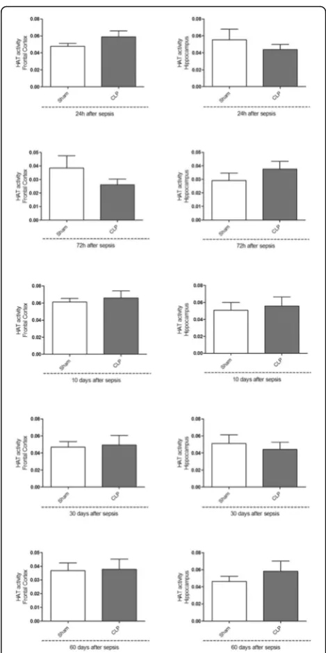
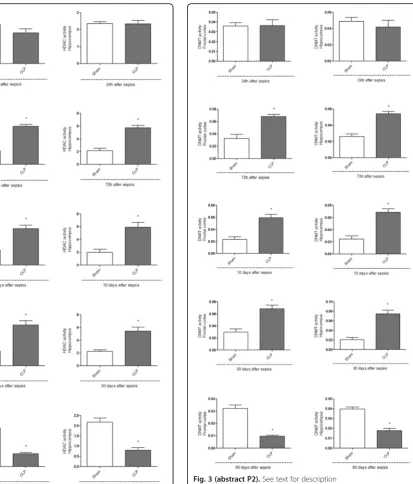
Male Wistar rats were subjected to sham or CLP and cerebral struc-tures were removed in 24h, 72h, 10, 30 and 60 days after sepsis. HAT, HDAC and DNMT enzymes activities were measured in frontal cortex and hippocampus in different times.
Results
No changes found in HAT activity (Fig. 1). Increased HDAC (Fig. 2) and DNMT activity (Fig. 3) was observed 72h, 10 and 30 days after sepsis and a significant reduction 60 day after.
Conclusions
It’s possible observe epigenetic alterations and deregulation in gene transcription in animal model of sepsis. Since in sepsis the presence of oxidative stress and the release of inflammatory mediators are well reported, these insults can lead to epigenetic changes, such as gene transcription, which may be related to exacerbation of the in-flammatory response. Environmental influences can modulate the epigenetic response and therefore, be a therapeutic strategy for the treatment of sepsis.
References
1. Margueron R, Trojer P, Reinberg D: The key to development: interpreting the histone code? Curr Opin Genet Dev. 2005, 15:163–176.
2. Reik W: Stability and flexibility of epigenetic gene regulation in mammalian development. Nature. 2007, 447:425–432.
3. Chen Y, Hong T, Wang S, Mo J, Tian T, Zhou X: Epigenetic modification of nucleic acids: from basic studies to medical applications. Chem Soc
Rev. 2017, 46(10):2844-2872. Fig. 1 (abstract P2).See text for description
Fig. 2 (abstract P2).See text for description
P3
Is antibiotic treatment always necessary for chronic critical ill patients?
Natalia Beloborodova, Irina Buyakova, Ekaterina Chernevskaya Federal Research and Clinical Center of Intensive Care Medicine and Rehabilitology, Laboratory of metabolism in critical state, Moscow, Russia Correspondence:Ekaterina Chernevskaya ([email protected])
Intensive Care Medicine Experimental2019,7(Suppl 2):P3
Background
Antibiotics are prescribed to almost all of chronic critical ill (CCI) pa-tients. This often leads to colonization of multi-resistant strains of mi-croorganisms and dramatic disturbances of gut microbiota. Metabolic activity of microbes can be assessed by the measurement of the levels of aromatic microbial metabolite (AMM) in serum, which are associated with the severity and mortality of ICU patients [1]. The modern trend is to reduce the antibacterial pressure. The aim of our study is to estimate the frequency of antibiotic use in CCI patients, to discuss the relationship between change in the neurological status and metabolic profile of AMM.
Materials and Methods
The study included 40 CCI patients with neurological disorders (stroke, traumatic brain injury, neurosurgical intervention for brain tu-mors). The level of AMM was measured in blood serum using GC-MS (Thermo Scientific). Biomarkers (PCT, S100) were measured using Elecsys immunoassay.
Results
Antibiotics (cephalosporins, aminoglycosides, fluoroquinolones, etc.) were prescribed on 74% of CCI patients in different cases (bacteriuria, leukocyturia, fever, etc.), but the retrospective analysis showed that levels of PCT were low (0.02 to 0.236 ng/ml). A significant increase the level of AMM, mainly due to para-hydrophenylacetic acid (p-HPhAA) was accompanied by the negative dynamics of the somatic and neurological status. Low serum levels of AMM correlated with positive clinical dynamics, a decrease of p-HPhAA level and an in-crease in para-hydroxybenzoic acid correlated with an improvement in neurological status. AMM`s data are compared with analysis of the gut microbiota using 16S rRNA sequencing in different groups of patients.
Conclusions
In chronic critical ill patients with neurological disorders, the pres-ence of indirect signs of infection is not an indication for antimicro-bial therapy, if biomarkers and AMM remain within the reference values, since antibiotics do not contribute to the improvement of the clinical and neurological condition. For this group of patients’, it is ne-cessary to develop new treatment strategies based on the correction of microbiota metabolism.
References
1. Beloborodova NV, Sarshor YN et al.: Involvement of Aromatic Metabolites in the Pathogenesis of Septic Shock. Shock. 2018, 50(3):273-279.
P4
Connection between biomarkers and aromatic metabolites in
сerebrospinal fluid in critically ill patients
Ekaterina Chernevskaya1, Natalia Beloborodova1, Tatyana Litvinova1, Irina Alexandrova2, Maria Getsina1, Alisa Pautova1
1
Federal Research and Clinical Center of Intensive Care Medicine and Rehabilitology, Laboratory of metabolism in critical state, Moscow, Russia;2Federal State Autonomous Institution“N .N. Burdenko National Scientific and Practical Center for Neurosurgery”, Moscow, Russia Correspondence:Ekaterina Chernevskaya ([email protected]) Intensive Care Medicine Experimental2019,7(Suppl 2):P4
Background
Some aromatic derivatives of tryptophane - 5-hydroxyindoleacetic acid (5-HIAA) and 3-indoleacetic acid (3-IAA) are under direct or undirect con-trol of the gut microbiota and may play an important role in gut–brain axis. It was found that in serum high levels of some aromatic microbial metabolites (AMM) of phenolic structure are associated with severity of
infection in critically ill patients with sepsis. Neuron specific enolase and S100 protein are biomarkers that reflect the neurotrophic and neurotoxic effects of neuron and glial cells. Procalcitonin (PCT) is a biomarker which is elevated in serum in the case of bacterial infection, however, the use-fulness of PCT measurement in CSF has shown conflicting results. Cut-off value of PCT in CSF for bacterial meningitis was lower than serum level of PCT [1]. High level of PCT in CSF may indicate the loss of integrity of the blood-brain barrier, but some microbial metabolites may also pene-trate the barrier in critical conditions. The aim of our study was to identify and qualify indolic and phenolic metabolites and evaluate correlation of certain biomarkers in the CSF in critically ill patients.
Materials and Methods
The study included 37 CSF samples taken from neurosurgical critic-ally ill patients with CNS infection, traumatic brain injury. The levels of AMM and 3-IAA were measured in the CSF using GC-MS (Thermo Scientific), biomarkers (PCT, S100, NSE, IL6) were measured by Elecsys immunoassay, 5-HIAA was measured by ELISA (Cloud-Clone Corp). Results
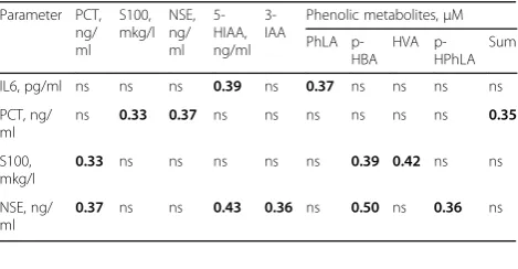
The median level of the sum of 6 aromatic metabolites (benzoic, phenyllactic (PhLA), p-hydroxybenzoic (p-HBA), p-hydroxyphenilacetic, homovanilic (HVA) and p-hydroxyphenillactic (p-HPhLA) acid) in the CSF was 4.4μM. The direct Spearman`s correl-ation between the level of PCT and sum of AMM (0.35, p<0.05) was revealed. The correlations between neurological biomarkers and some phenolic and indolic metabolites were also revealed (table 1). Conclusions
Connection between aromatic metabolites and neurological bio-markers (S100, NSE) indicates the potential involvement of phenolic and indolic metabolites in the pathogenesis of brain dysfunction.
Acknowledgements
Supported by the Russian Science Foundation Grant 15-15-00110
Reference
1. Zhang L, Ma L et al. Diagnostic Value of Procalcitonin for Bacterial Meningitis in Children: A Comparison Analysis Between Serum and Cerebrospinal Fluid Procalcitonin Levels. Clin Pediatr (Phila). 2018 Oct 29:9922818809477
P5
Association between site of infection and in-hospital mortality among patients with sepsis in the emergency departments of third-level hospitals in Medellin, Colombia
César Caraballo1,4, Johana Ascuntar1, Carolina Hincapié1, Camilo Restrepo1, Elisa Bernal2, Fabián Jaimes1,3
1
Grupo Académico de Epidemiologíıa Clínica (GRAEPIC), Universidad de Antioquia, Medellín, Colombia;2Servicio de Medicina Interna, Hospital Pablo Tobón Uribe, Medellín, Colombia;3Dirección de Investigaciones, Hospital San Vicente Fundación, Medellín, Colombia;4Center for Outcomes Research and Evaluation (CORE), Yale University School of Medicine, New Haven, CT, USA
Correspondence:Fabián Jaimes ([email protected]) Intensive Care Medicine Experimental2019,7(Suppl 2):P5
Table 1 (abstract P4).Correlations between biomarkers and aromatic metabolites (p<0.05)
Parameter РСТ, ng/ ml S100, mkg/l NSE, ng/ ml 5-HIAA, ng/ml 3-IAA
Phenolic metabolites,μM
PhLA p-HBA
HVA p-HPhLA
Sum
IL6, pg/ml ns ns ns 0.39 ns 0.37 ns ns ns ns
РСТ, ng/ ml
ns 0.33 0.37 ns ns ns ns ns ns 0.35
S100, mkg/l
0.33 ns ns ns ns ns 0.39 0.42 ns ns
NSE, ng/ ml
Background
The impact of infection site in patients with sepsis on hospital mor-tality have not been reliably estimated1. We aimed to determine, in patients presenting to the emergency department with sepsis or sep-tic shock, the association between the infection site and in-hospital mortality.
Materials and Methods
Multicenter prospective cohort in three emergency departments and critical care units of high complexity hospitals in Medellín (Colombia). We recruited patients older than 18 years admitted with sepsis or septic shock as the main diagnosis. The exposure variable was site of infection according to standardized CDC definitions and the primary outcome variable was in-hospital mortality. A hierarchical logistic regression model was fitted for adjusting for acknowledged prognostic factors as comorbidities, organ dysfunction and emergency treatments.
Results
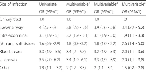
From 5022 eligible patients, 2510 were included in the study. The most frequent site of infection was the urinary tract with 27.8% of the cases, followed by pneumonia with 27.5% and intra-abdominal focus with 10.8% of the patients. In the 5.4% of the cases there was not a clear site of infection at admission. Using hierarchical logistic regression models with urinary tract as the reference, there were sig-nificant differences in mortality (Table 1).
Conclusions
There is an association between the different sites of infection and in-hospital mortality in patients with sepsis and septic shock and this should be considered in the prognostic models for these conditions.
Reference
1. Motzkus CA, Luckmann R. Does infection site matter? A systematic review of infection site mortality in sepsis. J Intensive Care Med. 2017;32:473-9.
P6
Risk factors for development of sepsis in a pediatric ICU
Gabriel do Amaral Cavalcante¹, Andrea Ramires Kairala², Rafael Augusto Faust Machado³, Bruno Mamede Lins Brasiliense³, Gabriela Jordão Vieira Gomes4 ¹Medicine department of UniCEUB, Brasília, Brasil; ²Instituto Hospital de Base, Brasília, Brasil; ³Brasil medicine department of UNICEPLAC, Brasília, Brasil;4Secretaria de Estado de Saúde do Distrito-Federal, Brasília, Brasil Correspondence:Gabriel do Amaral Cavalcante
Intensive Care Medicine Experimental2019,7(Suppl 2):P6
Objective
To determine and evaluate clinical aspects and the main risk factors for development of sepsis in patients admitted in a pediatric Inten-sive Care Unit (ICU).
Materials and methods
We performed a retrospective and observational study by evaluating electronic medical records of patients, aged between 0 and 12 years old, admitted among January and December of 2017 in a pediatric ICU at a tertiary care hospital in Distrito-Federal. Patients that died in 48 hours of the admission, immunocompromised or admitted in the unit already with sepsis were excluded.
Results
Three hundred and thirty-four children were admitted in 2017, 171 of those were included in the study. The majority were male (N: 102; 59.6%) and under 2 years old (N:107;62.6%). 16 died (9.4%). Most of them hospitalized for 6 to 12 days (N:67; 39.2%). The reason of the hospitalization were clinical (N:92; 53.8%), surgical (N:72; 42.1%) and trauma (N:7; 4.1%). Central venous access was made in 136 (78.9%) patients, followed by tracheal intubation (N:109; 63.7%), thorax drain-age (N:20; 11.7%) and 50% had long-standing bladder catheter. In our sample, 52 (30.4%) were diagnosed with sepsis and, among those, 8 evolved to septic shock. 24 (46.2%) of whom with sepsis had age between 28 days and 2 years. Also among those with sepsis, 75% had tracheal intubation, there being statistical correlation be-tween the procedure and sepsis (p-value: 0.043). The other proce-dures didn’t show correlation with the development of sepsis. The sites of the infection among those with sepsis were mainly pulmon-ary (67%), central nervous system (13%), urinpulmon-ary tract (3%), abdom-inal (8%), cardiovascular (3%) and others (6%). 5 patients with diagnosis of sepsis died (9.6%). There was found no relation between the diagnosis of sepsis and age, sex and death, value = 0.758) (p-value = 0.995) and (p-(p-value = 0.939) respectively.
Conclusions
In a pediatric ICU, besides the medical reason of the hospitalization, there are necessary invasive procedures that contribute to the devel-opment of infections of many levels; there are mechanisms of pro-tection and investigation that can lower sepsis incidence, also leading to an inferior death risk and incidence of complications dur-ing the staydur-ing in hospital.
P7
Early sepsis diagnosis: lesser internship time and death in a pediatric ICU
Gabriel do Amaral Cavalcante¹, Andrea Ramires Kairala², Rafael Augusto Faust Machado³, Bruno Mamede Lins Brasiliense³, Gabriela Jordão Vieira Gomes4
¹Medicine department of UniCEUB, Brasília, Brasil; ²Instituto Hospital de Base, Brasília, Brasil; ³Medicine department of UNICEPLAC, Brasília, Brasil; 4
Secretaria de Estado de Saúde do Distrito-Federal, Brasília, Brasil Correspondence:Gabriel do Amaral Cavalcante
Intensive Care Medicine Experimental2019,7(Suppl 2):P7
Objective
To demonstrate the importance of early diagnosis and treatment in patients with sepsis in a pediatric intensive care unit (ICU).
Materials and methods
A retrospective observational study by evaluating electronic medical records of patients, aged between 0 and 12 years old, admitted in 2017 in a pediatric ICU at a tertiary care hospital. Patients that died in 48 hours since admission, immunocompromised or already admit-ted with the diagnosis of sepsis were excluded.
Results
Three hundred and thirty-four children were admitted in 2017, 171 were included in the study. The majority were male (N: 102; 59.6%) and under 2 years old (N:107;62.6%). 16 died (9.4%). Most were hospitalized for 6 to 12 days (N:67; 39.2%). The reason of the hospitalization was clinical (N:92; 53.8%), surgical (N:72; 42.1%) and trauma (N:7; 4.1%). 52 (30.4%) patients were diag-nosed with sepsis. 24 (46.2%) of whom with sepsis were aged between 28 days and 2 years old. 5 (9.6%) of them died, which means 2.9% of total patients admitted in the ICU and 31% of total death registered in the unit in 2017 (p-value = 0.033), there-fore showing correlation between death and sepsis. The average Table 1 (abstract P5).Hierarchical Logistic regression and mortality
Site of infection Univariate Multivariable1
Multivariable2
Multivariable3
OR (95%CI) OR (95%CI) OR (95%CI) OR (95%CI)
Urinary tract 1.0 1.0 1.0 1.0
Lower airway 4 (2.7 - 6) 3.8 (2.6 - 5.8) 3.9 (2.6 - 5.8) 3.4 (2.2 - 5.2)
Intra-abdominal 3.1 (1.9 - 5) 3.2 (1.9 - 5.1) 3.1 (1.9 - 5.0) 1,9 (1.1 - 3.3)
Skin and soft tissues 1.6 (0.9 -2.9) 1.8 (0.9 -3.2) 1.8 (1.0 - 3.2) 2.6 (1.4 - 5.0)
Bloodstream 3.3 (1.9 - 5.5) 3.4 (2 - 5.7) 3.2 (1.9 - 5.3) 2.0 (1.1 - 3.6)
Unknown 3.5 (2.0 -6.2) 3.4 (1.9 -6.1) 3,3 (1,9 - 5,9) 2,0 (1,1 - 3,8)
Other 1.9 (1.1–3.2) 2 (1.2 - 3.5) 2 (1.1 - 3.4) 1.5 (0.8 - 2.8) 1
Adjusted for age, sex, Charlson
2Adjusted for model 1 plus IVF > 1500 first hour, antibiotics and blood cultures
first three hours
time from admission and diagnosis was 3.6 days; done in < 5 days in 43 patients. Septic shock was identified in 8 patients, the diagnosis time was < 5 days in all of them. Those with sepsis (N:25; 48.1%) remained > 13 days in the unit; those without sep-sis (N:86; 72.2%) remained <13 days in the unit (p-value = 0.025). With regard to the PIM score, patients with sepsis (19.4%) had it between 0 to 1,1; and among those without sepsis (34.5%) were at the same score range (p-value = 0.025). Therefore showing correlation of higher scores and the possibility of the sepsis diagnosis.
Conclusions
Sepsis is a serious public health issue; the diagnosis is clinical and the faster and aggressive the treatment begins, it alters drastically the prognosis and development of the disease. The primary objective of the first hours of diagnosis is lower the death risk and restore signs of hypoperfusion, lowering the death risk and time of internship.
P8
Evaluation of pathogenic agents and antimicrobial susceptibility of chronic suppurative otitis media at Kigali Universality teaching hospital
Marie F Kayitesi1, Claude M Muvunyi2, Evariste Mushuru3, Rajab Mugabo4 1ENT department, Butare University teaching hospital, Butare, Rwanda; 2
Microbiology Department, National referral laboratory, Kigali, Rwanda; 3Internal Medicine department, Butare University teaching hospital, University of Rwanda, Butare, Rwanda;4ENT Department King Faisal hospital, Kighali, Rwanda
Correspondence:Marie F Kayitesi ([email protected]) Intensive Care Medicine Experimental2019,7(Suppl 2):P8
Background
Chronic suppurative otitis media is a chronic inflammation of the middle ear and mastoid cavity, with more than 2 weeks of otorrhea. Various studies have shown that both positive and gram-negative bacteria, which differ according to the sites, are responsible for infection of middle ear. The knowledge of the prevailing flora and their susceptibility to antibiotics is an important step for an ap-propriate treatment
Materials and Methods
The current study was cross sectional survey involving enrolled 110 patients who consulted ENT Department at KUTH with active chronic suppurative otitis media or its complication, from November 2014 up to January 2015. The patient demographics, clinical presentation, microbiology and antibiotic sensitivity were collected using data col-lection sheet.
Results
The age of our population ranged between 2 and 89 years, the maximum was in the age range of 16- 30 years (55.5%). The pro-portion of male to female was almost similar, male constituted 50. 9% while females were 49.1%. The majority had discharge for more than 5 years. For the results of culture and sensitivity, 65.5% showed significant microbial growth of single organism, with majority being Staphylococcus aureus35%, followed by Klep-siella spp 15%, and Pseudomonas aeruginosa together with En-terobacter spp accounting for 10 % for each. S.aureus showed high sensitivity to ciprofloxacin and clindamycin, but it were re-sistant to penicillin. For overall of antimicrobial used, ciprofloxacin was revealed to be most effective antimicrobial drug against many organisms at 51.8%. Chloremphenicol was effective at 14.5% while cefotaxim and augmentin showed to be effective at 10% and 8.2% respectively.
Conclusions
There is variation in isolated organisms as well as antimicrobial drugs. For this reason, to know the exact sensitive antibiotic to a certain ear infection treated without success, it is advisable to do culture of dis-charge and sensitivity.
P9
Withdrawn
P10
Etiology and antimicrobial resistance patterns of neonatal sepsis at Mulago National Referral Hospital, Uganda
Josephine Tumuhamye1,4, Halvor Sommerfelt1, Freddie Bwanga2, James K Tumwine3, David Mukunya1,2, Victoria Nankabirwa1,4
1Centre for Intervention Science in Maternal and Child health (CISMAC) and Centre for International Health, University of Bergen, Norway; 2Department of Medical Microbiology, Makerere University Kampala, Uganda;3Department of Pediatrics and Child Health, Makerere University Kampala, Uganda,4Department of Epidemiology and Biostatistics, Makerere University Kampala, Uganda
Correspondence:Josephine Tumuhamye ([email protected]) Intensive Care Medicine Experimental2019,7(Suppl 2):P10
Background
Globally, approximately 2.5 million babies die in the first month of life[1]. Nearly all (99%) of these neonatal deaths occur in low income countries. The aim of this study was to describe the bacterial etiology and the antimicrobial resistance patterns of the isolated bacteria among newborns clinically suspected of having sepsis
Materials and Methods
A cross-sectional study was conducted at the Mulago national refer-ral hospital in Kampala, Uganda. Venous blood for culture was col-lected from 305 newborns with clinical signs of sepsis. Validated questionnaires on mobile devices were used to obtain sociodemo-graphic characteristics. An automated blood culture system was used (BD BactecTM) plus other conventional culture methods. Kirby Bauer disk diffusion method was used for antimicrobial susceptibility test-ing accordtest-ing to clinical laboratory standard institute. mecA PCR was conducted for confirmation of methicillin resistant Staphylococcus aureus(MRSA)
Results
The mean birth weight of the neonates was 3.1 kg (SD 0.6), 32% of them were≤7 days old and 55% were males. The proportion of pa-tients with a bacterial pathogen known to cause sepsis was 14% (95% CI; 10%-19%). This included 27Staphylococcus aureusisolates, Escherichia coli(6), Klebsiella pneumoniae(5),Streptococcus pneumo-niae(1),Neisseria spp(1),Enterobacter spp(1) andCitrobacter freundii (1). All the 5K.pneumoniaeisolates, 5/6E.coliisolates and 26/27 S.aur-eusisolates were resistant to ampicillin. Resistance to the most com-monly used aminoglycoside varied between species in that 6 (22%) of theS. aureus, one of theE. coliand two of the K. pneumoniae iso-lates were resistant to gentamicin. Among the twenty sevenS.aureus isolated, 20(74%) were MRSA, 19 (70%) were resistant to erythro-mycin, 10 (37%) were resistant to ciprofloxacin, 7 (26%) were resist-ant to trimethroprim-sulphamethoxazole and 8(30%) displayed erythromycin inducible clindamycin resistance (D-test positive). How-ever allS.aureusisolates were sensitive to vancomycin. Three Gram-negative enteric bacterial isolates were extended broad spectrum beta lactamase producers; 1 E. coliand 2K. pneumoniaebut were sensitive to imipenem.
Conclusions
S. aureuswas the most common bacterial isolate among newborns with clinical signs of sepsis at the national referral hospital. The high frequency of MRSA among these isolates is worrisome and questions the empirical management of neonatal sepsis. Erythromycin indu-cible clindamycin resistance further limits treatment options for MRSA infections
Reference
P11
Elevated levels of Nt-proBNP, proinflamatory cytokines, procalcitonin and lactate are associated with increased risk of mortality in Sepsis and Acute Renal Injury patients
Luis Huespe, Silvio Lazzeri, Carlos Mizdraji, Liu Ting, Santiago Ballejos, Lara Costa, Fabian Plano, Juan Melana, Tania Stoyanof, Victoria Aguirre, Monica Auchter, Juan Pablo Rodríguez
Intensive Care Unit, San Martín University Hospital and Biomolecular Research Laboratory, Faculty of Medicine. UNNE Rivadavia 1250 Corrientes (3400)-Argentina
Correspondence:Luis Huespe ([email protected]) Intensive Care Medicine Experimental2019,7(Suppl 2):P11
Background
Sepsis is a potentially fatal organ dysfunction caused by a dysregu-lated host response to infection. Acute kidney Injury is the most fre-quent complication in patients with septic shock and is an independent risk factor for death. Patients diagnosed with Sepsis-3 were included in a prospective observational protocol with the fol-lowing objectives: 1) Mortality at 28 and 90 days, 2) Acute Renal In-jury and causes of non-recovery at 7 days and 3) Type-5 Cardiorenal Syndrome.
Materials and Methods
All patients with Sepsis-3 were were included in the study (December 2017-December 2018.) Epidemiological data, SOFA, Nt-proBNP, proin-flamatory cytokines, procalcitonin, lactate, primary site of infection, microbiological culture, days of ventilación and standard care were determined. To identify the subgroup of patients with ARF, we used sepsis as an initial insult and the KDIGO criteria to determine creatin-ine increase≥0.3 mg / dl or 50% of the previous lower value within 48 hours of admission to the protocol, or urine volume <0.5 ml/kg/H in the same period. Patients with CKD or hemodialysis before admis-sion were excluded.
Septic shock was established in the initial protocol and NA drugs (5 μg/minute) were administrated. Aditionally, Nt-proBNP and echocar-diographic were determined. Blood samples were collected and mRNA of proinflammatory cytokines were measured by RT-qPCR. Results
From all patients (n=385) admitted to ICU, 54 of them were diagnosed with Sepsis-3. Average data showed: Age 44.2 years (20-81y), SOFA 7.3 (2-14), Nt-proBNP 5610 pg/dl (112-21890), Procalcitonin 17,68 ng/dl (0.7-100), Lactate 2.45 mmol (1.69-10.7) Ventilation 8,9 days. Patients with KDIGO criteria 22 of which 5 patients (18%) required hemodialysis, 18 patients (33.3%) had an Nt-proBNP> 1000 and mortality was 40%. If we compare it to this subgroup over the totality of the annual patients, mortality was 3.63%, renal replacement therapy 1.29%, septic shock 14.02% and sepsis-3 22.07%. The use of vasoactive drugs 10.03% and Cardiorenal Syndrome type-5 was 8.05%. All patients have elevated levels of interleukins 6,7,10 and 12
Conclusions
Acute Kidney Injury and non-recovery at seven days after the initial insult in patients with sepsis and septic shock increases the mortality at 90 days. The identification of a subgroup of patients is useful for directing therapeutics and biomarker determinations are necessary. The association of renal involvement and transient cardiac failure can make us suspect the presence of type 5 cardiorenal syndrome.
P12
Serum-induced cytotoxicity of patients with sepsis in cell culture HEK- Preliminary results for the development of a rapid AKI diagnostic test
Luis Huespe, Silvio Lazzeri, Carlos Mizdraji, Diego Farizano, Rodrigo Sanabria, Juan Melana, Tamara Barnes, Victoria Aguirre, Juan Todaro,Roberto Jabornisky, Monica Auchter, Juan Pablo Rodriguez Intensive Care Unit, San Martín University Hospital and Biomolecular Research Laboratory, Faculty of Medicine. UNNE Rivadavia 1250 Corrientes (3400), Argentina
Correspondence:Luis Huespe ([email protected]) Intensive Care Medicine Experimental2019,7(Suppl 2):P12
Background
Sepsis is a potentially fatal organ dysfunction caused by a dysregu-lated host response to infection. Acute kidney Injury is the most fre-quent complication in patients with septic shock and we hypothesize that the damage is due to toxins related to infection, which is why we use in vitro cultures with HEK -293 cell.
Materials and Methods
All patients with Sepsis-3 were included in the study (December 2017-December 2018.)
Patients with ARF, we used sepsis as an initial insult and the KDIGO criteria to determine AKI. Blood samples from patients Sepsis.3 (n 15) were obtained with the prior informed consent and the bioethical standards of the Hospital Committee. The biochemical tests were an-alyzed in quadruplicate andin vitrotest using HEK-293 cell line, in a humid atmosphere with 5% C02 and 37°C. Cell monolayer was grown up to 60% confluence using RPMI with 5% fetal bovine serum. Once the monolayers were obtained, culture medium was removed and washed 3 times with 1X PBS. Inverted optical microscopy was used at different magnifications 40-400X
Results
From all patients (n=385) admitted to ICU, 54 p with Sepsis-3. Aver-age data showed: Age 44.2 years (20-81y), SOFA 7.3 (2-14), p with KDIGO criteria 22 of which 5 patients (18%) required hemodialysis, 18 patients (33.3%) had an Nt-proBNP> 1000 and mortality was 40%. 15 p of them were analyzed in vitro test using HEK-293 cell line and subsequently, cells were treated as the following: A HEK-293 culture with incomplete RPMI medium (control without fetal bovine serum), B HEK-293 cell treated with non-septic serum, C HEK-293 cell treated with septic serum in concentrations (0.5%, 2.5%, 5%, 10% and 15%). Cell cultures incubated for 24 hs, and morphological analysis for monolayers to evaluate changes compatible with cellular death. Con-trol cell grown in incomplete RPMI medium cells treated with non-septic serum did not show significant differences between them and no cytoplasmatic or nuclear damage was observed. However, treated cells showed cell damage in direct relation to the amount of septic serum added to the culture.
Conclusions
Acute Kidney Injury and non-recovery at seven days after the initial insult in patients with sepsis and septic shock increases the mortality. This group demonstratein vitrodamage caused by cytoxin from the infectious focus. This result lead us to think that the development of a rapid technique to diagnose AKI with a cell culture laboratory.
P13
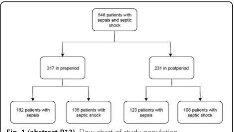
The effect of Intelligent Sepsis Management System on survival outcome in patients with sepsis and septic shock in accordance with Sepsis-3 definitions
Juhyun Song1, Daewon Park2, Sungwoo Moon1, Hyeri Seok2, Sejoong Ahn1
1Emergency Department, Korea University Ansan Hospital, Republic of Korea;2Infectious Diseases, Korea University Ansan Hospital, Republic of Korea
Correspondence:Juhyun Song ([email protected]) Intensive Care Medicine Experimental2019,7(Suppl 2):P13
Background
clinicians to screen, diagnose, and manage septic patients. The pur-pose of the present study was to assess the effect of i-SMS on com-pliance with the SSC bundles and survival outcome in patients with sepsis and septic shock diagnosed in ED. In addition, we tried to de-termine risk factors for 28-day mortality.
Materials and methods
We performed a pre-post study in patients with sepsis and septic shock. During the pre-period (January 1, 2016-Setember 25, 2017), patients were managed with routine customary process. During the post period (September 26, 2017-July 10, 2018), patients were man-aged with assistance from i-SMS upon arrival to ED.
Results
A total of 548 patients were included; 317 in preperiod and 231 in postperiod (Table 1 and Figure 1). After implementation of i-SMS, overall compliance with SSC recommendations improved from 26.8% to 52.8% (P <0.001) (Figure 2). There was no significant difference in 28-day mortality between preperiod (37.2%) and postperiod (32.0%) (P = 0.08) (Table 1). In Kaplan-Meier survival analysis and Log-rank test, there was no significant difference of survival curves between preperiod and postperiod (P = 0.666) (Figure 3). SOFA score and lac-tate levels were independent risk factors for 28-day mortality in all enrolled patients (Table 2).
Conclusions
Implementation of i-SMS significantly improved compliance with SSC recommendations among patients with sepsis and septic shock in ac-cordance with Sepsis-3 definitions, but did not improve short-term survival outcome.
Acknowledgements
The authors thank research nurse Hye-Yoon Jung and researcher Min-Sook Jung for their contributions to the project. We also thank Jae-Hyung Cha, PhD for lending statistical
support.
References
1. Angus DC and van der Poll T. Severe sepsis and septic shock. N Engl J Med. 2013;369(9):840-51.
2. Van Zanten AR, Brinkman S, Arbous MS, Abu-Hanna A, Levy MM, de Kei-zer NF. Guideline bundles adherence and mortality in severe sepsis and septic shock. Crit Care Med. 2014;Aug;42(8):1890-1898
3. McColl T, Gatien M, Calder L, Yadav K, Tam R, Ong M, Taljaard M, Stiell I. Implementation of an Emergency Department Sepsis Bundle and System Redesign: A Process Improvement Initiative. CJEM. 2017 Mar;19(2):112-121
4. Patocka C, Turner J, Xue X, and Segal E. Evaluation of an emergency department triage screening tool for suspected severe sepsis and septic shock. J Health Qual. 2014 Jan-Feb;36(1):52-61
5. Rhodes A, Evans LE, Alhazzani W, Levy MM, Antonelli M, Ferrer R, Kumar A, Sevransky JE, Sprung CL, Nunnally ME, et al. Surviving Sepsis Campaign: International Guidelines for Management of Sepsis and Septic Shock:2016. Crit Care Med. 2017; 45(3):486-552.
Table 1 (abstract P13).Baseline characteristics of enrolled patients and comparison between preperiod and postperiod
Pre (N = 317)
Post (N = 231)
P value
Age (years), median (IQR) 71 (60-79) 73 (62-82) 0.148
Male, no. (%) 158 (49.8) 115 (49.8) 0.996
Initial q-SOFA≥2, no. (%) 317 (100) 231 (100)
Site of infection, no (%)
Respiratory 198 (62.5) 135 (58.4) 0.315
Genitourinary 83 (26.2) 61 (26.4) 0.572
Gastrointestinal 22 (6.9) 17 (7.4)
Hepatobiliary 8 (2.5) 6 (2.6)
Central nervous 7 (2.2) 5 (2.2)
Cardiovascular 6 (1.9) 5 (2.2)
Skin and soft tissue 4 (1.3) 3 (1.3)
Other sites 11 (3.5) 8 (3.5)
Unknown 8 (2.5) 6 (2.6)
SOFA score, mean ± SEM 7.8 ± 0.4 8.0 ± 0.4 0.425
Septic shock, no. (%) 135 (42.6%) 108 (46.8%) 0.232
Disposition in ED, no (%)
Intensive care unit admission 107 (33.8) 75 (32.5) 0.417
General ward admission 116 (36.6) 83 (35.9)
Death in ED 15 (4.7) 11 (4.8)
Others 79 (24.9) 62 (26.8)
Length of stay (days), median (IQR) 9 (4-13) 10 (4-18) 0.436
Length of antibiotics (days), median 8 (3-13) 9 (4-16) 0.361
Compliance with recommendations, no. (%)
85 (26.8) 122 (52.8) <0.001
Broad-spectrum antibiotics≤3 hours 185 (58.4) 160 (69.3) 0.02
Blood culture before antibiotics 247 (77.9) 198 (85.7) 0.04
Appropriate fluid resuscitation 215 (67.8) 167 (72.3) 0.206
Lactate measurement≥2 within 6 hours
116 (36.6) 205 (88.7) <0.001
Time to 1st antibiotics (min), median (IQR) 121 (72-188) 112 (68-180) 0.349
7-day mortality, no (%) 44 (13.9) 30 (13.0) 0.315
14-day mortality, no (%) 87 (27.4) 62 (26.8) 0.412
P14
Diagnostic and prognostic value of interleukin-6, pentraxin-3, and procalcitonin levels among patients with sepsis and septic shock diagnosed at emergency department according to Sepsis-3 definitions
Juhyun Song1, Daewon Park2, Sungwoo Moon1, Hyeri Seok2, Sejoong Ahn1, Wonseok Choi2
1Emergency Department, Korea University Ansan Hospital, Republic of Korea; 2
Infectious Diseases, Korea University Ansan Hospital, Republic of Korea Correspondence:Juhyun Song ([email protected]) Intensive Care Medicine Experimental2019,7(Suppl 2):P14
Background
Sepsis is a global public health problem. Despite the advances in modern medicine, more than 5.3 million people die from sepsis an-nually, with an estimated overall mortality of about 30% [1-3]. Early diagnosis and appropriate treatment can improve survival outcome in patients with sepsis [4]. Despite pre-existing diagnostic criteria for sepsis, early diagnosis is usually challenging due to unknown source of infection and the vague definitions of sepsis syndrome [5]. C-reactive protein (CRP) and procalcitonin (PCT) have been widely used to facilitate the diagnosis of sepsis, but the clinical values of these are limited [6, 7]. A recent study reported that IL-6 level is a diagnos-tic marker of infection and a prognosdiagnos-tic marker, in patients with organ dysfunction [8]. Another study showed that PTX-3 discrimi-nates sepsis and septic shock patients from healthy controls in a medical intensive care unit (ICU) setting [9]. However, evidence con-cerning the clinical value of IL-6 and PTX-3 has been controversial in several studies. The purpose of the present study was to investigate both the diagnostic and prognostic value of IL-6, PTX-3, and PCT among patients with sepsis and septic shock diagnosed at an emer-gency department (ED) using the latest Sepsis-3 definitions. Materials and Methods
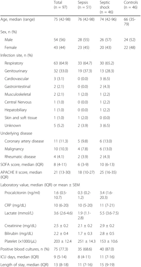
This study investigated biomarkers’ clinical value among patients with sepsis and septic shock. Among 143 enrolled subjects (51 with sepsis, 46 with septic shock, and 46 healthy volunteers), serum levels of IL-6, PTX-3, and PCT were measured (Table 1). Follow-up IL-6 and PTX-3 levels were measured among patients with initial septic shock within 24 hours of hospital discharge. Optimal cut-off values were obtained for sepsis and septic shock. Prognostic value was evaluated by Cox regression analysis and Kaplan-Meier survival analysis. Results
The median values (IQR) of IL-6 in controls, sepsis, and septic shock were 0.6 (0.2-1.6), 89.9 (45.2-272.6), and 1378.6 (256.4-11062.1) pg/ Fig. 2 (abstract P13).Compliance with recommendations before
and after implementation of intelligent sepsis management system among patients with sepsis and septic shock
Fig. 3 (abstract P13).Kaplan-Meier curve of 28-day mortality before and after implementation of intelligent sepsis management system in patients with sepsis and septic Shock
Table 2 (abstract P13).Univariate and Multivariate Cox Regression Analysis of Risk factors for 28-Day Mortality in patients with sepsis and septic shock
HR (95% CI) p value Adjusted HR (95% CI)
p value
Age 1.016 (1.006-1.028) 0.003 1.012 (1.000-1.024) 0.052
Male sex 1.002 (0.999-1.004) 0.12
SOFA score 1.282 (1.228-1.339) <0.001 1.265 (1.199-1.334) <0.001
Procalcitonin 1.004 (1.000-1.009) 0.079 1.000 (0.995-1.005) 0.954
Lactate 1.080 (1.053-1.108) <0.001 1.072 (1.032-1.112) 0.001
CRP 1.011 (0.998-1.025) 0.10 1.002 (0.985-1.019) 0.846
Time to 1stantibiotics 0.999 (0.998-0.999) 0.029 0.999 (0.998-1.000) 0.050
Septic shock 2.657 (1.327-5.317) 0.004 1.247 (0.817-1.903) 0.306
ml, respectively (Figure 1). Serum IL-6 levels could discriminate sepsis (AUC, 0.97-1.00, p<0.001; cut-off value, 5.89 pg/mL [sensitivity 97.0%, specificity 97.2%]) from controls and could discriminate septic shock (AUC, 0.85-0.95; cut-off value, 53.59 pg/mL [sensitivity 91.8%, specifi-city 63.2%]) from the others (controls and sepsis) (Table 2 and Figure 2). Twenty-eight-day mortality was significantly higher in the high IL-6 (≥53.59 pg/ml) group than in the low IL-6 (<53.59 ng/ml) group (p=0.002) (Figure 3). IL-6 was an independent risk factor for 28-day mortality among patients with sepsis and septic shock (HR, 1.0004; 95% CI, 1.0003-1.0005; p=0.024) (Table 3). PTX-3 level was not significant in the multivariate Cox regression analysis (HR, 1.003; 95% CI, 0.998-1.008; p=0.095) (Table 3). Both initial and follow-up PTX-3 levels of septic shock patients who died dur-ing admission were consistently significantly higher than those of septic shock patients who recovered (initial: p=0.004, follow-up: p<0.001) (Figure 4).
Conclusions
IL-6 level was superior to PTX-3 and PCT levels in both diagnostic and prognostic value for sepsis and septic shock diagnosed in the emergency department using Sepsis-3 definitions. IL-6 level was an independent risk factor for 28-day mortality.
Acknowledgements
The authors thank research nurse Hye-Yoon Jung and researcher Min-Sook Jung for their contributions to the project. We also thank Jae-Hyung Cha, PhD for lending statistical support.
References
1. Singer M, Deutschman CS, Seymour CW, Shankar-Hari M, Annane D, Bauer M, Bellomo R, Bernard GR, Chiche J-D, Coopersmith CM, et al. The Third International Consensus Definitions for Sepsis and Septic Shock (Sepsis-3). JAMA. 2016; 315(8):801-10.
2. Rhodes A, Evans LE, Alhazzani W, Levy MM, Antonelli M, Ferrer R, Kumar A, Sevransky JE, Sprung CL, Nunnally ME, et al. Surviving Sepsis Campaign: International Guidelines for Management of Sepsis and Septic Shock: 2016. Crit Care Med. 2017; 45(3):486-552.
3. Angus DC, van der Poll T. Severe sepsis and septic shock. N Engl J Med. 2013; 369(9):840-51.
4. De Backer D, Dorman T. Surviving Sepsis Guidelines: A Continuous Move Toward Better Care of Patients With Sepsis. JAMA. 2017; 317(8):807-8. 5. Biron BM, Ayala A, Lomas-Neira JL. Biomarkers for Sepsis: What Is and
What Might Be? Biomark Insights. 2015; 10(Suppl 4):7-17.
6. Kibe S, Adams K, Barlow G. Diagnostic and prognostic biomarkers of sepsis in critical care. J Antimicrob Chemother. 2011; 66 Suppl 2:ii33-40. 7. Henriquez-Camacho C, Losa J. Biomarkers for sepsis. Biomed Res Int.
2014; 2014:547818.
8. Takahashi W, Nakada TA, Yazaki M, Oda S. Interleukin-6 Levels Act as a Diagnostic Marker for Infection and a Prognostic Marker in Patients with Organ Dysfunction in Intensive Care Units. Shock. 2016; 46(3):254-60.
9. Hamed S, Behnes M, Pauly D, Lepiorz D, Barre M, Becher T.
Diagnostic value of Pentraxin-3 in patients with sepsis and septic shock in accordance with latest sepsis-3 definitions. BMC Infect Dis. 2017; 17(1):554.
Table 1 (abstract P14).Baseline Characteristics of Study Population
Total (n = 97)
Sepsis (n = 51)
Septic shock (n = 46)
Controls (n = 46)
Age, median (range) 75 (42-98) 76 (42-98) 74 (42-96) 66 (35-79)
Sex, n (%)
Male 54 (56) 28 (55) 26 (57) 24 (52)
Female 43 (44) 23 (45) 20 (43) 22 (48)
Infection site, n (%)
Respiratory 63 (64.9) 33 (64.7) 30 (65.2)
Genitourinary 32 (33.0) 19 (37.3) 13 (28.3)
Cardiovascular 3 (3.1) 0 (0.0) 3 (6.5)
Gastrointestinal 2 (2.1) 0 (0.0) 2 (4.3)
Musculoskeletal 2 (2.1) 1 (2.0) 1 (2.2)
Central Nervous 1 (1.0) 0 (0.0) 1 (2.2)
Hepatobiliary 1 (1.0) 0 (0.0) 1 (2.2)
Skin and soft tissue 1 (1.0) 1 (2.0) 0 (0.0)
Unknown 5 (5.2) 2 (3.9) 3 (6.5)
Underlying disease
Coronary artery disease 11 (11.3) 5 (9.8) 6 (13.0)
Malignancy 10 (10.3) 4 (7.8) 6 (13.0)
Rheumatic disease 4 (4.1) 2 (3.9) 2 (4.3)
SOFA score, median (IQR) 8 (4-11) 6 (3-9) 10 (6-13)
APACHE II score, median (IQR)
21 (13-30) 18 (10-27) 25 (16-35)
Laboratory value, median (IQR) or mean ± SEM
Procalcitonin (ng/ml) 1.6 (0.5-10.7)
0.3 (0.2-1.2)
3.4 (1.6-20.3)
CRP (mg/dL) 10 (6-20) 10 (5-20) 11 (7-21)
Lactate (mmol/L) 3.6 (2.6-4.6) 1.9 (1.1-2.8)
5.5 (3.6-7.5)
Creatinine (mg/dL) 2.5 ± 0.2 2.1 ± 0.2 2.9 ± 0.2
Bilirubin (mg/dL) 2.2 ± 0.4 1.7 ± 0.3 2.8 ± 0.5
Platelet (×1000/μL) 203 ± 12.4 251 ± 14.3 153 ± 10.6
Positive blood cultures, n (%) 75 (77.3) 35 (68.6) 40 (87.0)
ICU days, median (IQR) 9 (5-14) 8 (4-11) 11 (7-16)
Length of stay, median (IQR) 13 (8-18) 11 (7-16) 15 (9-19)
P15
Combined prognostic role of interleukin-35 and presepsin (sCD14 subtype) in clinical setting of sepsis and septic shock
Cinzia Peronace1, Maria Teresa Loria1, Carolina Mirello1, Aida Giancotti1, Paola Morelli1, Nadia Marascio1, Eugenio Garofalo2, Andrea Bruni2, Paolo Navalesi2, Angela Quirino1, Maria Carla Liberto1, Giovanni Matera1 1Department of Health Sciences, Operating Unit of Microbiology,
“Magna Graecia”University, Catanzaro, Italy;2Operating Unit of Intensive Care,“Magna Graecia”University, Catanzaro, Italy
Correspondence:Giovanni Matera ([email protected]) Intensive Care Medicine Experimental2019,7(Suppl 2):P15 Table 2 (abstract P14).Diagnostic Value of Interleukin-6 (IL-6),
Pentraxin-3 (PTX-3), and Procalcitonin (PCT) for Patients with Sepsis and Septic Shock in the Emergency Department
Severity AUC (95% CI) Cutoff value Sensitivity (%) Specificity (%) P value
IL-6 Sepsis 0.99 (0.97-1.00) 5.89 97.0 97.2 <0.001
(pg/ml) Septic shock 0.90 (0.85-0.95) 53.59 91.8 63.2 <0.001
PTX-3 Sepsis 0.97 (0.95-0.99) 6.02 92.6 97.4 <0.001
(ng/ml) Septic shock 0.84 (0.77-0.90) 12.05 93.2 60.7 <0.001
PCT Sepsis 0.91 (0.86-0.96) 0.23 77.7 94.9 <0.001
(ng/ml) Septic shock 0.86 (0.79-0.92) 0.83 75.0 83.1 <0.001
AUCarea under the curve,CIconfidence interval
Table 3 (abstract P14).Univariate and Multivariate Cox Proportional Model of Risk Factors for 28-day Mortality
HR (95% CI) p value Adjusted HR (95% CI) p value
Age 1.023 (0.994-1.052) 0.120
Male sex 1.029 (0.535-1.978) 0.932
GCS score 0.777 (0.692-0.873) <0.001 0.777 (0.695-0.869) <0.001
SOFA score 1.206 (1.076-1.353) 0.001 1.048 (0.901-1.219) 0.208
APACHE II score 1.198 (1.068-1.346) 0.001 1.031 (0.898-1.187) 0.231
Pentraxin-3 1.005 (1.001-1.009) 0.031 1.003 (0.998-1.008) 0.095
Interleukin-6 1.001 (1.000-1.002) 0.017 1.001 (1.000-1.002) 0.024
Procalcitonin 0.995 (0.981-1.009) 0.481
Lactate 1.167 (1.068-1.275) 0.001 1.135 (1.033-1.247) 0.009
CRP 1.011 (0.978-1.045) 0.525
Septic shock 2.657 (1.327-5.317) 0.004 1.249 (0.472-3.302) 0.240
HRhazard ratio,CIconfidence interval,GCSGlasgow coma scale,SOFAsequential organ failure assessment,APACHEacute physiology and chronic health evaluation,CRPC-reactive protein
Fig. 1 (abstract P14).Interleukin-6, Pentraxin-3, Procalcitonin, CRP, and Lactate Levels in Patients with Sepsis and Septic Shock Diagnosed in the Emergency Department according to Sepsis-3 Definitions. CRP, C-reactive protein
Fig. 2 (abstract P14).Receiver Operating Characteristic (ROC) Curves to Discriminate Sepsis (a) and Septic Shock (b) by Interleukin-6, Pentraxin-3, Lactate, and Procalcitonin Levels Measured in the Emergency Department
Fig. 3 (abstract P14).Kaplan-Meier Curve of 28-day Mortality in Patients with Sepsis and Septic Shock Stratified by the Optimal Cut-off Value of Pentraxin-3 (a) and Interleukin-6 (b) for Predicting Septic shock (28-day mortality by pentraxin-3: 16.9% vs 43.9%, 28-day mortality by interleukin-6: 5.6% vs 47.4%)
Background
The detection of sepsis-specific biomarkers ameliorate recognition and management of sepsis by improvement: diagnosis, monitoring response to treatment, and stratifying patients based on prognosis or underlying biological response. The diagnostic and prognostic role of novel biomarkers of sepsis is the aim of our study. In particular, we have investigated the kinetic of IL-35, presepsin and procalcitonin in critical septic patients with poor prognosis.
Materials and Methods
Fifty-nine critical septic patients admitted to ICU of the University Hospital of Catanzaro (Italy) were enrolled; a group of healthy volun-teers were also included. All studied subjects were stratified into sur-vivors and nonsursur-vivors, based on 28 days survival and according to microbiological results in blood culture positive and blood culture negative groups. Clinical data and blood samples were collected for quantitative analysis of IL-35, sCD14-ST and PCT at: i) time 0 (T0): time at which the first blood culture for analysis was collected; ii) time T minus 7 (T-7): 7 days before exitus or hospital discharge; iii) time T minus 2 (T-2): 2 days before exitus or hospital discharge. Data were subjected to statistical analysis by ANOVA plus PLSD test. A p value < 0.05 was considered statistically significant.
Results
Interleukin-35 levels were found significantly (p< 0.05) increased in the group of eventually dead patients, both at T0 and at T-2 times. By contrary, such different behavior between alive and dead patients did not achieve any significant difference at T-7; although the group of patients with a poor prognosis still exhibited a light increase of IL-35. RegardingsCD14-ST, our data confirmed that presepsin is a valu-able prognostic biomarker for the studied patients (p< 0.05). PCT ex-hibited a significantly lower levels in the eventually non-survivors only at time T0; by contrary both at times T-7 and T-2 PCT levels of dead patients were found significantly higher than survivors. Conclusions
Serial increase of IL-35 and sCD14-ST or presepsin reveals prognostic value for in hospital mortality compared to PCT as observed at T0. Recent study shown the intrisic multimodal co-existing pro-and anti-inflammatory mechanisms reflect specific immune reprogramming [1].
Reference
1. Denstaedt SJ, Singer BH, Standiford TJ. Sepsis and Nosocomial Infection:Patient Characteristics, Mechanisms, and Modulation. Front Immunol. 2018 9:2446. doi: 10.3389/fimmu.2018.02446
P16
Rapid identification ofListeria monocytogenesfrom vertebral joint abscess clinical sample using eSensor and Electrowetting technology
Angela Quirino1, Gallo Luigia1, Francesca Divenuto1, Maria Concetta Reale1, Cinzia Peronace1, Giorgio Settimo Barreca1, Angelo Giuseppe Lamberti1, Enrico Maria Trecarichi2, Carlo Torti2, Giorgio Gasparini3, Maria Carla Liberto1, Giovanni Matera1
1
Department of Health Sciences, Operating Unit of Microbiology, “Magna Graecia”University, Catanzaro, Italy;2Operating Unit of Infectious and Tropical Diseases,“Magna Graecia University”, Catanzaro, Italy; 3Operating Unit of Orthopaedic and Trauma Surgery,“Magna Graecia University”, Catanzaro, Italy
Correspondence:Giovanni Matera ([email protected]) Intensive Care Medicine Experimental2019,7(Suppl 2):P16
Background
Listeria monocytogenes (L. monocytogenes)is a facultative intracellular Gram-positive bacterium that infects humans through food. The broad clinical spectrum of Listeria monocytogenes infections includes frequent clinical forms, such as meningitis or bacteremia, and un-common manifestations, such as septic arthritis. Osteoarticular infec-tions due to L. monocytogenes have been reported very rare. We report on a case of osteoarticular abscess, which developed into a bacteremia both caused byL. monocytogenesin an older adult, using a rapid identification by innovative technology.
Materials and Methods
An 83-year-old man was admitted to our university hospital with a retroperitoneal abscess and acute low back pain suggestive of spon-dylodiscitis. He had no sign of meningoencephalitis or fever. A clin-ical sample from vertebral joint abscess and some blood cultures were sent to our laboratory for microbiological diagnosis. Sepsis bio-markers (procalcitonin and sCD14-ST) were also required. All of the samples were analyzed by conventional methods. Moreover, verte-bral joint abscess was analyzed by ePlex device, that identifies a broad panel of pathogens and resistance genes, using BCID-GP Panel (GenMarkDx, USA). The system relies on electrowetting technology to perform multiplexed nucleic acid extraction, amplification and di-gestion followed by the detection of analyte targets using eSensor technology. ePlex device, has been approved and commercialized for positive blood cultures only. We used this molecular technology dir-ectly on the sample of vertebral joint abscess. Positive blood cultures were assessed by molecular method using the FilmArray system (Bio-Merieux, Italy). Serum procalcitonin and presepsin were tested by ELFA and CLEIA methods respectively.
Results
ePlex system identified L. monocytogenesin about 90 minutes dir-ectly on sample of vertebral joint abscess. Moreover, L. monocyto-geneswas identified using the FilmArray blood culture ID Panel in about one hour on positive blood cultures. Sub-cultures, processed using conventional and proteomic methods, confirmed molecular diagnosis results.
Conclusions
Rapid detection of L. monocytogenes by innovative technology allowed the clinicians to establish an early antibiotic therapy. This, as well as other molecular techniques, may represents an integrated ap-proach to conventional methods for etiological diagnosis.
P17
After recovery from severe-sepsis, the lung carries significant quiescent lesions with potential recurrent pathogenicity Rodrigo B Souza, Ana MA Liberatore, Ivan HJ Koh
Department of Surgery, Escola Paulista de Medicina, Universidade Federal de São Paulo, São Paulo, Brasil
Correspondence:Ivan HJ Koh ([email protected]) Intensive Care Medicine Experimental2019,7(Suppl 2):P17
Background
The causal role of sepsis on the long-term impairment and survival remains unclear, but is a fact that survivors of sepsis are profoundly functionally impaired. The lung is the organ often affected in sepsis and course with high lethality by ARDS. Thus, this study aimed to evaluate the pulmonary status in survivors of a severe sepsis in search of remaining lesions with potential recurrence of pulmonary disease.
Materials and Methods
Under general anesthesia, Wistar rats (250g) were submitted to se-vere sepsis (iv. inoculation of 2 mLEscherichia coli108 CFU/ mL), that course with hypotension within 4-6 hours and with 50-60% mortality within 26 hours. Lung of the survivals was collected at 30 and 90 days under general anesthesia and were sacrificed after. (n=4/ period). The samples were submitted to histological study using HE dye and results were compared to lung of naïve (T0) and septic ani-mals of 6hours after challenge (n=4/period).
Results
alveolar wall, infiltrate of mono and polynuclear cells in most of al-veoli, multiple areas with hemorrhage, peri-bronchial leukocyte infil-trates, atherosclerosis, and intense BALT hyperplasia associated to obstruction of bronchioles and multiple areas of atelectasis. These findings demonstrated that healthy-looking animals were concealing a functionally impaired lung structure combined to an intense in-flammatory reaction state in course, denoting an impaired lung physiological condition to face with new deleterious stimuli in sepsis survivors.
Conclusions
A severely compromised state of lung following sepsis recovery might explain the ease for higher susceptibility for recurrent pul-monary diseases. These events are possibly related to the post-sepsis syndrome that courses with a persistent immunosuppres-sion, inflammation and catabolism. Clinical studies are needed to confirm these animal experimental findings. In the future, under-standing of mechanisms supporting these persistent lung injuries may afford intervention targets to ameliorate post-sepsis high mortality.
Acknowledgements
Grant acknowledgment: FAPESP 2017/21052-0
References
1. Won-Young Kim and Sang-Bum Hong: Sepsis and Acute Respiratory Dis-tress Syndrome: Recent Update. Tuberc Respir Dis 2016;79:53-57. 2. Manu Shankar-Hari & Gordon D. Rubenfeld: Understanding Long-Term
Outcomes Following Sepsis: Implications and Challenges. Curr Infect Dis Rep (2016) 18: 37.
3. Sorin Hostiuc, Dan Dermengiu, Mihai Ceaușu, Mugurel Constantin Rusu, George Cristian Curcă: Pathology and immunopathology of the lung in sepsis. Rom J Leg Med [19] 83-88 [2011].
P18
Can we do it better together? A Derbyshire countywide approach to sepsis care
Alina M. Paunescu1, Duncan Cameron2
1Emergency Medicine Consultant, University Hospitals of Derby and Burton NHS Foundation Trust, Derby, United Kingdom;2Transformation Team Lead, University Hospitals of Derby and Burton NHS Foundation Trust, Derby, United Kingdom
Correspondence:Alina M. Paunescu ([email protected]) Intensive Care Medicine Experimental2019,7(Suppl 2):P18
Background
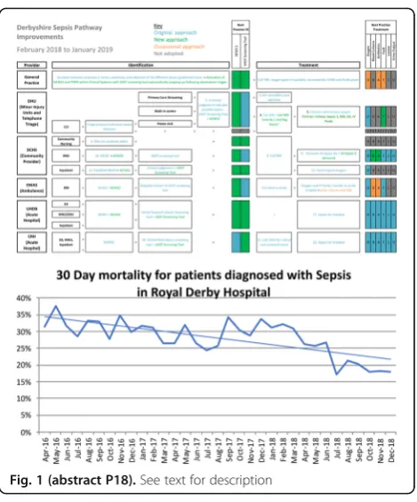
In the United Kingdom 70% of sepsis cases are derived from an in-fection developed in the community[1]. It is estimated that there is potential to reduce deaths by up to 10,000 per year by the optimization of care.
Mortality review of sepsis deaths in Royal Derby Hospital for No-vember 2017 revealed that 50% of patients died in the first couple of hours to 1 day following admission. Of these, 85% were previously treated in the community with antibiotics for dif-ferent periods of time, some of them with repeated visits to their General Practitioner
The findings of the reviews suggested communication deficits and inconsistent pathways between primary and secondary care. Materials and Methods
A Derbyshire wide project group was set up at the authors sug-gestion, including commissioners, the ambulance service, out of hours service, community providers, primary and secondary care. The final aim of the project was improving sepsis outcomes by adopting a standard approach to screening and treatment coun-tywide, at all levels of care. We have conducted process mapping of the pathways and procedures each provider followed when dealing with a potential sepsis case and identified where delays/ inconsistencies/ issues existed.
The agreed action plan:
Adopt the same escalation triggers and similar screening methods Reduce the time from identification to treatment especially in remote rural areas of Peak District Increase awareness/education of Sepsis and Derbyshire pathways across providers Improve sepsis treatment, review antibiotic protocols and patient education about antibiotics Develop post sepsis supportResults
Important steps have been achieved following our intervention (Table 1) starting with agreement on using same new early warn-ing scores (NEWS 2) and paediatric observation priority score (POPS) throughout Derbyshire. It is now possible for sepsis treat-ment to be initiated in the community long before the patient reaches hospital through education of community nurses and general practitioners’ agreement. While the initiative is ongoing due to the massive ramifications of the project, intermediate data showed that crude mortality from sepsis is on a continuous downward trend (Graph 1).
Conclusions
Developing a unified approach and increasing communication be-tween providers generated benefits for patient, practitioners and local health economy. While raw data analysis makes it easy to meas-ure patient outcomes, the benefits of creating a local peer learning resource and support network are not easily quantifiable and can only be predicted as positive.
Acknowledgements
1. United Kingdom, Derbyshire Sepsis Group, United Kingdom
2. Joanna Harisson, Urgent Care Analyst, University Hospitals of Derby and Burton NHS Foundation Trust, Derby,
Reference
1. Donald M Yealy, David T Huang Anthony Delaney, Marian Knight, Adrienne G Randolph, Ron Daniels and Tim Nutbeam, Recognizing and managing sepsis :what needs to be done?, BMC
Med.2015;13:98, Published online 2015 Apr 27, PMCID:PMC4410741, PMID:25927426
P19
Targeting the NO-sGC axis to monitor and treat vascular dysfunction and vasoplegia in sepsis
Forough Jahandideh1, Sareh Panahi1, Kimberley Macala1, Stephane L. Bourque1,2
1Department of Anesthesiology and Pain Medicine, University of Alberta, Edmonton, Canada;2Department of Physiology, University of Alberta, Edmonton, Canada
Correspondence:Stephane L. Bourque ([email protected]) Intensive Care Medicine Experimental2019,7(Suppl 2):P19
Background
Sepsis is a life-threatening condition caused by a dysregulated host response to infection. If unimpeded, sepsis can progress to septic shock, characterized by refractory hypotension and unresponsive vasculature (i.e. vasoplegia) resulting in tissue hypoperfusion and eventually organ failure [1]. The overall mortality rate of septic shock is more than 50%. The vascular dysfunction and refractory hypotension in septic shock are caused, at least in part, by excess reactive oxygen species (ROS) generation and dysregulated nitric oxide (NO) produc-tion (via inducible NO synthase upregulaproduc-tion and increased scavenging by excess ROS) [2,3,4]. Moreover, excess ROS oxi-dizes and hence damages soluble guanylate cyclase (sGC), the downstream mediator of NO, resulting in the impaired organ blood flow [5,6]. We believe the loss of sGC-mediated vaso-dilation is a critical determinant of reduced organ blood flow in sepsis. The objective of this study is to assess the state of vascular dysfunction and altered blood flow patterns as sepsis progresses to septic shock. We also sought to assess the
effi-cacy of (i) the sGC activator cinaciguat or (ii) the superoxide dismutase mimetic tempol in improving hemodynamics and survival in a murine model of sepsis.
Materials and Methods
The experimental protocols described herein have been ap-proved by the University of Alberta Animal Care and Use Com-mittee in conformance with the FASEB Statement of Principles for the use of Animals in Research and Education. Male C57Bl/6 mice were instrumented with fiber-optic pressure sensors for direct blood pressure monitoring. Flow probes were placed around the left common carotid, superior mesenteric, and right renal arteries to monitor blood flow to the brain, gut, and kid-ney, respectively. After baseline recordings, sepsis was induced by an intraperitoneal injection of a fecal slurry. Thirty minutes after induction of sepsis, mice were treated with the cinaciguat (15μg/kg IV) or tempol (30mg/kg, IV).
Results
The blood pressure in septic mice reduced significantly over time (44% ± 4% reduction after 4 h of fecal slurry injection) compared to control mice (Figure 1A), but no significant changes in heart rate were noted (Figure 1B). Blood flow in the carotid, superior mesenteric, and renal arteries reduced at 47 ± 4%, 71 ± 8%, and 57 ± 13% respectively, 4 hours after fecal slurry injection (Figures 2A, B, and C, respectively). Vessel activity, assessed by monitoring constrictor and relaxation re-sponses to bolus doses of phenylephrine (10 μg/kg body weight) and sodium nitroprusside (5 μg/kg body weight) was reduced by 36 ± 9% and 53 ± 7% respectively, in septic mice compared to controls, suggesting impaired regional vascular function. These data suggest certain organs are more suscep-tible to vascular dysfunction and hypoperfusion with the pro-gression of sepsis. While a low dose (15μg/kg IV) of cinaciguat provided the best survival outcomes in our model, our prelimin-ary data have also shown that administration of tempol (30mg/ kg, IV), 30min after injection of fecal slurry, prolongs survival and mitigates the decline in blood pressure and superior mes-enteric artery blood flow.
Conclusions
The proposed therapeutic is expected to reduce organ damage and improve blood flow to organs without causing systemic vasodilation and hypotension in sepsis.
References
1. Singer M, Deutschman CS, Seymour CW, Shankar-Hari M, Annane D, Bauer M, Bellomo R, Bernard GR, Chiche JD, Coopersmith CM, Hotchkiss RS, Levy MM, Marshall JC, Martin GS, Opal SM, Rubenfeld GD, van der Poll T, Vincent JL, Angus DC. The third international consensus definitions for sepsis and septic shock (sepsis-3). JAMA. 2016;315:801-810.
2. Kimmoun A, Ducrocq N, Levy B. Mechanisms of vascular
hyporesponsiveness in septic shock. Curr Vasc Pharmacol. 2013;11:139-149. 3. Muhl H, Bachmann M, Pfeilschifter J. Inducible NO synthase and
antibacterial host defense in times of Th17/Th22/T22 immunity. Cell Microbiol. 2011;13:340-348.
4. Wong CM, Au CL, Tsang SY, Lau CW, Yao X, Cai Z, Chung AC. Role of inducible nitric oxide synthase in endothelium-independent relaxation to raloxifene in rat aorta. Br J Pharmacol. 2017;174:718-733.
5. Gutterman DD, Chabowski DS, Kadlec AO, Durand MJ, Freed JK, Ait-Aissa K, Beyer AM. The human microcirculation: Regulation of flow and be-yond. Circ Res. 2016;118:157-172.
6. Bateman RM, Sharpe MD, Ellis CG. Bench-to-bedside review: Microvascu-lar dysfunction in sepsis–hemodynamics, oxygen transport, and nitric oxide. Crit Care. 2003;7:359-373.
P20
Primary peritonitis with rapid evolution to abdominal sepsis in an immunocompetent patient
Daniel N. de Almeida¹, Vitória S. Freitas¹, Pamela M. Ureta² 1
Centro Universitário Serra dos Órgãos, UNIFESO, Teresópolis, Brazil; 2Universidad Nacional de la Matanza, UNLAM, Buenos Aires - Argentina Correspondence:Daniel N. de Almeida
Intensive Care Medicine Experimental2019,7(Suppl 2):P20
Background
We present the case of a 55-year-old Argentinian woman, with a clinical of peritonitis with 24h of evolution to sepsis, character-ized initially by acute diffuse abdominal pain associated with diarrhea without mucus or blood. Patient denied nausea, evolving to abdominal distention, absence of peristalsis and signs of peri-toneal irritation, negative catharsis and absence of flatulence with hemodynamic decompensation.
Case Report
In the physical examination, she was oriented in time and space, cooperative, Glasgow 15/15, no fever (36ºC), hypotensive (80/ 60mmHg), tachycardic 112 bpm, with respiratory rate at 18 breaths per minute, abdomen distended, painful at superficial palpation and deep peritoneal resistance, absence of intestinal sounds, symmetric extremities and venous return lower than 2 seconds. The following complementary exams were made: elec-trocardiogram and chest X-Ray, with the imaging of the X-Ray being interpreted as septic shock and validated by an intensive care unit professional[1].
During the 10 weeks hospitalized, the patient underwent various procedures following the diagnosis, including reanimation with vasopressors and this medication caused ischemia and necrosis of phalanges later, exploratory laparotomy, bilateral pleural effu-sion, pericardial effueffu-sion, acute cholecystitis, distal ileum fistula, blood transfusion, non-invasive ventilation and nephrostomy. She also presented various complications such as intestinal ischemia, surgical wound infection, respiratory insufficiency and hepatic dysfunction.
The patient started with piperacillin, tazobactam and vancomycin, then imipenem with vancomycin. 7 weeks later, the patient was disconnected from the non-invasive ventilation, with no intercur-rences and slowly evolving to improve. Two weeks after that she started oral feeding, with associated k108 tube for better caloric income. The patient was treated with antibiotics for 8 days (Colistine), 35 days (Linezolid), 26 days (Ciprofloxacin) and was suspended from the parenteral nutrition since 10 weeks of admission.
In the context of sepsis in abdominal focus with SOFA 2, tom-ography showed delayed right renal excretion associated with retropielocalicial dilation. Three days later the abdominal fluid was aspirated and passed tests revealing: Creatinine (urine) 37 mg/dL, Urea 819 mg/dL, Sodium 110 mmol/L and Potassium 22 mmol/L. In addition to culture of abdominal fluid showing be-sides the Klebsiella pneumoniae, the presence of Enterococcus faeciumjustifies the exchange of Imipenem to Linezolid[2]. Conclusion
Since the case qualifies as a rare occasion of primary peritonitis with rapid evolution to abdominal sepsis in a healthy individual, it stands to question what were the factors involved. Moreover, it questions what would be the gold-standard treatments to minimize the need for additional procedures, and their complications.
References
1. Fátima Kotleski Thomaz de Lima A., Prevedello Franco R. - Acta Veterinaria Brasilica: SÍNDROME DA RESPOSTA INFLAMATÓRIA SISTÊMICA (SRIS), UM DESAFIO DIAGNÓSTICO, v.3, n.4, p.123-131, 2010
2. Janssens U, et al. Value of SOFA (Sequential Organ Failure Assessment) score and total maximum SOFA score in 812 patients with acute cardiovascular disorders [abstract]. Crit Care 2001;5(Suppl 1):P225. Fig. 1 (abstract P19).See text for description