!∀# !∃%
# !&∋∋ ∋()(∋
i
Isolated Lumbar Extension Exercise as an
Intervention for Chronic Low Back Pain
James Daniel Steele
Centre for Health, Exercise & Sport Science, Southampton Solent University, Southampton, Hampshire, UK
A thesis submitted in partial fulfilment of the requirements of Nottingham Trent
University and Southampton Solent University for the degree of Doctor of
Philosophy
ii
Copyright Statement
iii
ABSTRACT
iv
Acknowledgements
Firstly I’d like to take the time to thank my wonderful girlfriend and love of my life Emma.
Without her providing the initial impetus all those years ago to break out of my boring job
and choose to study something I was passionate about I’m not sure I’d be here writing this
today. Her patience and support while I have been a student for what seemed like forever
has been immeasurable. Don’t worry, now I can go get a real job and start taking care of
you!!
I’d like to thank the late Arthur Jones for his contributions to this area through both
Nautilus and MedX as in all likelihood again I probably wouldn’t be here studying this particular area if it were not for his efforts.
My supervisors deserve particular thanks also. Dr Stewart Bruce-Low has maintained a constant supportive role through both the highs and the lows of this process. He has invested far more time to in this process than his responsibility entailed. Hopefully my accomplishment will be reward for his effort – oh, that and the awesome trip to Dubai ;-) – Dr Dave Smith receives my thanks for remaining down to earth through this whole process and keeping me grounded –that and being the most efficient ‘Grammar Nazi’ I’ve ever met. He made me realise that even during the PhD there is time to enjoy yourself
and relax. Dr David Jessop is a biomechanical wizard, and he’s ‘dead good’ on MatLab
also. He often pointed me in the right direction after hours of frustration.
v
My friends have been there throughout the whole process too. Sean – ‘Me and him
are….buddies’ – for being my best friend and constant inspiration through his own
achievements. James for being my friend, colleague and official coffee time partner, providing laughs and serious discussion on the world in its entirety. The guys who have enabled me to keep up my hobbies in roleplaying and wargaming throughout the process
– Ross, Lea, Adam, Ian, Dave, Simon, John, Ant – that escapism has been much needed.
I’d like to thank the staff here at Solent and everyone else who helped out with the work
here particularly Tim, Grant, and James for accommodating my perpetual presence in the labs, Scott for his insights on research methods, and Dr Neil Osborne for his efforts in screening participants and providing critical feedback on manuscripts.
Finally I’d also like to thank the research community at Solent, including Profs,
vi
CONTENTS
Section Pg.
No.
ABSTRACT iii
Acknowledgements iv
List of Tables xiii
List of Figures xiv
Glossary xvi
1.0 INTRODUCTION 1
1.1 Defining Low Back Pain within a Multifactorial Model 1
1.2 Prevalence, Costs and Impact 3
1.3 Lumbar Spine Anatomy 6
1.4 Mechanisms of Pain within LBP 8
1.5 A Multifactorial Perspective of Low Back Pain 14
2.0 LITERATURE REVIEW 17
2.1 Introduction and Overview 17
2.2 A Reappraisal of the Deconditioning Hypothesis in Low Back Pain: Review of Evidence from a Triumvirate of Research Methods on Specific Lumbar Extensor Deconditioning
19
2.2.1 Overview 19
2.2.2 Introduction 20
2.2.2.1 Defining the Disuse/Deconditioning Hypothesis –‘Disuse’
OR ‘Deconditioning’?
20
2.2.2.2 Specific Disuse/Deconditioning of the Lumbar Extensors 24
vii
2.2.3 Strength and Endurance of the Lumbar Extensor Musculature in LBP 26 2.2.3.1 Considerations for Studies of Strength and Endurance of the
Lumbar Extensor Musculature
26
2.2.3.2 Isolated Lumbar Extension Studies 29
2.2.3.3 Summary of Strength and Endurance Studies of the Lumbar Extensor Musculature
31
2.2.4 Imaging and Histochemical Studies of the Lumbar Extensor Musculature in LBP
34
2.2.4.1 Considerations for Imaging and Histochemical Studies of the Lumbar Extensor Musculature
34
2.2.4.2 Imaging Studies of the Lumbar Musculature 36 2.2.4.3 Histochemical Studies of the Lumbar Musculature 40 2.2.4.4 Summary of Imaging and Histochemical Studies of the
Lumbar Extensor Musculature
42
2.2.5 Electromyography Studies of Fatigue in the Lumbar Extensor Musculature in LBP
46
2.2.5.1 Considerations for Electromyographic Fatigue Analysis of the Lumbar Extensor Musculature
46
2.2.5.2 Fatigability Studies of the Lumbar Musculature 49 2.2.5.3 Summary of Electromyographic Studies of Fatigue of the
Lumbar Extensor Musculature
52
2.2.6 Prospective Studies of Lumbar Extensor Deconditioning in LBP 54 2.2.6.1 Prospective Evidence from Strength and Endurance in LBP 55 2.2.6.2 Prospective Evidence from MRI & EMG in LBP 58
2.2.6.3 Summary of Prospective Studies 59
2.2.7 Discussion 67
2.2.8 Conclusion 71
2.3 A Review of the Specificity of Exercises Designed for Conditioning the Lumbar Extensors
viii
2.3.1 Overview 72
2.3.2 Introduction 73
2.3.3 Methods 75
2.3.3.1 Exercises Designed for the Lumbar Extensors 75
2.3.3.2 Validity of Outcome Measures 76
2.3.4 Results 79
2.3.4.1 Acute Activation of the Lumbar Extensors through Specific Exercise
79
2.3.4.2 Chronic Adaptation to Specific Exercise Training for the Lumbar Extensors
86
2.3.5 Discussion 91
2.4 Clinical use of Specific Exercise for the Isolated Lumbar Extensors in Chronic Low Back Pain
96
2.4.1 Overview 96
2.4.2 Introduction 97
2.4.3 Methods 101
2.4.4 Results 101
2.4.4.1 Pain, Disability and Clinical Meaningfulness of Outcomes 116 2.4.4.2 Manipulation of Resistance Training Variables for use of
ILEX
122
2.4.4.3 Studies of ILEX and other Specific Exercise Approaches 125
2.4.5 Discussion 128
2.4.6 Conclusion 133
2.5 Synthesis and Overarching Rationale for the Thesis 134
2.6 Areas of Empirical Study 136
2.6.1 Range of Motion and Isolated Lumbar Extension Exercise in Chronic Low Back Pain
136
2.6.2 Gait Variability and Isolated Lumbar Extension Exercise in Chronic Low Back Pain
ix
2.6.3 Intervertebral Disc Hydration and Isolated Lumbar Extension Exercise in Chronic Low Back Pain
140
2.7 Summary of Literature Review 143
2.8 Proposed Research Questions and Hypotheses 144
3.0 METHODS 145
3.1 Study Design 145
3.1.1 Study Design; Study 1 and Study 2 145
3.1.2 Study Design; Study 3 145
3.2 Participants 146
3.2.1 Participants; Study 1 and Study 2 147
3.2.2 Participants; Study 3 149
3.3 Equipment 150
3.3.1 Equipment; Study 1 and Study 2 150
3.3.2 Equipment; Study 3 151
3.4 Participant Testing 153
3.4.1 Participant testing; Study 1 and Study 2 153
3.4.1.1 Three Dimensional Motion Analyses 153
3.4.1.2 Biomechanical Model 154
3.4.1.3 Marker Set Up 154
3.4.1.4 Kinematic Data 155
3.4.2 Participant Testing: Study 3 158
3.5 Participant Training 160
3.6 Data Analysis 161
3.5.1 Data Analysis; Study 1 161
3.5.2 Data Analysis; Study 2 162
x
4.0 RESULTS & DISCUSSION 164
4.1 Isolated Lumbar Extension Exercise in Chronic LBP: Comparison of Limited Range of Motion and Full Range of Motion Isolated Lumbar Extension Exercise
164
4.1.1 Results 164
4.1.1.1 Participants 164
4.1.1.2 Isolated Lumbar Extension Strength 165
4.1.1.3 Lumbar and Standing Range of Motion 166
4.1.1.4 Pain and Disability 166
4.1.2 Discussion 167
4.2 Isolated Lumbar Extension Resistance Exercise Effects Upon Gait Variability in Chronic Low Back Pain Participants
173
4.2.1 Results 173
4.2.1.1 Participants 173
4.2.1.2 Baseline Kinematic Data 173
4.2.1.3 Effects of Intervention upon Kinematic Variables 176
4.2.2 Discussion 178
4.3 The Effects of Isolated Lumbar Extension Resistance Training Upon Disc Hydration Measured Indirectly through Seated Stadiometry in Participants with Chronic Low Back Pain
110
4.3.1 Results 184
4.3.1.1 Participants 184
4.3.1.2 Seated Stadiometry 184
4.3.1.3 Isolated Lumbar Extension Strength 185
4.3.1.4 Pain and Disability 185
4.3.2 Discussion 186
5.0 CONCLUSIONS 191
5.1 Future Directions 195
xi
5.1.2 Study 2 197
5.1.3 Study 3 198
5.1.4 General 199
6.0 REFERENCES 202
7.0 APPENDICES 262
7.1 Published Work
7.1.1 Peer Reviewed Journal Articles 7.1.2 Conference Presentations
7.2 Top Lesson(s) Learnt from Undertaking the Ph.D 7.3 Ethics Documentation
7.3.1 Southampton Solent University Ethics Approval – Study 1 & 2 7.3.2 NHS Research Ethics Service Approval – Study 1 & 2
7.3.3 Southampton Solent University Ethics Approval – Study 3 7.4 Participant Documentation
7.3.1 Study 1 & 2 Participant Information Sheet 7.3.2 Study 1 & 2 Informed Consent Form 7.3.3 Study 3 Participant Information Sheet 7.3.4 Study 3 Informed Consent Form 7.3.5 Participant Referral Form
7.5 MedX Equipment Operation Guidelines 7.6 Summary Tables from Deconditioning Review
7.5.1 Summary of studies testing strength and endurance of the lumbar extensor musculature in LBP
7.5.2 Summary of imaging and histochemical studies of the lumbar extensor musculature in LBP
xii
7.5.4 Summary of prospective studies of lumbar extensor musculature deconditioning in LBP
xiii
LIST OF TABLES
Table Pg. No.
Table 1. Summary of studies testing strength and endurance of the lumbar extensor musculature in LBP
32
Table 2. Summary of imaging and histochemical studies of the lumbar extensor musculature in LBP
43
Table 3. Summary of studies testing fatigability with EMG of the lumbar extensor musculature in LBP
53
Table 4. Summary of prospective studies of lumbar extensor musculature deconditioning in LBP
61
Table 5. Summary of studies examining ILEX in CLBP upon pain, disability and GPOs
120
Table 6. Participant Baseline Demographics (Study 1) 164
Table 7. Change in lumbar and standing ROM 166
Table 8. Change in VAS and ODI (Study 1) 167
Table 9. Participant Baseline Demographics (study 2) 173
Table 10. Pre and Post Kinematic data 177
Table 11. Participant Baseline Demographics (Study 3) 184
Table 12. Seated stadiometry result from each time point. 184
xiv
LIST OF FIGURES
Figure Pg. No.
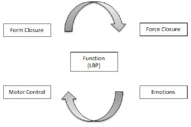
Figure 1. Integrated multifactorial framework of function (Lee & Vleeming, 1998)
15
Figure 2. Disuse Syndrome Model, from Verbunt et al. (2003) 20 Figure 3. Deconditioning Syndrome Model – Adapted* Disuse Syndrome
Model, adapted from Verbunt et al. (2003)
23
Figure 4. Example of a restraint system used to allow isolated lumbar extension (ILEX) exercise and testing to be performed (Reprinted with permission from MedX Corporation)
99
Figure 5. Power Analysis to determine participant numbers 146 Figure 6. CONSORT diagram to illustrate participant numbers for
enrolment, allocation, follow-up and analysis stages for study 1
148
Figure 7. CONSORT diagram to illustrate participant numbers for enrolment, allocation, follow-up and analysis stages for study 2
149
Figure 8. Flow of participants through study 3 150
Figure 9. Custom built wall mounted adjustable postural rods. 152
Figure 10. Schematic of seated stadiometry setup. 152
Figure 11. Three dimensional motion camera set up 154
Figure 12. Marker arrangement 155
Figure 13. Example of limited ROM using the mid 50% of participants individual full ROM, in this case 72o.
160
Figure 14. Pre and post mean isometric ILEX strength curves plotted by quartile across the ROM.
165
Figure 15. Waveform patterns, Winters CV, CVp and CVo 175
Figure 16. Example participant data showing pre and post individual trial waveforms and pre and post waveform variability transformed to zero (CVp)
177
xv
xvi
GLOSSARY1
Throughout the thesis when ‘the author’ is used it refers to James Steele, the author of the thesis
Important terms and acronyms highlighted here are in alphabetical order
1RM – One repetition maximum
BMD– Bone mineral density
BMI –Body mass index
CBT– Cognitive behavioural therapy
Chronic LBP (CLBP)– LBP having lasted for >12 weeks
CNS– Central nervous system
CSA –Cross sectional area
CT –Computed tomography
CVo - Variability of the mean offset in ensemble average of a time-series waveform
CVp– Variability of the pattern in ensemble average of a time-series waveform
Deconditioning– Decrease in function
Disc Degeneration– A physiologic process associated with aging. More severe
degeneration and/or structural abnormality may be indicative of a pathological process or injury and is commonly present in those suffering from chronic LBP
Disuse– Decrease in physical activity levels
EMG– Electromyography
EMG fatigue indices - Methods of analysing the EMG signal for determination of
fatigability e.g. root mean square amplitude, mean, median or mode frequency slopes, initial frequencies etc.
ES – Erector spinae
Gait Variability– Abnormal variation in gait parameters
GPO – Global perceived outcome
1 Two copies of the glossary are provided with one for removal by reader to use as reference and bookmark
xvii
Isolated Lumbar Extension (ILEX)– Lumbar extension performed when the
pelvis has been appropriately restrained to prevent movement
LBP– Low back pain
Lumbar Extensor Musculature – Lumbar erector spinae and multifidus
MCIC– Minimal clinically important change
MF - Multifidus
MMF– Momentary muscular failure
MOS –Medical outcomes study
MRI – Magnetic resonance imaging
Multifactorial Framework - An integrated multifactorial framework of function
considering all potentially interrelated deficiencies in function in LBP under the areas of; form closure (structure), force closure (force produced by myofascial action), motor control (neural patterning and control), and emotions (psychological and psycho-social factors)
MVC –Maximal voluntary contraction
Non-specific LBP – LBP for which it is not possible to identify a specific cause of
pain
ODI– Oswestry disability index
PNS– Peripheral nervous system
QL –Quadratus lumborum
Resistance Training– An exercise modality performed with the goal of
conditioning the muscles (i.e. increasing strength, endurance or hypertrophy) using external resistance (i.e. free weights, bodyweight exercise, variable resistance machines, hydraulic resistance machines, and pneumatic resistance machines)
RMDQ –Roland Morris disability questionnaire
ROM– Range of motion
Seated Stadiometry– Measurement of seated stature for indirect determination
of spinal height and disc hydration
xviii
Stad1st– The first measurement taken during each seated stadiometry trial
StadAvg– The average of the ten measurements taken during each seated
stadiometry trial
StadShrink– The difference between the first and last of the 10 measurements
taken during each seated stadiometry trial
Trunk Extension (TEX)– A compound movement involving both hip extension
and lumbar extension
TSK – Tampa scale for kinesiophobia
VAS– Visual analogue pain scale
Winters CV– Method typically used to calculate the variability in ensemble
xix
GLOSSARY
Throughout the thesis when ‘the author’ is used it refers to James Steele, the author of the thesis
Important terms and acronyms highlighted here are in alphabetical order
1RM – One repetition maximum
BMD– Bone mineral density
BMI –Body mass index
CBT– Cognitive behavioural therapy
Chronic LBP (CLBP)– LBP having lasted for >12 weeks
CNS– Central nervous system
CSA –Cross sectional area
CT –Computed tomography
CVo - Variability of the mean offset in ensemble average of a time-series waveform
CVp– Variability of the pattern in ensemble average of a time-series waveform
Deconditioning– Decrease in function
Disc Degeneration– A physiologic process associated with aging. More severe
degeneration and/or structural abnormality may be indicative of a pathological process or injury and is commonly present in those suffering from chronic LBP
Disuse– Decrease in physical activity levels
EMG– Electromyography
EMG fatigue indices - Methods of analysing the EMG signal for determination of
fatigability e.g. root mean square amplitude, mean, median or mode frequency slopes, initial frequencies etc.
ES – Erector spinae
Gait Variability– Abnormal variation in gait parameters
GPO – Global perceived outcome
Isolated Lumbar Extension (ILEX)– Lumbar extension performed when the
xx
LBP– Low back pain
Lumbar Extensor Musculature – Lumbar erector spinae and multifidus
MCIC– Minimal clinically important change
MF - Multifidus
MMF– Momentary muscular failure
MOS –Medical outcomes study
MRI – Magnetic resonance imaging
Multifactorial Framework - An integrated multifactorial framework of function
considering all potentially interrelated deficiencies in function in LBP under the areas of; form closure (structure), force closure (force produced by myofascial action), motor control (neural patterning and control), and emotions (psychological and psycho-social factors)
MVC –Maximal voluntary contraction
Non-specific LBP – LBP for which it is not possible to identify a specific cause of
pain
ODI– Oswestry disability index
PNS– Peripheral nervous system
QL –Quadratus lumborum
Resistance Training– An exercise modality performed with the goal of
conditioning the muscles (i.e. increasing strength, endurance or hypertrophy) using external resistance (i.e. free weights, bodyweight exercise, variable resistance machines, hydraulic resistance machines, and pneumatic resistance machines)
RMDQ –Roland Morris disability questionnaire
ROM– Range of motion
Seated Stadiometry– Measurement of seated stature for indirect determination
of spinal height and disc hydration
SF36 –Short form 36 health questionnaire
xxi
StadAvg– The average of the ten measurements taken during each seated
stadiometry trial
StadShrink– The difference between the first and last of the 10 measurements
taken during each seated stadiometry trial
Trunk Extension (TEX)– A compound movement involving both hip extension
and lumbar extension
TSK – Tampa scale for kinesiophobia
VAS– Visual analogue pain scale
Winters CV– Method typically used to calculate the variability in ensemble
1. INTRODUCTION
1.1 Defining Low Back Pain
Low back pain (LBP) is pain localized to the lumbar area ranging from the inferior ribcage to the waistline (12th thoracic/1st lumbar to 5th lumbar/1st sacral vertebrae) and often
includes radiating leg pain such as sciatica. It is often labelled ‘non-specific’1 and has
been described by the National Institute for Health and Clinical Excellence (NICE, 2009)
as “…tension, soreness and/or stiffness in the lower back region for which it is not
possible to identify a specific cause of the pain” (pp. 4). It is estimated that in as much as 85% of LBP cases no specific patho-anatomical diagnosis can be found (White & Gordon, 1982; Nachemson et al., 2000). Non-specific LBP is further categorised as acute, sub-acute or chronic. Acute pain occurs suddenly and lasts <6 weeks, sub-sub-acute pain lasts between 6-12 weeks, whereas chronic pain develops gradually, lasting >12 weeks and is often recurring (Frymoyer, 1988). The ‘natural history’ of LBP is described as the majority of low back injuries and acute LBP cases recovering before chronicity develops, and the enormous costs associated with LBP are due to the sub-group of those with chronic LBP2
(NICE, 2009). Thus chronic LBP is looked upon by some as an entirely different entity from low back injury and acute LBP (Balague et al., 2012). However, while there are a variety of further co-morbidities associated with, and which develop with, chronic LBP, such as psycho-social factors, it should be noted that logically all “...chronic back pain always starts as acute back pain” (Adams et al., 2010, pp. 967).Indeed, in contrast to the
common notion of LBP’s natural history involving recovery of most acute LBP, evidence suggests that a considerable proportion (69% to 75%) of low back injury and acute LBP develops into chronic LBP (Papageorgiou et al., 1996; Croft et al., 1998), often with increasing frequency and severity (Donelson et al., 2012). Though most people visiting
1
As will be noted in a later section of this introduction, there are a number of possible sources of pain causing mechanisms in LBP although it is often difficult to ascribe in individual cases using diagnostic techniques what is specifically causing pain. This is one of the reasons for a high proportion of LBP being labelled ‘non-specific’ i.e. there is absence of evidence
(not meaning evidence of absence) for a specific pain mechanism. However, a small proportion of LBP (1%-4%) is identified as ‘Red Flag LBP’ including serious causes of pain such as tumour or malignancy, cauda equina syndrome, infection, or spinal fracture (Downie et al., 2013). These types of LBP are beyond the scope of this thesis to consider.
their physician for LBP complaints will cease to continue consulting them after one month, many continue to experience pain one year after (Papageorgiou et al., 1996; Croft et al., 1998) which suggests patient dissatisfaction with lack of improvement from the care received. This also reinforces that many who suffer from an acute low back injury or acute LBP will likely go on to have persistent chronic symptoms.
Chronic LBP is a multifactorial condition with a wide variety of associated physical dysfunctions including but not limited to; limited lumbar range of motion (ROM; Holmes et al., 1996; Nelson et al., 1995), gait abnormality (Lamoth et al., 2004; Anders et al., 2005; Lamoth et al 2006a; Lamoth et al 2006b; Lamoth et al., 2008; Carpes et al., 2008; Tsao &
1.2 Prevalence, Costs and Impact
“Back pain is one of the most costly conditions for which an economic analysis has
been carried out in the UK”
(Maniadakis & Gray, 2000, pp. 95).
(i.e. Medicare and Medicaid) has increased significantly suggesting the majority of increased direct costs from health care utilisation have been supported through tax funded systems. Though different health and social care systems are not directly comparable (Dagenais et al., 2008) it is evident that the prevalence of LBP is a major contributor to costs placed upon the health services in western civilised societies.
Typical treatments used for LBP contribute to the high direct costs associated with LBP (Katz, 2006). For example, Katz (2006) highlighted that General Practitioner (GP) and Physician visits are estimated to cost ~£100 each increasing to around ~£6000 for medical admissions. Surgery costs significantly more ranging from ~£21000 to ~£55500, yet, surgery can often be avoided through more cost effective means (as will be highlighted below). Van Tulder et al. (1995) estimated that total direct medical costs from treatment constituted $367 million of which $200 million was accounted for by hospital care costs. This is likely due to the higher costs of hospital care ($3856; ~£5782; per inpatient). However, although outpatient care is relatively cheaper ($199; ~£298; per outpatient), the high rates of LBP incidence to be highlighted mean this adds up considerably.
102, table. 7.16) reports for 1998 and 2008 highlight similar rates of LBP over these ten years. However, the 2000 report illustrates that the ten years preceding it (1988-1998) saw a sharp rise in sickness and invalidity benefit for LBP. In Contrast, US estimates of claims due to LBP decreased by 34% between 1987 and 1995 (Borenstein, 2000) but there is no indication of whether prevalence changed at all. The data of Freburger et al., (2009), however, show that US prevalence may have increased slightly (~6.3%). Whether this truly reflects increased prevalence of LBP, or merely less willingness to tolerate, it is unclear (ONS, 2000). It is difficult to confirm either explanations through observational data on prevalence, though the ONS Health of Adult Britain survey, whilst echoing data on increased sickness and invalidity benefit claims, presented data showing that prevalence barely changed between 1971 and 1981 (ONS, 1997).
anatomically predisposed to LBP, potentially explaining its high prevalence across a range of populations. This wide-spread prevalence across culture gaps suggests that a physical factor, independent of cultural and psychosocial influences, may be predominantly implicated in the multifactorial nature of LBP.
It is apparent that, despite difficulty accurately identifying true prevalence due to different methods, most studies highlight LBP as an issue extending across a wide range of populations. The individual burden, as well as economic costs that such rates of prevalence present is clear and as such understanding LBPs etiology as well as identifying effective treatments is vital to reducing this.
1.3 Lumbar Spine Anatomy
The anatomy of the lumbar spine is complex with bony elements consisting of the vertebrae, the intervertebral discs between the vertebral bodies, the ligaments reinforcing and passively supporting the vertebrae as well as the musclulature actively supporting and providing movement, and finally the spinal cord and nerves innervating the local musculature (Drake et al., 2008). From a evolutionary comparative anatomy perspective the present lumbar morphology of Homo sapiens represents a gross structure encompassing a wide and short pelvis, long flexible lumbar column and both comparatively large hip extensors (gluteal and hamstring musculature) and small lumbar extensors (erector spinae and multifidus; Lovejoy, 2007; Steele, 2013a).
The lumbar spine consists of the 5 vertebrae from L1 to L5 and encompassing the L5-S1 lumbopelvic junction, though, in a small number of modern humans (~3-5%) the presence of a 6th lumbar vertebrae has been noted (Lovejoy, 2007). The vertebrae consist of the
elements of the vertebral arch serve as attachments for muscles and ligaments, levers for muscular contraction to act against, and articulations with adjacent vertebrae (Drake et al., 2008).
The vertebrae articulate with one another through two joint types; the sympheses between the vertebral bodies (intervertebral discs), and the synovial joints between the articular processes (zygapophyseal or facet joints). The intervertebral discs join adajacent vertebrae by means of a thin layer of hyaline cartilage (known as the endplate) and are composed of the nucleus pulposus (the gelatinous center providing hydrostatic properties to changes in pressure) and the annulus fibrosus (composed of the outer fibers of the lamellae which are differentially oriented in adjacent lamellae; Adams et al., 2010). The facet joints are where the two articular processes meet and are enclosed by a thin articular capsule. A number of ligaments also provide passive stability to the vertebrae including the anterior and posterior longitudinal ligaments surrounding the vertebral body, and the interspinous ligaments including the ligament flava and spinuous ligaments (Drake et al., 2008).
The complexity of the structures within the lumbar spine presents a number of potential mechanisms for pain originating from the area. Indeed many of the structures noted have been evidenced to be implicated in LBP.
1.4 Mechanisms of Pain within LBP
As highlighted, chronic LBP is multifactorial and a variety of symptoms/dysfunctions associated with it might cause pain due to stress they exert upon structures of the lower back known to elicit a pain response (Farfan & Gracovetsky, 1984). Pain is defined by the International Association of the Study of Pain (IASP) as “an unpleasant sensory and emotional experience associated with actual or potential tissue damage or described in
terms of such damage.” (IASP, 1994). Further, pain can be broken down into three components (Loeser, 1980); nociception, pain itself, and emotional suffering. Nociception refers to the process by which the nervous system senses noxious stimuli, pain itself is a central output relating to the perception of this sensation, and suffering is an emotional response to the pain output, or a false perception of a pain output. Such additional clarification of the definition implies that, though pain itself is a subjective experience, in general it has an organic source, or a potential organic source. However, despite there being a range of sources potentially responsible for pain perception (Salzberg, 2012), the possibility of pain characterised as emotional suffering arising without an organic source, or indeed absence of pain despite tissue damage, cannot be dismissed entirely.
perception including prior experience, the context of the noxious stimuli, and also other physiological phenomena that may alter the nociceptive input (Price, 2000; DeLeo & Winkelstein, 2002; Apkarian et al. 2005). This has led to a biopsychosocial perspective being adopted to better explain the complex relationships between nociception, pain and suffering (Engel, 1980, Turk & Okifuji, 2002; Gatchel et al., 2007). Affective disorders can have significant impact upon pain response particularly when combined with actual tissue damage or other chronic pain states (Carragee et al. 2000). Evidence suggests that chronic pain states such as LBP are characterised by sensitisation of the central nervous system (CNS; DeLeo & Winkelstein, 2002; Steiger et al., 2012a) and also the peripheral
nervous system (PNS) (Hirsch et al. 1963; Edgar, 2007). Recent research examining structural differences in gray matter between age-matched chronic symptomatic and asymptomatic participants revealed chronic pain states are characterised by subcortical reorganisation (reduced brainstem and somatosensory cortex gray matter), suggested as a result of on-going nociception (Schmidt-Wilcke et al., 2006). Indeed neurons maintain a significant degree of plasticity allowing phenotypic changes which subsequently can alter the course of the pain response (Woolf & Salter, 2000).
range of individual responses in LBP and the marked differences in presence or absence of lumbar spine abnormalities in diagnostic tests and associated perception of pain.
Specific abnormalities, and the likelihood the structures they refer to are indeed potentially pain causing, should also be considered. Weishaupt et al. (1998) identified that disc bulging or protrusion, and degeneration, were more common than other more severe abnormalities such as herniation, end plate abnormalities, nerve root compression and facet joint osteoarthritis in asymptomatic individuals. Holt (1968), in a paper highlighting the issue of false positive results from discography in asymptomatic participants, dismissed the finding in his data that although discography showed cases of degeneration in 37% of asymptomatic participants, discography produced LBP or sciatic symptoms in all cases of herniation. Walsh et al. (1990) replicated this study also including a symptomatic group as comparison. They found significantly greater abnormal discographs and pain responses in the symptomatic group; however they did not report any distinction between degeneration and herniation. The intervertebral disc itself has a nociceptive nerve supply (Hirsch et al., 1963) that penetrates deep into the nucleus pulposus. This appears to be hyperalgesic in participants with degenerated discs compared to that of normal discs (Edgar, 2007). Perhaps, considering these findings, the degree of disc deformation imparts some influence on pain (Holt, 1968). In asymptomatic participants
Schmorl’s nodes (Intervertebral disc protrusion through the endplate and into the vertebral
degrees of degeneration, structural abnormality, or a combination of the both are more consistently apparent in participants with chronic LBP than in those who are asymptomatic in a dose dependent manner (Cheung et al., 2009; de Schepper et al., 2010). Even if not all abnormalities can be ascribed as the source of chronic LBP, any degenerative changes also heighten the risk for more severe disc degeneration or injury and thus potentially pain and suffering (Adams et al., 2010).
Direct nerve or disc injuries are not the only mechanisms which can initiate a pain response. Bogduk (1997) suggests that for a structure to be deemed a potential cause of pain it must meet four factors: have a nerve supply; be identified to cause pain similar to that seen in clinical scenarios in normal volunteers; be susceptible to painful disease or injury; and be demonstrated as a source of pain using diagnostic techniques. Facet joint pain is prevalent in chronic LBP and based on these criteria is well established to be a potential source of pain (Manchikanti et al. 1999; Manchikanti et al. 2004). In addition the ligamentous tissue of the lumbar region may be a potential source for pain as abnormalities are more common in symptomatic participants (Fujiwara et al. 2000) and they are shown to contain a rich free nerve supply suggesting injury could contribute to pain (Hirsch. 1963; Kiter et al. 2010). However, the lumbar musculature may not necessarily be a direct source of pain due to the persistence of muscle abnormalities after symptoms have resolved in first case acute LBP (Hides et al. 1996). Though, due to its active role in providing stability, the musculature may indirectly be responsible for instigating injury and pain causing mechanisms in the other structures (Donisch & Basmajian, 1972; Bogduk & Twomey, 1987; Panjabi et al., 1989; Crisco & Panjabi, 1991; Cholewicki et al., 1997; Solomonow et al., 1998; Moseley et al., 2002; MacDonald et al., 2006; Rosatelli et al., 2008).
Bogduk & Twomey, 1987; Panjabi et al., 1989; Crisco & Panjabi, 1991; Cholewicki et al., 1997; Solomonow et al., 1998; Moseley et al., 2002; MacDonald et al., 2006; Rosatelli et al., 2008), it might be hypothesised that in the normally functioning lumbar spine, nerve root compression or other injury should not occur unless something has caused instability and altered joint biomechanics e.g. abnormal muscle function and/or injury. DeLeo and Winkelstein (2002) hypothesise that injury initiates a cascade of events, which may not be linear, but that ultimately contribute to a pain response and may alter over time due to the interrelated influence of such events. Indeed the non-linearity of such events (Brinkmann, 1985; Butler et al., 1990; Kirkaldy-Willis and Burton, 1992) may be due to the non-linear effect of distal primary injury upon proximal secondary injury/pain mechanisms (Whiting &
Zernicke, 1998). Panjabi’s (2006) hypothesis of the non-linear sequential events after
sub-failure injury concisely describes how these events might be highly variable.
Whiting & Zernicke (1998) in discussing injury and pain highlight the important point that mechanisms should not be confused with the related albeit different concept of predisposing or contributory factors (i.e. factors that increase the chance of a mechanisms initiation). Most LBP is likely the result of some form of injury initiating a pain mechanism, the specific injury mechanisms being an intermediary factor (though it is acknowledged that pain may arise independent of injury and an organic nociceptive source). What should be considered, as mentioned, are the factors contributing to the initiation of these subsequent mechanisms. It seems the majority of LBP appears to have an organic pain causing origin. Although, LBP may be psychosomatic (perhaps better defined in these
cases as ‘suffering’ considering Loeser’s (1980) aspects of pain) in some instances, and
and that in the context of an existing condition, treating the causative mechanism or predisposing factor might also result in favourable outcomes. In the case of addressing such downstream consequences shown to be initially caused by injury, we should ask whether or not it is more prudent to instead address what caused injury in the first place in order to allow healing to occur.
1.5 A Multifactorial Perspective of Low Back Pain
As noted a number of potentially pain causing mechanisms exist in LBP, though, it is not always possible to directly attribute pain to a specific source through diagnostic means. Because of this it is not always possible to monitor improvement in an underlying cause, pain mechanism or diagnosis. As such, changes in function are often used to indicate improvement.
multifactorial nature, in addition to suggesting that the efficacy of treatments should be evaluated by considering a range of functional outcomes.
Figure 1. Integrated multifactorial framework of function (Lee & Vleeming, 1998)
The population specifically considered in this thesis are those suffering from LBP that are categorised as non-specific (NICE, 2009). It has been suggested that there is potential benefit of sub-categorisation of LBP based upon symptom presentation (Lebouef-Yde et al., 1997; Hepple & Robertson, 2006). In light of the mulitfactorial nature of LBP sub-categorisation might offer considerable value. Many clinicians consider it a valid concept that should direct treatment when considering non-specific chronic LBP; however, there is little validating evidence to support it3 (Kent & Keating, 2004). Indeed research, although
suggesting that imaging abnormalities are more common in symptomatic as opposed to asymptomatic participants, has found that they rarely bear any differential relationship to individual clinical outcomes (i.e. pain and perceived disability) thus offering little prognostic value for determining treatment (McNee et al., 2011; Shambrook et al., 2011; Endean et al., 2011). Though it may not yet offer value in directing treatment choice within a clinical setting, conceptualisation using a multifactorial framework instead represents a useful tool in directing research into etiology and for considering a range of outcomes regarding
3 Despite the problems with sub-categorisation and determination of pain causing symptoms in non-specific LBP it should
2. LITERATURE REVIEW
2.1 Introduction and Overview
Considering that LBP appears to be most predominantly caused by injury or degeneration to one or many of the structures of the lumbar spine, its stability and ability to resist external loading seems a promising predisposing factor to examine as playing a role in low back injury risk and LBP. Indeed as noted, the extensor musculature have been demonstrated to be predominantly involved in maintaining such stability (Donisch & Basmajian, 1972; Bogduk & Twomey, 1987; Panjabi et al., 1989; Crisco & Panjabi, 1991; Cholewicki et al., 1997; Solomonow et al., 1998; Moseley et al., 2002; MacDonald et al., 2006; Rosatelli et al., 2008) and their condition could impact upon low back injury risk and pain. Further, how to target and condition the musculature in the most efficacious manner
may also be justified in examination. Therefore the following sections of this literature review chapter examine the following;
1. The etiological role of lumbar extensor muscle deconditioning in LBP.
2. Which exercise based approaches might be most efficacious in conditioning the lumbar extensor musculature.
3. What impact such exercise based approaches addressing the lumbar extensor musculature have upon pain and disability in symptomatic persons.
These sections are presented as independent systematic reviews whereby an initial independent introduction is provided for each section followed by methods, including search strategy and inclusion criteria, results of the literature review, and a discussion and conclusion offered. The first three sections have been previously published in peer reviewed journals with the present author as first author. These sections therefore are presented in the same format as their published versions4 and thus are written in a
manner independent from the remainder of the thesis.5 However, at the end of the
4 With the exception of the heading and referencing formats being amended to the required format for this thesis.
literature review chapter a section synthesising the evidence and conclusions from these independent reviews is offered providing an overarching rationale for the thesis. From this the specific areas of empirical research are presented and literature reviewed offering justification for their investigation within the context of the overarching thesis rationale, in addition to the research questions and hypotheses this thesis aims to address.
2.2 A Reappraisal of the Deconditioning Hypothesis in Low Back Pain: Review of
Evidence from a Triumvirate of Research Methods on Specific Lumbar Extensor
Deconditioning6
2.2.1 Overview7
‘Disuse’ and ‘Deconditioning’ in relation to low back pain (LBP) are terms often used
interchangeably. Discussions of ‘disuse’ refer to general physical inactivity, which
evidence suggests does not differ between symptomatic and asymptomatic persons.
‘Deconditioning’ refers to a decrease in function, commonly both cardiovascular/aerobic
fitness and muscular strength/endurance again noting little difference. However, examination of decreased function relating specifically to lumbar extensor musculature deconditioning has yet to be examined corroborating all possible methods. Thus, this review attempts to reappraise the deconditioning hypothesis in LBP specifically considering lumbar extensor deconditioning. A literature review was conducted examining both cross sectional and prospective data on specific lumbar extensor deconditioning and
LBP. A narrative approach and ‘snowballing’ style literature search was used involving
initial use of PubMed and Google Scholar databases searching up to December 2012. Included where studies utilising the following three research methods allowing specific induction of the role of such deconditioning; 1) strength/endurance testing of the isolated lumbar extensor musculature, 2) imaging and histochemical examination of the lumbar extensor musculature, and 3) fatigue testing of the lumbar extensor musculature using electromyography. Despite issues interpreting individual studies due to methods, the majority of evidence suggests LBP is associated with decreased strength/endurance, atrophy, and excessive fatigability of the lumbar extensors. Prospective studies also suggest lumbar extensor deconditioning may be a common risk factor predicting acute low back injury and LBP. The hypothesis of specific lumbar extensor deconditioning as being a causal factor in LBP is presently well supported. It is by no means the only causative
6 Note that this section has been previously published as an independent article by the author; Steele et al., 2013.A Reappraisal of the Deconditioning Hypothesis in Low Back Pain: Review of Evidence from a Triumvirate of Research
Methods on Specific Lumbar Extensor Deconditioning. Current Medical Research and Opinion. 30(5), pp 865-911.
factor and further research should more rigorously test this hypothesis addressing the methodological issues highlighted regarding previous studies. However, its role suggests specific exercise may be a worthwhile preventative and rehabilitative approach.
2.2.2 Introduction
2.2.2.1 Defining the Disuse/Deconditioning Hypothesis - ‘Disuse’ OR ‘Deconditioning’?
The ‘Disuse Syndrome’ was originally described by Bortz II (1984) and more recently has
been reviewed by Verbunt et al. (2003; 2010). The rationale behind Disuse Syndrome is that pain causes low levels of physical activity (i.e. avoidance behaviour or guarded movement; Main and Watson, 1996) which contribute to deconditioning and chronicity in low back pain (LBP), and cause the further interrelated physical and psychological changes shown in figure 2. In essence, it proposes that injury and pain precede
deconditioning and potentially many of LBP’s symptoms, leading to a ‘vicious cycle.’
Figure 2. Disuse Syndrome Model, from Verbunt et al. (2003)
participants, suggesting that lack of physical activity due to the presence of pain or injury may not contribute to the presence of deconditioning or LBP (Verbunt et al., 2001). Indeed a more recent study has also highlighted that physical activity levels appear to not change as a result of LBP, even as it develops from acute into chronic LBP (Bousema et al., 2007). This suggests that symptomatic chronic LBP participants may not suffer from development of disuse after the initial incidence of LBP. It seems possible, therefore, that
the direction of temporal relationships in the ‘Disuse Syndrome’ model may be unfitting in
its usual presentation (figure 2). This is not to suggest that deconditioning as a result of existing pain and its related behaviours is not a possibility, indeed the presence of injury has been shown to affect muscular function and could therefore instigate deconditioning itself, or at the least further enhance its development (Mannion, 1999; Panjabi, 2006).
Instead, ‘deconditioning’ (i.e. defined as a decrease in function) may be first implicated as
a potential cause of low back injury and pain, as opposed to LBP leading to ‘disuse’ and
then to ‘deconditioning’. Indeed Verbunt et al. (2003) attempt to distinguish between
‘disuse’ and ‘deconditioning’; however in both their definitions they inevitably invoke
general physical inactivity (‘disuse’) as being responsible for ‘deconditioning’ and that this
inactivity is the result of pain. Here we instead differentiate between ‘disuse’ and
‘deconditioning’ and pose that the disuse syndrome model does not consider what first
causes or increases the probability of injury or pain occurring (which may lead to further
‘disuse’) in the first place.
and altered joint biomechanics, subsequently causing injuries and instigating mechanisms by which pain results. Then, at this point, the cycle by which a reduction in activity levels further promote chronicity, and the changes associated with it, may begin to have an influence.
The model may therefore require an addition that considers the initial injury in the first place (figure 3). A high percentage of low back injury and acute LBP develops into chronic LBP (Papageorgiou et al., 1996; Croft et al., 1998) and so it seems logical that something must affect the risk of low back injury in the first instance. Indeed Adams and colleagues (2010) have recently commented on the pertinent fact that all ‘chronic back pain always
starts as acute back pain.’ Thus, logically, something must first be responsible for the
Figure 3. Deconditioning Syndrome Model – Adapted* Disuse Syndrome Model, adapted from Verbunt et al. (2003)
Many prior reviews on the topic of deconditioning in LBP have utilised a broad focus encompassing decrease in function of both cardiovascular/aerobic fitness as well as muscular strength/endurance (Verbunt et al., 2003; 2010; Wittink et al., 2000; Smeets et al., 2006). These reviews suggest that deconditioning of these kinds is not apparent in those with chronic LBP. However, the studies considered have utilised varied methods of examining this association, many of which are not entirely specific and these are explained throughout this article. The lack of consideration of the specific study methodologies used by previous authors has perhaps contributed to the vague
distinctions between ‘disuse’ and ‘deconditioning’ as well as the shift in focus from
physical risk factors towards a more cognitive based appraisal of LBP and the effects of rehabilitation (Steiger et al., 2012a). A more specific analysis of the literature, focusing
limitations of prior techniques, might therefore be found to yield contrasting conclusions regarding the presence of deconditioning in LBP and the models relationships.
2.2.2.2 Specific Disuse/Deconditioning of the Lumbar Extensors
The important role of the lumbar extensor musculature, the Erector Spinae (ES; i.e. iliocostalis lumborum and longissimus thoracis) and both deep and superficial lumbar Multifidus (MF), in providing stability to the lumbar spine, has been alluded to in numerous studies (Donisch & Basmajian, 1972; Bogduk & Twomey, 1987; Panjabi et al., 1989; Crisco & Panjabi, 1991; Cholewicki et al., 1997; Solomonow et al., 1998; Moseley et al., 2002; MacDonald et al., 2006; Rosatelli et al., 2008). Prior reviews of the literature have indicated that, although it is difficult to distinguish which muscles provide the greatest relative contribution to spinal stability, their importance in co-operatively contributing to lumbar spinal stability is clear (MacDonald et al., 2006; Cholewicki & Van Vliet, 2002). The relative contribution of individual muscles will vary depending on the specific task being performed (Ladin et al., 1989); however, MacDonald et al. (2006) explain that all lumbar extensors can contribute towards stability of the intervertebral segments through compression of the vertebral unit and increased joint stiffness. Clearly the lumbar extensor musculature playing such an important role in providing stability to the lumbar spine suggests that deconditioning and dysfunction in these muscles could lead to changes in stability and biomechanics. This change in biomechanics may result in increasing passive tissue stresses and potentially impart an injury or pain response in structures of the lumbar spine (Farfan & Gracovetsky, 1984).
2.2.2.3 Aim and Approach of this Review
The focus upon ‘disuse’ and ‘deconditioning’ in a general sense has led to much
therefore is to test this hypothesis by examining the evidence reporting the nature of the relationship between specific deconditioning of the lumbar extensors and LBP from a triumvirate of research methods including:
Strength/endurance testing of lumbar extensor musculature,
Imaging and histochemical examination of the lumbar extensor musculature
Fatigue testing of the lumbar extensor musculature using electromyography
Three sections will follow, each covering the three research methods highlighted as they have been used in cross sectional examination of symptomatic chronic LBP participants compared with asymptomatic healthy participants. A fourth section shall examine prospective studies that have sought to examine the effect of deconditioning using these methods upon development of LBP in asymptomatic participants. Throughout, any methodological concerns and considerations with studies shall be highlighted initially and noted whilst discussing such studies. In light of those methodological issues discussed in each section it will also be noted which types of studies were excluded from consideration (however, those excluded are still summarised within the full summary tables in the appendices8). Given the broad scope of this review a narrative approach utilising a ‘snowballing’ style literature search (Greenhalgh & Peacock, 2005) was used initially
involving PubMed and Google Scholar databases searching up to December 2012
utilising search terms including combinations and synonyms of ‘low back pain’ low back
injury’ ‘lumbar’ ‘back’ ‘spine’ ‘extensors’ ‘lumbar extension’ trunk extension’ ‘erector
spinae’ ‘multifidus’ ‘iliocostalis lumborum’ ‘longissimus thoracis’ ‘strength’ ‘endurance’
‘atrophy’ ‘cross sectional area’ ‘fat infiltration’ ‘muscle density’ ‘histochemistry’ ‘fibre type’
‘electromyography’ ‘fatigability’ etc. In addition previous reviews and any located articles
reference lists were searched. This was selected as the best way to locate, examine and synthesise the maximum amount of information in the various sections covered, thus initial inclusion criteria were based upon applicability to this particular area of discussion,
and whether studies had utilised the above noted research methods, before applying specific exclusion criteria (noted in each section of this review).
2.2.3 Strength and Endurance of the Lumbar Extensor Musculature in LBP
2.2.3.1 Considerations for Studies of Strength and Endurance of the Lumbar Extensor
Musculature
is more important than the other with regards to LBP (McGill, 2007). Indeed Mannion (1999) has commented that the hypothesis of fatigability as being associated with LBP is essentially analogous to the hypothesis of insufficient strength (both being manifestations of lumbar extensor deconditioning relevant to the deconditioning hypothesis).
Numerous studies have sought to identify the relationship between functional measures of strength and endurance of the lumbar extensor muscles. However, it should be highlighted that the validity of a number of methods of tests for strength and/or endurance of the lumbar spine are questionable due to methodological difficulties; a primary concern being whether sufficient pelvic restraints have been utilised. Essentially, tests have either been performed to examine trunk extension (TEX), OR, isolated lumbar extension (ILEX; testing utilising pelvic stabilisation through use of a semi seated position with rear pelvic restraint and a belt across the thighs). In considering lumbar extensor deconditioning TEX studies require careful reflection along with corroboration of more valid test measures i.e. ILEX. If the pelvis is not stabilised during testing of extension then it is impossible to determine the actual source of measured extension torque during tests of strength and may involve the hip extensors (Kankaanpaa et al., 1998a; Kankaanpaa et al., 1998b; San
Juan et al., 2005; Da Silva et al., 2009; Lariviere et al., 2010a; Clark et al., 2002; 2003)
contributing to overstate torque measures (Smidt et al., 1983; Petersen et al., 1987; Graves et al., 1992a), due to the longer moment arms over which the gluteus and
hamstrings exert force, and their relatively larger cross-sectional areas (Farfan, 1975). At most only 3o of pelvic rotation (Inanami, 1991), likely a result of soft tissue compliance,
occurs during ILEX testing of this kind. Lack of pelvic restraint perhaps partly explains the inconsistent reproducibility of TEX endurance tests (Alaranta et al., 1994; Moffroid et al., 1994; Mayer et al., 1995; McGill et al., 1999; Latimer et al., 1999) as compared with the consistency of ILEX strength and endurance testing (Graves et al., 1990a; Robinson et al.,
1992a; Udermann et al., 2003; Hager et al., 2006). Indeed, despite the aforementioned
tests of ILEX strength and TEX endurance (Perez et al., 2007). This highlights that the tests may utilise different musculature.
Although tests of ILEX are more valid representations of lumbar extensor function due to TEX being a compound movement requiring additional rotation of the pelvis through the hip extensor musculature (Kankaanpaa et al., 1998a; Kankaanpaa et al., 1998b; Clark et
al., 2002; 2003; Smidt et al., 1983; Petersen et al., 1987; Graves et al., 1992a; Farfan,
1975; Inanami, 1991; Fulton, 1993; Pollock et al., 1993; Smith et al., 2008), a large number of studies have made use of tests measuring TEX. Smidt et al. (1983) also explain that consideration of both tests of TEX and ILEX are indeed valuable when interpreted together as they allow both a deductive, and further an inductive, approach to
identify the so called ‘weak link’ within the kinetic chain and thus we initially considered
both studies in this review. Beimborn & Morrissey (1988) reviewed early literature on trunk muscle performance in LBP suggesting a consistent association with reduced TEX strength in symptomatic participants, as well as further studies. These TEX studies have been summarised in the appendices9 provided and appear to show inconsistent
associations; some results supporting a link between TEX strength/endurance and LBP some which do not.
The inconsistency of both TEX tests of strength and endurance should not be surprising as hip extensor deconditioning appears to not be associated with chronic LBP (Kamaz et al., 2007) and as explained, without appropriate restraint of the pelvis the musculature of the hip extensors will serve to confound results. However, despite hip extensor deconditioning having apparently little association with LBP it seems some other aspect of TEX, perhaps ILEX, may be associated with it. As TEX is composed of both hip and lumbar extension it therefore seems logical that tests should attempt to remove the involvement of the hip extensors to examine ILEX. Thus, of key importance and inclusion to this review are studies that have used appropriate methods of testing ILEX. As shall
also be noted, previous surgery may have implications for the results of studies examining the deconditioning hypothesis (Weber et al., 1997; Rantanen et al., 1993; Sihvonen et al., 1993; Motosuneya et al., 2006) and ideally participants with prior surgery should be excluded. However, only one study utilising ILEX has controlled for this factor yet may suffer from its own shortcoming of small sample size. Thus in the following section all ILEX studies have been examined with this limitation noted.
2.2.3.2 Isolated Lumbar Extension Studies
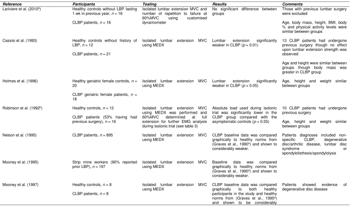
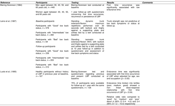
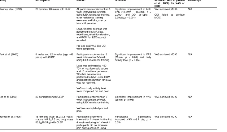
The validity of the extension test used is of great importance in examining the association between strength, endurance and LBP, therefore studies that have considered this are potentially more useful in answering the question of whether specific lumbar extension deconditioning is associated with LBP. Unfortunately in comparison with studies of TEX, studies of ILEX are relatively scarce. However, studies utilising testing that appropriately restrains the pelvis consistently report significantly reduced ILEX strength in symptomatic chronic LBP participants compared with asymptomatic controls (Cassisi et al., 1993; Holmes et al., 1996; Robinson et al., 1992b). Other studies (Nelson et al., 1995; Mooney
et al., 1995; Mooney et al., 1997; Boyce et al., 2008) have further reported reduced strength results from symptomatic participants compared to normal values obtained from healthy asymptomatic controls in other research (Graves et al., 1990a).
There is, however, only one study by Lariviere et al. (2010a) of ILEX using valid restraints
that does not support the link between specific deconditioning and chronic LBP. Lariviere et al. (2010a) reported no difference between asymptomatic and symptomatic chronic LBP
hypothesis). Other larger studies that have supported the link between ILEX weakness and chronic LBP have used in some instances upwards of 100 symptomatic participants and demonstrate reduced strength compared to healthy norms (Nelson et al., 1995; Mooney et al., 1995). In addition and in particular, the study by Nelson et al.(1995) of 895 chronic LBP participants suggested that a range of diagnoses existed in their sample
(Patients’ diagnoses included non-specific chronic LBP, degenerative disc/arthritic
disease, lumbar disc syndrome or spondylolisthesis/spondylolysis) and thus was likely quite representative of the typical heterogeneity of chronic LBP populations. Age, stature and body mass were also similar between groups in the study by Lariviere et al. (2010a),
however this was also reportedly the case for a number of other studies supporting the association (Cassisi et al., 1993; Holmes et al., 1996; Robinson et al., 1992b) and so is
unlikely to explain the difference in results.
One limitation of studies supporting the link between reduced ILEX strength and LBP is that these studies either did not report whether they excluded (Holmes et al., 1996; Nelson et al., 1995; Mooney et al., 1995; Mooney et al., 1997; Boyce et al., 2008), or chose not to exclude (Cassisi et al., 1993; Robinson et al., 1992b), participants who had
undergone previous surgery. Lariviere et al. (2010a) did exclude those having undergone
previous lumbar surgery and thus this may explain the different results found by these investigators. As has been noted, previous surgery can have potentially deleterious consequences to the lumbar extensor musculature anatomy (Weber et al., 1997; Rantanen et al., 1993; Sihvonen et al., 1993; Motosuneya et al., 2006) and so might be thought to interfere with ILEX strength in symptomatic participants. Although a number of TEX studies have excluded those with previous surgery, with some supporting and some refuting10 an association between deconditioning and LBP, we must consider the inherent
limitations of this approach already highlighted when specifically concerned with the lumbar extensors. There is certainly potential for further research to clarify whether differences in ILEX strength do indeed exist independent of previous lumbar surgery.
A final concern is the lack of statistical comparison with healthy controls groups in some studies (Nelson et al., 1995; Mooney et al., 1995; Mooney et al., 1997; Boyce et al., 2008). Though these results are consistent with those that have conducted statistical comparisons (Cassisi et al., 1993; Holmes et al., 1996; Robinson et al., 1992b) this is a
weakness and again something to be ensured in future research.
It is noted that only one study reported upon tests of isolated lumbar extension endurance (Lariviere et al., 2010a). However due to the inherent relationship between strength and
endurance it seems logical that the reported reduced lumbar extension strength in chronic LBP would be indicative of a reduced endurance also. The limitations discussed above also apply to this aspect of the study however, and there is further scope for research specifically examining this.
2.2.3.3 Summary of Strength and Endurance Studies of the Lumbar Extensor Musculature
Of the studies examined, those employing sufficient pelvic restraints as their means of assessing lumbar extension have consistently reported results that lend support to the association of specific lumbar extensor deconditioning with chronic LBP (Cassisi et al., 1993; Holmes et al., 1996; Robinson et al., 1992b; Nelson et al., 1995; Mooney et al.,
1995; Mooney et al., 1997; Boyce et al., 2008) with only one exception (Lariviere et al., 2010a). It seems clear that when valid testing of ILEX is used, most evidence suggests a