Officially Associated with
“The Egyptian Society of Oral Implantology”
An Official Publication of
“Ivano-Frankivsk National Medical University, Ukraine”
Case Report
IMPACTED INVERTED MAXILLARY THIRD MOLAR WITH UNUSUAL
SYMPTOMS
Vaishali Das, * RD Das, ** Anuradha Nemane ***
* Associate Professor, Yerala Medical Trust, Kharghar, Mumbai, Maharashtra, India
**Professor & Head, Department of Prosthodontics, YMTDC, Kharghar, Navi Mumbai, Maharashtra, India ***Reader, Department of Prosthodontics, YMTDC, Kharghar, Navi Mumbai, Maharashtra, India
________________________________________________________________________
ABSTRACT
Horizontally placed impacted maxillary wisdom teeth with occlusal surface facing the tuberosity is rather an unusual occurrence. A very few such cases have been reported in the literature. I report one such rare case of a 38 years old female with bizarre complaints of severe pain and numbness over left side of the face in the temperomandibular joint region extending along the zygomatic region associated with blocked and fullness of the left ear and blurring of vision. Its rare that impacted third molar teeth pressurizes the eustachian tube which connects the middle ear space to the back of the nose. Considering all the factors and after weighing risks and benefits, she was treated surgically for relieving her symptoms after careful evaluation.
KEYWORDS: Impacted; inverted; surgical removal
INTRODUCTION
Impacted teeth are generally nonfunctional ,malposed, may or may not be associated with pathology.[1,2] An impacted tooth is the one which is prevented from it's normal path of eruption in the dental arch due to lack of space in the dental arch or obstruction in the eruptive pathway of the tooth.[3] Maxillary third molars are frequently impacted teeth in the permanent dentition.[4-7] In the maxilla ,the third molars may be displaced to the floor of the sinus or may be inverted vertically or horizontally complicating its removal.[5-8] Deviation of the third molars is attributed to their unusual proliferation before the development of the tooth germ.[9] In very rare cases, the impacted third maxillary third molars can pressurize the Eustachian tube leading to
symptoms like clogged ear feeling, recurrent middle ear infection and heaviness of the affected area.[3]
CASE REPORT
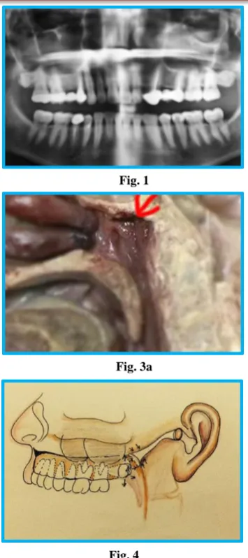
An unusual case of impacted maxillary third molars presented to me at Gokuldham medical centre, Mumbai. A 38 years old female patient presented with pain and numbness on the left side her face including the temporal and zygomatic region since April 2011. Twitching of the left eye was also a complaint. She consulted a neurophysician, who started her on tablet; Prothiadine 50mg, HS. This eased her a bit for a year. However, the numbness reduced but earache and ear symptoms worsened. A consultation with an otolaryngologist was in vain. Later, a general dentist adviced her for an orthopantomogram, (Fig. 1) and identified impacted maxillary third molars but refuted it as a cause of pain and was adviced to continue with the neurophysician's treatment. By April 2012, she developed trismus. Neurophysician re-evaluated and opined it as psychosomatic but nevertheless adviced MRI brain angiography which was within normal limits. Oral physiotherapy was given. By now, she realized that the hearing in her left ear had diminished and also she complained of slight blurring of vision. Opinion of ophthalmologist got glaucoma diagnosed and treatment was started for the same. In January 2013, she visited an Oral and Maxillofacial surgeon, who adviced her condylectomy after giving her a diagnosis of temperomandibular joint disorder. CT scans for the head including the maxilla and mandible was done. The scans did not reveal any major temperomandibular joint disease. Though proximity of the tooth to the Eustachian tube was expected, it was not reported. In March 2013, patient presented to me at Gokuldham Medical Centre, Mumbai with the
Maxillary third molar with unusual symptoms Das V, Das RD, Nemane A
same complaints. Careful review of the entire history coupled with thorough intraoral and extra oral examination was done. Intraoral examination revealed tenderness over the maxillary tuberosities bilaterally with degree of severity more on the left. Radiological evidence was evaluated. After weighing the benefits over the risks, I decided to go ahead with surgical intervention for the maxillary third molars. Guarded prognosis with regards to relief of pain and other symptoms was given. All possible complications including tuberosity fracture, displacement of the teeth into maxillary sinus or infratemporal space[5] and excessive bleeding were explained to the patient.
CLINICAL MANAGEMENT
Patient was taken up for surgery under general anesthesia by standard protocol. Decision to extract both the maxillary third molars was taken with the consent of the patient. Standard Ward's
incision was taken bilaterally. Tooth was deroofed by removal of bone in the third molar region. Guttering of the bone was done with a round bur number 8. The tooth was luxated and delivered. The maneuver was extremely difficult owing to the inaccessibility bilaterally and the occlusal table facing the tuberosity. Wound was irrigated thoroughly with betadine solution to clear off the debris and primary closure was achieved with 4-0vicryl sutures.
POSTOPERATIVE
Immediately postoperatively though painful, patient reported of the opening up of the clogged ear and drastic relief from heaviness of the left side of the face. No complications were observed besides the usual postoperative symptoms. After the uneventful recovery period of eighteen months postoperatively, patient is still free of her earlier symptoms. She is off prothiadine (50mg ) since then. She also reported of being free of repeated ear infections and can travel by air without severe earaches. Headaches are also cured. Though, she complains of slight pain in the temperomandibular region on the left side occasionally.
DISCUSSION
The decision for surgical intervention for the
Fig. 1 Fig. 2
Fig. 3a
Fig. 3b
maxillary third molars is done after a thorough assessment of history, symptoms and weighing against the difficulty index along with the possible intra and post operative complications.[2] Additional symptoms of numbness, clogged ear and blurred vision were a cause of concern and misled diagnosis. The surgical intervention in this case was more complicated than other types of the impacted teeth because of it's abnormal crown root position. Occlusal table facing the tuberosity made it greatly inaccessible and required exhaustive bone removal. Whether the treatment portocol should be conservative or surgical will depend upon the pathological changes occurred or not occured in the follicle or as in this case patient's need to overcome the uncomfortable symptoms associated. The maxillary third molars can be classified based upon their anatomical position according to their relative depth in the bone.[10] They can also be classified with respect to the long axis of maxillary second molars. The third method of classification is it's relationship to the maxillary sinus.[10] The case presented here thus was a Class C, with sinus approximation and horizontally inverted. On thorough research, very few cases have been cited in the literature. Eustachian tube connects middle ear cavity to nasopharynx, it's length being nearly 36mm in adults. It performs the function of pressure regulation, protection, and clearance (Fig. 2). Relationship of the Eustachian tube with the posterior maxilla can be appreciated in the cadaveric cross section through the same (Fig. 3a). Same is demonstrated in a model of the cross section of the nasal cavity and surrounding structures (Fig. 3b). CT scan sections also demonstrate the proximity of the tube to posterior aspect of maxilla. Usually fullness of Eustachian tube is felt because of pressure difference between the the air pressure in the middle ear and the pressure outside across the eardrum. At times, there could be physical blocking of the tube giving a feeling of fullness.[3] In the presented case, the proximity of the impacted, inverted maxillary third molar to the Eustachian tube seems to be the most probable reason of the presenting symptoms to develop. The position and bulk of the third molar with it's path of eruption towards the soft palate seemed to contribute to pressurizing the Eustachian tube as illustrated (Fig. 4) [Illustration is only for
understanding of the probable pathophysiology]. This is a retrospective diagnosis. Though, Eustachian tube blockages due to several other reasons are known to cause migraines, clogged ear feeling, pain in the eyes, overall heaviness in the same region.[3] The twitching of the eye muscles in this particular case reduced post-surgically, for which no justification besides relief from stress and overall pressure symptoms can be given.
CONCLUSION
On thorough review of literature available, it is clear that impacted inverted maxillary third molars are very rare and till August 2013 only few cases were reported. No literature supporting inverted horizontal maxillary third molar with such vague symptoms like numbness over the zygomatic region, clogged ear with frequent middle ear infection is yet reported. Negative pressure over the Eustachian tube seems to be the most probable cause. Important aspect of management of such teeth is to carefully weigh the risks and benefits associated with the surgical removal of the same. Use of fibro-optic endoscope in dentistry can greatly reduce morbidity. Generally, impacted teeth without any obvious pathology are considered to be harmless and hence a conservative treatment is recommended. But at times, patients like, in this case are referred to psychiatrists for psychosomatic reasons or a neurophysician for unexplained and vague pain, or to otolaryngologists for ear symptoms. It is, therefore, mandatory to carefully evaluate and analyze the signs and symptoms and investigations before referring the patient. Conservative treatment as leaving the tooth inside the bone can be followed when the impacted inverted teeth are asymptomatic and free of any pathology. Whereas, surgical intervention should be instituted at the earliest after thorough evaluation, review and diagnosis when patient is symptomatic.
CONFLICT OF INTEREST & SOURCE OF FUNDING
The author declares that there is no source of funding and there is no conflict of interest among all authors.
BIBLIOGRAPHY
Maxillary third molar with unusual symptoms Das V, Das RD, Nemane A
of American Dental Association 1973;87(11);186-8.
2. Held 1979, Inverted maxillary molar. Dental radiography 1979;52(4):87.
3. Anatomy and physiology of the Eustachian tube system. Charles Bluestone. Head and neck surgery. Otolaryngology 4th ed. 2006 Lippincott Williams and Williams.
4. Williams CD. Inverted embedded incisor. Dental Radiography and Photography 1957;30:78.
5. Winkler T, Von wowern N, Bittlmann S. Retrieval of an upper third molar from the infra temporal space. Journal of Oral Surgery 1977;35:130-132.
6. Yuvraj. Inverted maxillary third molar impaction - a case report. People's Journal of Scientific Research. 2011;4:57-58.
7. Pai V, Kundanbal M, Sequier PS, Rao. Inverted and impacted maxillary and mandibular third molars,a very rare case. Journal of oral health community Dentistry 2008;2(1):8-9.
8. Chhabra S, Chhabra N, Dhillon G. Inverted and impacted Maxillary third molar removal by lateral transposition method. International Journal of Experimental Dental Science 2012;1(1):26-9.
9. Alshamrani SM. Inverted and impacted maxillary third molars. Report of two cases. Odontostomatologic tropicale 2001;94:15-7. 10. Pell GJ, Gregory GT. Report on a 10 years