O R I G I N A L R E S E A R C H
Evaluation of endothelial/Descemet membrane
complex of eye bank donor corneas using
enhanced depth imaging optical coherence
tomography
This article was published in the following Dove Press journal: Clinical Ophthalmology
Zeba A Syed1
Gustavo Rosa Gameiro1 Marco Ruggeri1
Amr Elsawy1,2
Ibrahim Sayed-Ahmed1 Vatookarn Roongpoovapatr1 Mohamed Abdel-Mottaleb2 Mohamed Abou Shousha1
1Department of Ophthalmology, Bascom
Palmer Eye Institute, University of Miami Miller School of Medicine, Miami, FL,
USA;2Department of Electrical and
Computer Engineering, University of Miami College of Engineering, Coral Gables, FL, USA
Objective:We present a novel method for screening eye bank donor corneas using high definition optical coherence tomography (HD-OCT). This technology allows for the
quanti-fication of endothelial/Descemet membrane (En/DM) complex thickness ex vivo.
Design: Prospective interventional study.
Participants:Fifty-two corneal grafts from 27 donors were included in this study. Twenty additional control eyes and 11 eyes with Fuchs’ endothelial corneal dystrophy were also evaluated for comparison.
Methods:A custom built, high speed HD-OCT device (Envisu R2210, Bioptigen, Buffalo Grove, IL, USA) was used to obtain images, and custom-made graph-based segmentation software was used to automatically deconstruct corneal images into micro-layers. HD-OCT imaging was used to scan through the sealed sterile case of donor corneas stored in McCarey-Kaufman medium to image their En/DM complex through the center of the cornea.
Results: This technology allowed for quantification of En/DM complex thickness in all donor corneas through the sealed sterile container used to transport graft tissue. Mean En/ DM complex thickness of donor corneas was 17±4μm. The difference between donor cornea En/DM thickness and that of control subjects (16±2 μm) was not statistically significant (p=0.3), suggesting that the transport container and media do not affect measurements. There was a significant difference between En/DM thickness of Fuchs’endothelial corneal dystro-phy eyes (25±5μm) and both donor corneas (p<0.0001) and control subjects (p<0.0001).
Conclusions:We have described a new technique to measure En/DM complex thickness in eye bank donor corneas stored in a sealed sterile case. This may represent a novel adjunctive approach to screen corneal grafts for early endothelial disease.
Keywords:corneal transplant, Fuchs’endothelial corneal dystrophy, corneal graft screening, microscope-integrated OCT
Introduction
Quantification of Descemet membrane (DM) thickness has proven to be clinically useful in vivo. DM thickening has been documented in several forms of posterior corneal pathology including Fuchs’ endothelial corneal dystrophy,1 posterior polymorphous corneal dystrophy,2 and corneal graft rejection.3 Although slit lamp evaluation and specular examination are standard methods to evaluate corneal tissue prior to transplantation,4to the best of our knowledge there is currently no method described in
Correspondence: Mohamed Abou Shousha Department of Ophthalmology, Bascom Palmer Eye Institute, University of Miami Miller School of Medicine, 900 NW 17th Street, Miami, FL 33136, USA
Tel +1 305 326 6000
Email [email protected]
Clinical Ophthalmology
Dove
press
open access to scientific and medical research
Open Access Full Text Article
Clinical Ophthalmology downloaded from https://www.dovepress.com/ by 118.70.13.36 on 20-Aug-2020
the literature to screen eye bank donor corneas ex vivo using endothelial/Descemet membrane (En/DM) complex thickness. An important cause of late graft failure after full-thickness corneal transplant is Fuchs’ endothelial corneal dystrophy in the donor tissue.5 It is therefore of utmost importance to carefully select healthy grafts, as this will influence the outcome of the transplant. While examination of donor cornea prior to transplantation will usually exclude tissue with overt guttae, the evolution from the first emer-gence of guttae to significant edema typically takes many years.6It is therefore important to introduce a new screen-ing tool to detect early Fuchs’endothelial corneal dystrophy in donor tissue in order to exclude disease-prone grafts.
High definition anterior segment optical coherence tomo-graphy (HD-OCT) has been previously shown to detect char-acteristics of the En/DM complex and to measure its thickness in vivo with high accuracy.1The goal of this study is to report a novel method to ex vivo evaluate the En/DM complex in eye bank donor corneas using HD-OCT. We also report normative data of En/DM complex thickness in normal grafts and com-pare these measurements to in vivo En/DM thickness of a group of normal subjects and a cohort of patients with Fuchs’ dystrophy. HD-OCT of donor tissue represents a potential novel technique to screen corneal grafts for early Fuchs’dystrophy, therefore preventing the transplantation of grafts prone to late failure.
Materials and methods
Inclusion criteria
Fifty-two corneal grafts from 27 donors from the Florida Eye Bank were included in this study. Additionally, 20 control eyes from 20 patients were evaluated using the same imaging modality to determine normative En/DM thickness data. Eleven eyes of 7 patients with Fuchs’ endothelial corneal dystrophy were also evaluated for comparison. Approval for this study was obtained from the University of Miami Institutional Review Board (IRB). For imaging performed on corneal grafts, no signed consents were available from donors. Given that the images were obtained non-invasively with essentially no potential for personal identification, written consent was not obtained for the control and Fuchs’ eyes. However, verbal informed consent was confirmed for these participants, a process approved by our IRB.
Imaging technique
We used a custom built, high speed HD-OCT device (Envisu R2210, Bioptigen, Buffalo Grove, IL, USA). The
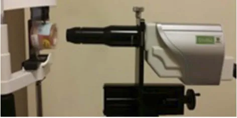
light source was a 3-module superluminescent diode (Broadlighter, T840-HP, Superlumdiodes Ltd, Moscow, Russia). The axial resolution of the system was 3 μm in tissue. Further details of the HD-OCT device have been previously decribed.1,7HD-OCT imaging was used to scan through the sealed sterile case of donor corneas stored in McCarey-Kaufman medium to image their En/DM com-plex (Figure 1). The system was used in enhanced depth imaging (EDI) configuration to obtain images of the pos-terior cornea with high contrast.8EDI HD-OCT images of the En/DM complex were obtained by adjusting the refer-ence arm of the OCT system to position the zero delay line posterior to the cornea. With this configuration, an inverted image of the cornea was produced where the anterior cornea was at the bottom of the image and the posterior cornea at the top, allowing for clearer identifi ca-tion of the En/DM complex.
We obtained 3 mm×3 mm B-scan (15 frames per B-scan to improve signal strength) images through the center of the donor corneas. Custom-made graph-based segmentation software was used to automatically deconstruct the corneal image into micro-layers based on edge detection, and frames were registered and averaged (Figure 2). Segments were manually edited as needed and surfaces reconstructed using bi-cubic interpolation. The En/DM region was then segmen-ted to produce En/DM thickness data.
HD-OCT images of 20 control eyes from 20 patients were also captured and used to obtain in vivo normal En/DM thickness data. As proof of concept, 11 eyes of 7 patients with Fuchs’ endothelial corneal dystrophy were imaged for comparison.
Statistical analysis
Statistical analyses were performed using SPSS software version 22.0 (SPSS, Chicago, IL, USA). Means of
vari-Figure 1 Imaging of a donor cornea through a sealed and sterile case. Abbreviation:HD-OCT, high definition optical coherence tomography.
Clinical Ophthalmology downloaded from https://www.dovepress.com/ by 118.70.13.36 on 20-Aug-2020
ables were compared between groups using independent sample t-tests. Two sided p-values less than 0.05 were considered statistically significant. Values are presented as means ± standard deviation.
Results
Patient age distributions
This study included 52 corneal grafts from 27 donors from the Florida Eye Bank. The average age of donors was 49.9 ±15.2 years. Additionally, 20 control eyes from 20 patients were evaluated (40.6±17.1 years), as well as 11 eyes of 7 patients with Fuchs’endothelial corneal dystrophy (67.5 ±9.3 years).
HD-OCT imaging and segmentation
A graph-based segmentation technique was used to auto-matically deconstruct the corneal image into micro-layers based on edge detection (Figure 2).
En/DM complex thickness in donor tissue
Using EDI HD-OCT, it was possible to obtain En/DM thickness data for all included eye bank donor corneas kept inside their sterile sealed case through the storage medium. Mean En/DM complex thickness of donor cor-neas was 17±4μm.
Comparison of En/DM complex thickness
in donor tissue, control eyes, and Fuchs
’
eyes
The difference between donor cornea En/DM thickness and that of control subjects (16±2μm) was not statistically sig-nificant (p=0.3). The difference between donor cornea En/
DM thickness and that of Fuchs’endothelial corneal dystro-phy eyes (25±5μm) was statistically significant (p<0.0001). The difference between the En/DM complex in control sub-jects and Fuchs’ eyes was also statistically significant (p<0.0001). The difference between these thickness mea-surements is presented graphically in Figure 3.
We had also obtained central corneal thickness (CCT) measurements using ultrasound pachymetry of control eyes and Fuchs’eyes. Average CCT in control eyes was 540.6±28.5μm, whereas average CCT in Fuchs’eyes was 566.7±57.6 μm. The difference between CCT of control eyes and Fuchs’ eyes was not statistically significant (p=0.0998).
Discussion
Although corneal tissue is screened prior to transplantation using slit lamp evaluation and specular examination,4these techniques alone may be limited in their ability to evaluate the overall health of graft tissue. For example, endothelial cell function cannot be predicted by measuring endothelial cell density; 14% of clear grafts may have an endothelial cell density <500 cells/mm2.9Furthermore, current screening tech-niques are typically unable to detect early Fuchs’endothelial corneal dystrophy in donor grafts,5an important cause of late graft failure. As such, additional tools to evaluate corneal grafts may allow for a more acute prediction of tissue survival. Thickening of the En/DM complex is a well-known early
finding in Fuchs’endothelial corneal dystrophy,10and there-fore screening grafts for En/DM complex thickening may be a potential strategy to exclude corneal tissue prone to failure.
While most centers currently employ OCT technology offering axial resolutions up to 4–7 microns, they are limited by their horizontal scan width.11 EDI HD-OCT
A
B
C
Figure 2HD-OCT images from(A)donor graft;(B)control eye; and(C)Fuchs’endothelial corneal dystrophy eye. Below each HD-OCT image is the result of segmentation showing the isolated En/DM complex demarcated with red arrows. Images are displayed with the zero delay at the bottom.
Abbreviations:HD-OCT, high definition optical coherence tomography; En/DM, endothelial/Descemet membrane.
Clinical Ophthalmology downloaded from https://www.dovepress.com/ by 118.70.13.36 on 20-Aug-2020
technology allows for more detailed imaging of individual corneal layers, and would therefore offer more precision in measuring the thickness of the En/DM complex. Aside from OCT, there is currently no available technology to reliably measure the En/DM complex thickness in vivo or ex vivo that would not require tissue destruction (as would be the case with histopathological evaluation).
In this report, we show that EDI HD-OCT can be used to measure thickness of the En/DM complex of eye bank donor corneas stored in McCarey-Kaufman medium through a sealed sterile case. DM thickening is present in Fuchs’ endothelial corneal dystrophy,1posterior polymorphous cor-neal dystrophy,2and corneal graft rejection,3and therefore may reflect the overall health of the En/DM tissue. Further research is needed to determine the clinical applicability of the En/DM thickness metric, whether it predicts graft survi-val, and how it compares to other measures such as endothe-lial cell count in influencing graft success.
We also demonstrate that ex vivo En/DM mean thickness of eye bank donor corneas is not significantly different from the in vivo En/DM thickness of age matched normal subjects. This finding validates the technique we used to study eye bank donor corneas. There are many sources of artifact in the use of HD-OCT, including media opacities.12As we do not currently have data on the influence of the donor cornea container on HD-OCT image quality, the similarity between En/DM thickness of donor corneas and normal subjects suggests that this methodology produces accurate measure-ments despite the presence of a container.
Interestingly, we did show a significant difference between En/DM complex thickness of control and Fuchs’ eyes, but there was no significant difference in CCT values. There are several possible explanations for this
finding. First, milder forms of Fuchs’ without frank cor-neal edema may still have En/DM thickening. Alternatively, because the numbers of control and Fuchs’ eyes were relatively small, this study may not have had the power to detect differences in CCT.
There are several limitations to this study. First, we compared the En/DM thicknesses of both ex vivo and in vivo corneas, therefore ignoring the possible role of cor-neal deturgescence in McCarey-Kaufman medium. However, as the thickness of En/DM was not statistically significant between donor tissue and control subjects, we may conclude that the storage medium did not largely affect this measure-ment. Secondly, we did not have specular microscopic data to correlate with En/DM thickness measurements. However, a previous study measured DM thickening using HD-OCT in eyes with Fuchs’dystrophy, and found a significant correla-tion with CCT. On the other hand, endothelial cell counts obtained using specular microscopy did not correlate with DM thickness or CCT, suggesting that DM thickness may be a more sensitive indicator.13Finally, while we had a sizeable number of corneal grafts in this study, the number of control eyes and corneas with Fuchs’dystrophy were smaller. While we did not detect a difference in En/DM between grafts and control eyes, it is possible that larger sample sizes would have influenced this result.
35
Mean Endot helia/Dexemet Membrane
Compled Thickness
(
µ
m)
30
25
20
15
10
5
0
Donor Corneas Control Corneas Fuchs’ Corneas
Figure 3Mean En/DM complex thickness (μm) in grafts versus control eyes and eyes with Fuchs’endothelial corneal dystrophy. Abbreviation:En/DM, endothelial/Descemet membrane.
Clinical Ophthalmology downloaded from https://www.dovepress.com/ by 118.70.13.36 on 20-Aug-2020
In summary, we have described a new technique that accurately measures En/DM complex thickness in eye bank donor corneas stored in a sealed sterile case. This may repre-sent a novel adjunctive approach to screen corneal grafts for early Fuchs’dystrophy. Further research is necessary to deter-mine whether En/DM thickness predictors graft survival outcomes.
Acknowledgments
Florida Lions Eye Bank for donor corneas that were included in this study. This study was supported by a NEI K23 award (K23EY026118), NEI core center grant to the University of Miami (P30 EY014801), and Research to Prevent Blindness (RPB). The funding organizations had no role in the design or conduct of this research.
Disclosure
United States Non-Provisional Patent (Application No. 14/ 247903) and United States Provisional Patent (Application No. 62/445,106) (MA). United States Non-Provisional Patents (Application No. 8992023 and 61809518), and PCT/US2018/013409. Patents and PCT are owned by University of Miami and licensed to Resolve Ophthalmics, LLC. MA is an equity holder and sit on the Board of Directors for Resolve Ophthalmics, LLC. Mr Amr Elsawy is listed as a coinventor for a patent 14/247903 licensed to Resolve Ophthalmics. Dr Mohamed Abou Shousha reports grants from National Eye Institute, during the conduct of the study. In addition, Dr Abou Shousha is listed as a coinventor for a patent United States Non-Provisional Patent (Application No. 14/247903) licensed to Resolve Ophthalmics, LLC, a patent United States Provisional Patent (Application No. 62/445,106) (MA). United States Non-Provisional Patents (Application No. 8992023 and 61809518), and PCT/US2018/013409. licensed to Resolve
Ophthalmics, LLC. The authors report no other conflicts of interest in this work.
References
1. Shousha MA, Perez VL, Wang J, et al. Use of ultra-high-resolution optical coherence tomography to detect in vivo characteristics of
Descemet’s membrane in Fuchs’ dystrophy. Ophthalmology.
2010;117:1220–1227. doi:10.1016/j.ophtha.2009.10.027
2. Sekundo W, Lee WR, Aitken DA, Kirkness CM. Multirecurrence of corneal posterior polymorphous dystrophy. An ultrastructural study.
Cornea. 1994;13:509–515.
3. Abou SM, Yoo SH, Sayed MS, et al. In vivo characteristics of corneal endothelium/descemet membrane complex for the diagnosis of
cor-neal graft rejection.Am J Ophthalmol. 2017;178:27–37. doi:10.1016/
j.ajo.2017.02.026
4. Saini JS, Reddy MK, Sharma S, Wagh S. Donor corneal tissue
evaluation.Indian J Ophthalmol. 1996;44:3–13.
5. Christopoulos V, Garner A. Emergence of cornea guttata in donor
tissue: a cause of late graft failure.Eye (Lond). 1993;7:772–774.
doi:10.1038/eye.1993.181
6. Wilson SE, Bourne WM. Fuchs’dystrophy.Cornea. 1988;7:2–18.
7. Shousha MA, Karp CL, Canto AP, et al. Diagnosis of ocular
surface lesions using ultra-high-resolution optical coherence
tomography. Ophthalmology. 2013;120:883–891. doi:10.1016/j.
ophtha.2012.10.025
8. Spaide RF, Koizumi H, Pozzoni MC. Enhanced depth imaging
spectral-domain optical coherence tomography.Am J Ophthalmol.
2008;146:496–500. doi:10.1016/j.ajo.2008.05.032
9. Lass JH, Sugar A, Benetz BA, et al. Endothelial cell density to predict
endothelial graft failure after penetrating keratoplasty.Arch Ophthalmol.
2010;128:63–69. doi:10.1001/archophthalmol.2010.128.63
10. Elhalis H, Azizi B, Jurkunas UV. Fuchs endothelial corneal
dystrophy.Ocul Surf. 2010;8:173–184.
11. Thomas BJ, Galor A, Nanji AA, et al. Ultra high-resolution anterior segment optical coherence tomography in the diagnosis and
manage-ment of ocular surface squamous neoplasia. Ocul Surf.
2014;12:46–58. doi:10.1016/j.jtos.2013.11.001
12. Tappeiner C, Barthelmes D, Abegg MH, Wolf S,
Fleischhauer JC. Impact of optic media opacities and image
compression on quantitative analysis of optical coherence
tomography. Invest Ophthalmol Vis Sci. 2008;49:1609–1614.
doi:10.1167/iovs.07-1264
13. Abou Shousha M, Yoo SH, Wang J, Perez VL. Novel technique to evaluate fuchs endothelial dystrophy: ultra-high-resolution optical coherence tomography versus specular microscopy. The Annual Meeting of the American Society of Cataract and Refractive Surgery (ASCRS); April 2010; Boston, MA.
Clinical Ophthalmology downloaded from https://www.dovepress.com/ by 118.70.13.36 on 20-Aug-2020
Clinical Ophthalmology
Dove
press
Publish your work in this journal
Clinical Ophthalmology is an international, peer-reviewed journal cover-ing all subspecialties within ophthalmology. Key topics include: Optometry; Visual science; Pharmacology and drug therapy in eye dis-eases; Basic Sciences; Primary and Secondary eye care; Patient Safety and Quality of Care Improvements. This journal is indexed on PubMed
Central and CAS, and is the official journal of The Society of Clinical Ophthalmology (SCO). The manuscript management system is completely online and includes a very quick and fair peer-review system, which is all easy to use. Visit http://www.dovepress.com/ testimonials.php to read real quotes from published authors.
Submit your manuscript here:https://www.dovepress.com/clinical-ophthalmology-journal
Clinical Ophthalmology downloaded from https://www.dovepress.com/ by 118.70.13.36 on 20-Aug-2020