Clinical Reasoning:
An unusual pattern of optic disc swelling
and visual loss
Amadeo R. Rodriguez, MD
Jason J.S. Barton, MD, PhD, FRCPC
SECTION 1
A 9-year-old boy was seen for bilaterally decreased vision. The patient noted gradual decrease in his vi-sual acuity over a few months with difficulty seeing the blackboard. Otherwise, he was asymptomatic with no headaches. His examination showed visual acuities of 20/60 in his right eye and 20/400 in his left eye. For color vision, pseudo-isochromatic plates were 15/16 right eye and 0/16 in the left eye. There was a left relative afferent pupil defect. Ocular
motility was normal. Funduscopy showed swelling of both optic discs affecting mainly the upper and lower aspects with sparing of the horizontal part of the disc, suggesting band atrophy. Maculae were nor-mal (figure 1).
Questions for consideration:
1. What is the differential diagnosis?
2. What initial investigations would help you in nar-rowing the differential?
GO TO SECTION 2
Figure 1 Fundus photographs and Goldmann visual field Address correspondence and
reprint requests to Dr. Amadeo R. Rodriguez, St. Joseph’s Healthcare CAHS Campus, 2757 King Street East, Hamilton, Ontario, Canada L8G 5E4 [email protected]
RESIDENT & FELLOW SECTION
SECTION 2
Optic disc swelling with visual loss is always a worri-some presentation. Optic neuritis in children is usu-ally bilateral and associated with optic disc swelling more frequently than in adults.1The course is usually acute with ocular pain, instead of the painless, insid-ious presentation in this patient. Neuroretinitis, which is the association of optic disc swelling and a macular star (a ring of hard exudates in the macula), is usually due to infectious diseases such as cat-scratch disease, Lyme disease, syphilis, or toxoplas-mosis.2Ischemia to the anterior optic nerve results in disc swelling and typically presents with segmental swelling affecting either the upper or lower half of the disc.3However, outside of the setting of severe blood loss or hypotension, ischemic optic neuropa-thy is rare in children. Arterial hypertension can also present with bilateral disc swelling and also visual loss if there is secondary macular edema. However, reti-nal hemorrhages or exudates are usually present.4 Toxic, metabolic, nutritional, and hereditary optic neuropathies tend to present with central or cecocen-tral scotomas since they tend to selectively affect the papillomacular bundle,5but not disc edema with the
exception of acute methanol toxicity. Infiltration of the anterior optic disc can cause swollen optic discs and visual loss. It usually occurs from leukemia, lym-phoma, or conditions such as sarcoidosis. Compres-sion of the optic nerve can also cause disc swelling. In our case with bilateral involvement, bilateral nerve compression or chiasmal involvement would be necessary to account for this presentation. Finally, papilledema from increased intracranial pressure should always be kept in mind in patients with disc swelling, although it does not commonly cause early acuity loss.6
In any patient with disc edema and visual loss, formal perimetry can be very useful in localizing the problem and narrowing the differential. In this case, Goldmann perimetry showed an almost complete bi-temporal hemianopia. In addition to perimetry, pa-tients with bilateral optic disc swelling require neuroimaging and lumbar puncture for measures of opening pressure and CSF content to address the above conditions.
Questions for consideration:
1. What would you do next? 2. What do you expect to find?
GO TO SECTION 3
SECTION 3
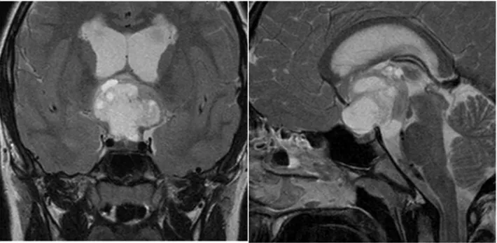
Asymmetric acuity and color vision loss, the presence of relative afferent pupillary defect, and bitemporal hemianopia all suggest asymmetric compression of the anterior visual pathway, namely the intracranial optic nerves and optic chiasm. An urgent head MRI showed a large suprasellar tumor extending into the third ventricle and causing hydrocephalus (figure 2). Compression of the visual pathway requires ur-gent intervention to avoid permanent visual loss. The patient underwent urgent neurosurgery. The tumor
arose from the optic chiasm itself and pathology con-firmed a pilocytic astrocytoma. Complete resection was not possible because of the risk of further dam-age to the visual pathway and therefore, the child was started on chemotherapy. A right occipital ventricu-loperitoneal shunt was inserted.
Questions for consideration:
1. How would you explain the pattern of optic disc swelling?
2. What is the prerequisite for this to occur?
GO TO SECTION 4
SECTION 4
This patient presented with hydrocephalus and bilat-eral papilledema. Increased intracranial pressure is transmitted to the optic nerve through the subarach-noid space and results in impaired axonal transport at the lamina cribrosa.7Even though it is common for papilledema to begin in the upper or lower aspect of the disc, in this case the presence of pallor along the horizontal portion suggests previous axonal loss from the chiasmal tumor (i.e., primary atrophy). Damage to the retinal ganglion cells axons anywhere along their course results in retrograde degeneration that becomes evident on funduscopy 4 to 6 weeks after injury. The horizontal band or bowtie atrophy is due to the compromise of the nasal fibers that decussate in the optic chiasm.8Thus papilledema is expressed mainly in the upper and lower aspects of the disc, which contain the arcuate fibers originating from the temporal aspect of the fundus.
DISCUSSIONIn this patient, there is a rare combi-nation of signs. The presence of bilateral optic atro-phy suggests chronicity since atroatro-phy takes at least several weeks to develop. The bilateral bowtie pattern of atrophy localizes the lesion to the optic chiasm. As the tumor continued growing and extending into the third ventricle, it resulted in increased intracranial pressure, hydrocephalus, and papilledema. Papill-edema is confined to the region containing axons that are not destined to decussate in the chiasm, be-cause atrophied axons cannot swell. This particular type of swelling is known as twin peaks papilledema as it resembles 2 hills (the swollen peaks) separated by a valley (band atrophy).9,10If papilledema persists it may also result in further axonal loss extending to the previously swollen aspects of the nerve, a secondary atrophy that will appear more diffuse without the localizing value of band or bowtie atrophy.
Additional asymmetric compression of the optic nerves explains the acuity loss, reduced color vision, and the left relative afferent pupil defect.
Detailed history and careful ophthalmoscopic ex-amination provide valuable information about the disease course, the possible location of the lesion, and the underlying mechanism. This case is also a re-mainder that optic disc swelling should not be ex-pected in the setting of optic atrophy. This concept is particularly important in patients with chronic pap-illedema and diffuse axonal loss: even if the intracra-nial pressure is significantly increased, there may be no changes in the appearance of the optic disc.
DISCLOSURE
Dr. Rodriguez received a speaker honorarium from Biogen Idec. Dr. Bar-ton serves on the editorial board of theJournal of Neuro-ophthalmologyand has received/receives research support from the NIH [NIMH 1R01 MH069898 (PI), the CIHR [MOP-77615 (PI), MOP-81270 (PI), and MOP-85004 (PI)] and from the Natural Sciences and Engineering Re-search Council [RGPIN 355879-08 (PI)].
REFERENCES
1. Morales DS, Siatkowski RM, Howard CW, Warman R. Optic neuritis in children. J Pediatr Ophthalmol Strabis-mus 2000;37:254 –259.
2. Brazis PW, Lee AG. Optic disc edema with a macular star. Mayo Clinic Proc 1996;71:1162–1166.
3. Rucker JV, Biousse V, Newman NJ. Ischemic optic neu-ropathies. Curr Opin Neurol 2004;17:27–35.
4. Van Stavern GP. Optic disc edema. Semin Neurol 2007; 27:233–243.
5. Newman NJ, Biousse V. Hereditary optic neuropathies. Eye 2004;18:1144 –1160.
6. Miller NR, Newman NJ, Biousse V, Kerrison JB, eds. Walsh and Hoyt’s Clinical Neuro-ophthalmology, 6th ed, vol 1. Philadelphia: Lippincott Williams & Wilkins; 2005. 7. Hayreh SS. Optic disc edema in raised intracranial pres-sure: V: pathogenesis. Arch Ophthalmol 1977;95:1553– 1565.
8. Unsold R, Hoyt WF. Band atrophy of the optic nerve. Arch Ophthalmol 1980;98:1637–1638.
9. Paul TO, Hoyt WF. Funduscopic appearance of papill-edema with optic tract atrophy. Arch Ophthalmol 1976; 94:467– 468.
10. Czarnecki JS, Weingeist TA, Burton TC, Thompson HS. “Twin peaks” papilledema: the appearance of papilledema with optic tract atrophy. Can J Ophthalmol 1976;11: 279 –281.
DOI 10.1212/WNL.0b013e3181d5618c
2010;74;e43-e46
Neurology
Amadeo R. Rodriguez and Jason J.S. Barton
Clinical Reasoning: An unusual pattern of optic disc swelling and visual loss
This information is current as of March 15, 2010
Services
Updated Information &
http://n.neurology.org/content/74/11/e43.full
including high resolution figures, can be found at:
References
http://n.neurology.org/content/74/11/e43.full#ref-list-1
This article cites 9 articles, 0 of which you can access for free at:
Subspecialty Collections
http://n.neurology.org/cgi/collection/visual_loss
Visual loss
http://n.neurology.org/cgi/collection/visual_fields
Visual fields
http://n.neurology.org/cgi/collection/optic_nerve
Optic nerve
following collection(s):
This article, along with others on similar topics, appears in the
Permissions & Licensing
http://www.neurology.org/about/about_the_journal#permissions
its entirety can be found online at:
Information about reproducing this article in parts (figures,tables) or in
Reprints
http://n.neurology.org/subscribers/advertise