Continuous assessment of electrical
epileptic activity in acute stroke
E. Carrera, MD; P. Michel, MD; P.-A. Despland, MD; M. Maeder-Ingvar, MD; C. Ruffieux, PhD; D. Debatisse, MSc; J. Ghika, MD; G. Devuyst, MD; and J. Bogousslavsky, MD
Abstract—Objective:To determine the incidence and risk factors of electrical seizures and other electrical epileptic activity using continuous EEG (cEEG) in patients with acute stroke.Methods:One hundred consecutive patients with acute stroke admitted to our stroke unit underwent cEEG using 10 electrodes. In addition to electrical seizures, repetitive focal sharp waves (RSHWs), repetitive focal spikes (RSPs), and periodic lateralized epileptic discharges (PLEDs) were recorded.Results:In the 100 patients, cEEG was recorded for a mean duration of 17 hours 34 minutes (range 1 hour 12 minutes to 37 hours 10 minutes). Epileptic activity occurred in 17 patients and consisted of RSHWs in seven, RSPs in seven, and PLEDs in three. Electrical seizures occurred in two patients. On univariate Cox regression analysis, predictors for electrical epileptic activity were stroke severity (high score on the National Institutes of Health Stroke Scale) (hazard ratio [HR] 1.12;p⫽0.002), cortical involvement (HR 5.71;p⫽0.021), and thrombolysis (HR 3.27; p⫽0.040). Age, sex, stroke type, use of EEG-modifying medication, and cardiovascular risk factors were not predictors of electrical epileptic activity. On multivariate analysis, stroke severity was the only independent predictor (HR 1.09; p ⫽ 0.016). Conclusion: In patients with acute stroke, electrical epileptic activity occurs more frequently than previously suspected.
NEUROLOGY 2006;67:99–104
Patients with acute stroke treated in a stroke unit show a relative reduction in mortality.1 Recent guidelines2-4 have put forward recommendations for the organization of stroke units and defined the use-fulness of several acute diagnostic tests, including brain CT and MRI, laboratory examinations, duplex and transcranial ultrasonography, and monitoring of blood pressure, EKG, oxygen saturation, and body temperature. These guidelines also provide strate-gies for the treatment of acute stroke, including general care and specific therapies, such as recanali-zation or prevention of complications.
However, the role of continuous EEG (cEEG) mon-itoring in the stroke unit has not been adequately assessed, despite the high incidence of clinical sei-zures, which ranges from 2 to 33% in the acute phase of stroke,5 and the potentially harmful effect of sei-zures on acute ischemic tissue.6The information re-garding the usefulness of cEEG in acute stroke was obtained in studies on severe stroke patients admit-ted to intensive care units. Electrical seizures were detected in 9 to 15% of patients, depending on pa-tient selection and the cEEG technique.7-9
In this study, we sought to determine the inci-dence and risk factors of electrical seizure and epi-leptic electrical activity in acute stroke patients admitted to our stroke unit.
Methods. Subject selection. We prospectively recruited 100 consecutive patients admitted to our stroke unit. Inclusion
cri-teria were acute symptoms and signs consistent with ischemic or hemorrhagic stroke. Exclusion criteria were subarachnoid or posttraumatic hemorrhage; venous thrombosis; structural le-sions, such as arteriovenous malformation rupture; and electro-lytic or metabolic disorders affecting the EEG, such as hepatic or renal failure. Patients with preexisting epileptic disorders were excluded.
Procedure. On admission, brain CT with perfusion se-quences and precerebral angio-CT were performed in the emer-gency department on all patients. In our stroke unit, where acute stroke patients usually stay for 24 hours or longer, de-pending on stroke severity, general condition, and complica-tions, the patients underwent cEEG. Typically, cEEG monitoring was started in the morning and was stopped the following day. When premature cEEG interruption occurred, the cause was reported by the nurse team. Intracranial and extracranial Doppler ultrasonography and blood pressure and EKG monitoring were performed on all patients, and MRI with diffusion-weight imaging, perfusion, T1 and T2 sequences, and transthoracic and transesophageal echocardiography were per-formed on selected patients. Neurologic examination using the National Institutes of Health Stroke Scale (NIHSS) score was performed on admission to the emergency department and at least twice daily in our stroke unit, including at the beginning and end of the cEEG recording and at discharge. Clinical epi-leptic seizures before admission or during hospitalization were recorded. The causes of ischemic strokes were determined using the TOAST10 classification. EEG protocol. The cEEG was
re-corded using 10 electrodes according to the International 10-20 system with an eight-channel subset (Fp2-C4, C4-O2, Fp2-T4, T4-O2, Fp1-C3, C3-O1, Fp1-T3, and T3-O1). The impedances of the silver-silver/chloride electrodes, which were glued to the scalp with collodion, were kept below 5 k⍀. The cEEG was acquired using SystemPLUS software (Micromed, Mogliano, Italy) and a sampling frequency of 256 Hz. Filter settings were 1 and 70 Hz. The cEEG trace was continuously displayed at the bedside.
From the Departments of Neurology (E.C., P.M., P.-A.D., M.M.-I., D.D., J.G., G.D., J.B.) and Biostatistics (C.R.), University Hospital, Lausanne, Switzerland. Disclosure: The authors report no conflicts of interest.
Received October 10, 2005. Accepted in final form March 16, 2006.
Address correspondence and reprint requests to Dr. Emmanuel Carrera, Department of Neurology, BH 13, 1011 Lausanne-CHUV, Switzerland; e-mail: [email protected]
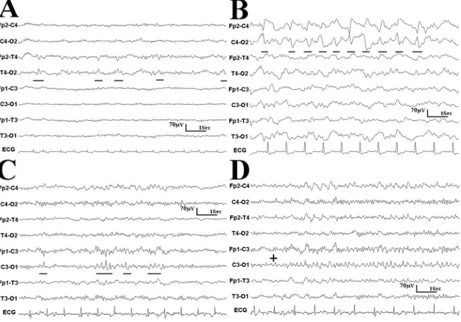
Epileptic activity was classified according to the following cri-teria (figure.)
1. RSHWs: Repetition of sharp waves of uniform morphology, duration, and localization, but without a definable and quantifi-able interval between consecutive waveforms.
2. RSPs: Repetition of spikes of uniform morphology, duration, and localization, but without a definable and quantifiable interval between consecutive waveforms.
3. PLEDs: Lateralized repetitive sharp waves, spikes, or sharply contoured waves at regular or nearly regular intervals and without a clear evolution in frequency or location.7
4. Electrical seizures: Rhythmic discharges or spikes lasting at least 10 seconds with a definite evolution in frequency location or morphology.7
Epileptic activity was recorded at the bedside during moment-to-moment online observation and during systematic review at the end of the recording by a board-certified electroencephalographer (E.C.). Each epileptic activity was confirmed by two independent board-certified electroencephalographers (M.M., P.A.D.) blinded to the clinical condition.
The prespecified primary endpoint was the occurrence of elec-trical epileptic activity according to the defined criteria.
Data were analyzed using commercially available statistical software (STATA 8.2). Cox proportional hazards regression analy-sis was used to investigate relationships between baseline charac-teristics (age, sex, cardiovascular risk factors, NIHSS score on admission, duration of cEEG recording, and interval from stroke onset to cEEG monitoring), and the occurrence of electrical epilep-tic activity. The significance level was set as 0.05. Variables that yielded a univariatep⬍0.1 were then included in a Cox propor-tional multivariate hazards analysis.
Results. Patient population. The 100 consecutive pa-tients consisted of 58 men and 42 women. The mean age was 68.7 years (range 31 to 94) and the mean (SD) NIHSS score on admission was 10.8 (7.1). There were 91 patients with ischemic strokes and nine with hemorrhagic strokes. Of the ischemic stroke patients, two presented with gener-alized tonic-clonic seizures at stroke onset and one with recurrent partial-complex seizures. Six of the 100 patients (three with and three without electrical epileptic activity) died during hospitalization; these six patients had a high NIHSS score (scores of 12, 20, 28, 21, 12, and 20) on admis-sion. Of the 100 patients, 17 were treated with EEG-modifying medication before or during cEEG. During EEG monitoring, one patient (Patient 16) was treated with
phe-nytoin 100 mg/day three times daily and clonazepam 2 mg/day for recurrent partial-complex seizure at stroke on-set. The other 16 patients received psychotropic agents because of preexisting psychiatric pathologies, agitation, or sleep disorders. No patients received antiepileptic treat-ment for electrical epileptic activity without clinical signs. No patients had undergone neurosurgical intervention be-fore EEG monitoring.
cEEG monitoring. The mean duration of cEEG moni-toring was 17 hours 34 minutes (range 1 hour 12 minutes to 37 hours 10 minutes). The cEEG was prematurely inter-rupted in 18 of the 100 patients due to agitation (n⫽10), spontaneous electrode failure (n ⫽ 2), computer failure (n⫽2), or the performance of acute tests, such as brain CT or MRI (n⫽4).
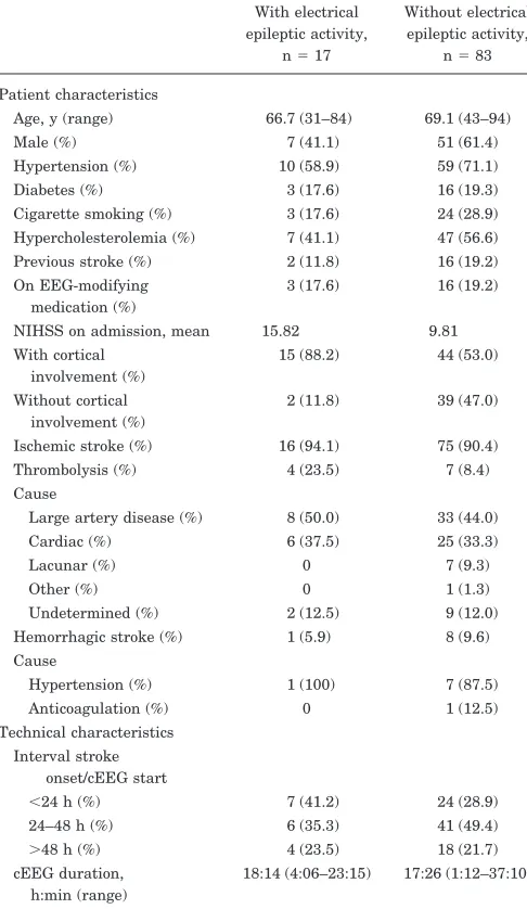
Electrical epileptic activity. Of the 100 patients, 17 had electrical epileptic activity. The baseline characteris-tics of the patients with and without electrical epileptic activity are presented in table 1. Electrical epileptic activ-ity consisted of RSHWs in seven patients, RSPs in seven, and PLEDs in three. The details of the individual patients with electrical epileptic activity are presented in table 2. Two patients, one with PLEDs (Patient 3) and one with RSPs (Patient 7), had focal electrical seizures during cEEG monitoring. No patient had generalized electrical epileptic activity or electrical status epilepticus. Among the patients with electrical epileptic activity, one had a hemorrhagic stroke limited to the right lenticular nucleus (Patient 4). Of the three patients with clinical seizures at stroke onset, one (Patient 16) had RSPs and the other two no electrical epilep-tic activity on the cEEG. No patient had a clinical seizure during EEG recording or after during hospitalization.
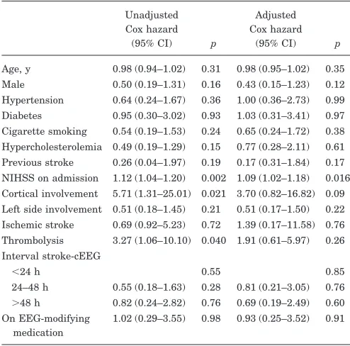
Hazard ratios (HRs), estimated using univariate Cox regression, are shown in table 3. Three factors were found to significantly increase the hazard of electrical epileptic activity: these were the NIHSS score on admis-sion (HR 1.12; 95% CI 1.04 to 1.20; p⫽ 0.002), cortical involvement (HR 5.71; 95% CI 1.31 to 25.01;p⫽0.021), and thrombolysis (HR 3.27; 95% CI 1.06 to 10.10; p ⫽
0.040). Age, sex, cardiovascular risk factors, stroke type
Figure. Examples of the different epi-leptic electrical activities. (A) Repetitive focal sharp waves focalized in the tem-poral area (T4) in a patient with right superficial middle cerebral artery (MCA) stroke (Patient 11, table 2). (B) Periodic lateralized epileptic discharges focalized in the central area (C4) in a patient with right complete MCA stroke (Patient 4). (C) Repetitive focal spikes focalized in the central area (C3) in a patient with left complete MCA stroke (Patient 7). (D) Beginning of a focal electrical seizure (C3) in the same pa-tient (Papa-tient 7).
(ischemic vs hemorrhagic), side of lesion, stroke etiology, EEG-modifying medication, and the interval from stroke onset to cEEG monitoring were not predictors of electri-cal epileptic activity. On multivariate Cox regression analysis, including the NIHSS score on admission, corti-cal involvement, and thrombolysis, the NIHSS score on admission (HR 1.09; 95% CI 1.02 to 1.18;p⫽0.016) was the only independent predictor of electrical epileptic activity.
Discussion. In this prospective study, based on cEEG in acute stroke patients admitted to a stroke unit, we found that electrical epileptic activity, in-cluding ictal and interictal abnormalities, was present in 17% of acute stroke patients. In addi-tion, stroke severity (assessed by the NIHSS score) on admission was the only independent predictor of electrical epileptic activity during cEEG.
The incidence of electrical epileptic activity in our study was higher than suspected on clinical
grounds using “standard” EEG11 in acute stroke patients or continuous EEG in critically ill pa-tients admitted to the intensive care unit.7,8 This may be due to the criteria used to classify the electrical epileptic activity in our study. cEEG in acute stroke patients is an emerging technique, and there is no definite consensus regarding the epileptic elements associated with clinical or elec-trical seizures. In our study, we grouped PLEDs, RSPs, and RSHWs together with electrical sei-zures. We included patients with PLEDs because PLEDs have been shown to be associated with seizures.11-13 The exact significance of PLEDs in the development of seizures is, however, controver-sial because some patients may present PLEDs in the absence of clinical or electrical seizures.14,15 In addition, and in contrast to other studies of EEG monitoring in acute stroke patients,7,8 we also re-ported RSHWs and RSPs because they were also shown, together with PLEDs, to be associated with the development of epilepsy in a study of epileptic EEG findings in acute stroke patients11 and in an-other study of unselected nonepileptic patients
re-ceiving EEG examination.16 We suggest that
RSHWs and RSPs may be indicators of interictal epileptic activity and therefore follow or precede seizures.11,17 However, further studies are needed to confirm the prognostic value of these different elements in terms of seizure development and clin-ical outcome. A continuum between these different “epileptiform” elements may be suggested. In our study, both patients with epileptic seizures had interictal epileptic activity (RSPs in one patient and PLEDs in the other). Another possible reason for the high prevalence of these types of electrical activity in our study is the duration of the cEEG monitoring. Compared with standard EEG in acute stroke patients with or without clinical seizures,11 cEEG recording was performed for a mean dura-tion of 17 hours 34 minutes in our study, increas-ing the probability of detectincreas-ing electrical epileptic activity and electrical seizures. Our findings em-phasize the potential usefulness of cEEG in detect-ing electrical epileptic activity in the stroke unit compared with standard EEG, which is usually performed on patients with suspected clinical seizures.18-20 Standard EEG may fail to detect epi-leptic activity and potentially give misleadingly fa-vorable information. The optimal duration of cEEG monitoring in acute stroke patients, however, re-mains to be defined with a larger sample size and probably depends on the characteristics of the pa-tient. In a previous study of critically ill patients,7 20% of electrical seizures were detected after more than 24 hours of cEEG monitoring.
In the present study, initial stroke severity, as-sessed by the NIHSS score on admission, was the only independent predictor of electrical epileptic activity. In the literature, stroke severity was found to be a predictor of clinical poststroke sei-zures in some studies,21-24but not all.25Recent elec-Table 1Characteristics of patients with and without electrical
epileptic activity
With electrical epileptic activity,
n⫽17
Without electrical epileptic activity,
n⫽83
Patient characteristics
Age, y (range) 66.7 (31–84) 69.1 (43–94) Male (%) 7 (41.1) 51 (61.4) Hypertension (%) 10 (58.9) 59 (71.1) Diabetes (%) 3 (17.6) 16 (19.3) Cigarette smoking (%) 3 (17.6) 24 (28.9) Hypercholesterolemia (%) 7 (41.1) 47 (56.6) Previous stroke (%) 2 (11.8) 16 (19.2) On EEG-modifying
medication (%)
3 (17.6) 16 (19.2)
NIHSS on admission, mean 15.82 9.81 With cortical
involvement (%)
15 (88.2) 44 (53.0)
Without cortical involvement (%)
2 (11.8) 39 (47.0)
Ischemic stroke (%) 16 (94.1) 75 (90.4) Thrombolysis (%) 4 (23.5) 7 (8.4) Cause
Large artery disease (%) 8 (50.0) 33 (44.0) Cardiac (%) 6 (37.5) 25 (33.3)
Lacunar (%) 0 7 (9.3)
Other (%) 0 1 (1.3)
Undetermined (%) 2 (12.5) 9 (12.0) Hemorrhagic stroke (%) 1 (5.9) 8 (9.6) Cause
Hypertension (%) 1 (100) 7 (87.5) Anticoagulation (%) 0 1 (12.5) Technical characteristics
Interval stroke onset/cEEG start
⬍24 h (%) 7 (41.2) 24 (28.9) 24–48 h (%) 6 (35.3) 41 (49.4) ⬎48 h (%) 4 (23.5) 18 (21.7) cEEG duration,
h:min (range)
18:14 (4:06–23:15) 17:26 (1:12–37:10)
NIHSS⫽National Institutes of Health Stroke Scale.
trophysiologic studies using cEEG in patients admitted to an intensive care unit7,8 did not find stroke severity to be a predictor of electrical epi-leptic seizures. In our study, the independent ef-fect of stroke severity may be due to patient selection. Rather than including severely disabled patients admitted to an intensive care unit with a high NIHSS score, all patients with acute stroke were recruited, irrespective of stroke severity. These findings suggest that, in a stroke unit, cEEG should be mainly performed on those pa-tients with a high NIHSS score at onset. In these patients, cEEG may help to detect subtle seizures or differentiate coma, stupor, and decreased level of consciousness secondary to electrical seizures from those of other origins, such as brain edema or stroke recurrence.26 Cortical involvement is com-monly seen in patients with poststroke clinical sei-zures22,23,27,28. In our study, cortical involvement of stroke was found to be of predictive value for elec-trical epileptic activity by univariate analysis, but not in the multivariate model. The fact that corti-cal involvement lost its prognostic value in the multivariate analysis does not mean that cortical location of stroke is without clinical relevance. The
Table 2Patients with electrical epileptic activity
No./sex/
age CVRF Medication Stroke
type Diagnostic Side Etiology Thrombolysis Clinical
seizure at
stroke
onset NIHSS
score at
admission NIHSS
score at
discharge Type of
baseline
epileptic
activity Time to
first
epileptic
event Electrical
seizure Time to
seizure Frequency* Focal
slowing†
1/F/80 HTA,DM ⫺ I Complete MCA L Cardioembolic ⫺ ⫺ 20 D RSPs 2:18:34 ⫺ ⫹ ⫹
2/F/79 — ⫺ I Complete MCA R Cardioembolic ⫺ ⫺ 28 D PLEDs 4:24:48 ⫹ 4:24:48 ⫹⫹ ⫹⫹
3/F/84 HTA ⫺ H Deep lenticular R HTA ⫺ ⫺ 15 9 PLEDs 0:00:05 ⫺ ⫹⫹ ⫹
4/F/70 HTA ⫺ I Complete MCA R Cardioembolic ⫺ ⫺ 22 28 PLEDs 4:28:00 ⫺ ⫹⫹ ⫹
5/F/50 — ⫺ I ACA and complete MCA
R LAD ⫹ ⫺ 17 13 RSHWs 1:44:12 ⫺ ⫹ ⫹⫹
6/M/57 HTA,HC ⫺ I ACA R LAD ⫺ ⫺ 10 9 RSHWs 0:46:05 ⫺ ⫹ ⫹
7/M/68 HTA ⫺ I Complete MCA L LAD ⫹ ⫺ 24 6 RSPs 4:28:45 ⫹ 8:04:37 ⫹ ⫹
8/F/66 CS ⫺ I Superficial MCA L LAD ⫺ ⫺ 5 4 RSPs 0:05:58 ⫺ ⫹ ⫹
9/M/54 HTA,HC ⫺ I Complete MCA R LAD ⫹ ⫺ 18 17 RSPs 1:02:47 ⫺ ⫹ ⫹⫹
10/M/60 DM ⫺ I PCA and complete MCA
L Cardioembolic ⫺ ⫺ 20 D RSPs 0:04:37 ⫺ ⫹⫹ ⫹⫹
11/F/73 HTA,HC ⫺ I Superficial MCA R LAD ⫺ ⫺ 14 3 RSHWs 18:52:23 ⫺ ⫹ ⫹
12/F/75 HTA,HC ⫺ I Bilateral PCA B Cardioembolic ⫺ ⫺ 17 12 RSPs 5:38:45 ⫺ ⫹ ⫹
13/M/79 HTA ⫹ I Watershed ACA-MCA
and PCA-MCA
R Undet ⫺ ⫺ 14 16 RSHWs 4:59:12 ⫺ ⫹ ⫹
14/M/64 HC, CS,
DM
⫺ I Deep MCA R LAD ⫺ ⫺ 18 20 RSHWs 7:58:12 ⫺ ⫹⫹ ⫹⫹
15/F/31 CS,HC ⫹ I Superficial MCA L Undet. ⫺ ⫺ 6 0 RSHWs 1:31:15 ⫺ ⫹ ⫹
16F/60 — ⫹ I ACA and complete MCA
R LAD ⫹ ⫹ 16 11 RSPs 1:04:45 ⫺ ⫹ ⫹⫹
17/M/84 HTA,HC ⫺ I Superficial MCA R Cardioembolic ⫺ ⫺ 5 5 RSHWs 11:53:34 ⫺ ⫹ ⫹
* Frequency:⫹ ⫽rare;⫹⫹; frequent;⫹⫹⫹ ⫽constant. † Focal slowing:⫹ ⫽minor;⫹⫹ ⫽major.
CVRF⫽cardiovascular risk factor; NIHSS⫽National Institutes of Health Stroke Scale; HTA⫽hypertension; DM⫽diabetes mellitus; I⫽ischemic stroke; MCA⫽middle cere-bral artery; H⫽hemorrhagic stroke; D⫽death; PLEDs⫽periodic lateralized epileptic discharges; HC⫽hypercholesterolemia; ACA⫽anterior cerebral artery; LAD⫽large ar-tery disease; RSHWs⫽repetitive sharp waves; B⫽bilateral; RSPs⫽repetitive spike waves; CS⫽cigarette smoking; PCA⫽posterior cerebral artery; undet.⫽undetermined.
Table 3Cox proportional hazards analysis of risk factors for electrical epileptic activity after acute stroke
Unadjusted Cox hazard
(95% CI) p
Adjusted Cox hazard
(95% CI) p
Age, y 0.98 (0.94–1.02) 0.31 0.98 (0.95–1.02) 0.35 Male 0.50 (0.19–1.31) 0.16 0.43 (0.15–1.23) 0.12 Hypertension 0.64 (0.24–1.67) 0.36 1.00 (0.36–2.73) 0.99 Diabetes 0.95 (0.30–3.02) 0.93 1.03 (0.31–3.41) 0.97 Cigarette smoking 0.54 (0.19–1.53) 0.24 0.65 (0.24–1.72) 0.38 Hypercholesterolemia 0.49 (0.19–1.29) 0.15 0.77 (0.28–2.11) 0.61 Previous stroke 0.26 (0.04–1.97) 0.19 0.17 (0.31–1.84) 0.17 NIHSS on admission 1.12 (1.04–1.20) 0.002 1.09 (1.02–1.18) 0.016 Cortical involvement 5.71 (1.31–25.01) 0.021 3.70 (0.82–16.82) 0.09 Left side involvement 0.51 (0.18–1.45) 0.21 0.51 (0.17–1.50) 0.22 Ischemic stroke 0.69 (0.92–5.23) 0.72 1.39 (0.17–11.58) 0.76 Thrombolysis 3.27 (1.06–10.10) 0.040 1.91 (0.61–5.97) 0.26 Interval stroke-cEEG
⬍24 h 0.55 0.85
24–48 h 0.55 (0.18–1.63) 0.28 0.81 (0.21–3.05) 0.76 ⬎48 h 0.82 (0.24–2.82) 0.76 0.69 (0.19–2.49) 0.60 On EEG-modifying
medication
1.02 (0.29–3.55) 0.98 0.93 (0.25–3.52) 0.91
lack of independent significance was probably due to the fact that patients with the highest NIHSS score, especially those with complete middle cere-bral artery stroke, had a cortical lesion. Further studies with a larger sample size are needed to compare patients with isolated subcortical lesions or isolated cortical lesions with the same NIHSS score. IV recombinant tissue plasminogen activa-tor (rt-PA) thrombolysis was found to be of predic-tive value for electrical epileptic activity in the univariate analysis, but not in the multivariate analysis. Because only 11 patients received throm-bolysis (four of whom presented electrical epileptic activity), no definitive conclusion can be drawn about the epileptogenic effect of rt-PA. However, our results suggest it may have a potential epilep-togenic effect and warrant a careful follow-up of rt-PA–treated patients, especially those with sec-ondary worsening possibly due to electrical
sei-zures. In animal models, an increased
excitotoxicity after IV administration of t-PA has been suggested to occur by amplification of intra-cellular calcium conductance29 or activation of ma-trix metalloproteinases (MMPs), for example, MMP-9.30,31 In our study, in contrast to observa-tions in other clinical22,25,32,33 or electrophysiologic8 studies, hemorrhagic stroke was not predictive of epileptic activity. The small sample size (N ⫽ 9) of patients with hemorrhagic stroke may be responsi-ble for our results. Age, sex, cardiovascular risk factors, stroke cause, or interval from stroke onset to cEEG had no predictive value for epileptic activity.
Our study has several limitations. The small pa-tient sample size, especially in the hemorrhagic stroke subgroup, is the main limitation because only 17 patients with electrical epileptic activity were re-corded. In addition, the rare patients with stroke requiring invasive treatment or ventilation initially were admitted to the intensive care unit rather than the stroke unit. They were therefore not included in our study or were only included once their condition had improved sufficiently to be managed in our stroke unit. Regarding the technique, only 10 elec-trodes were used, which may have limited the detec-tion of electrical seizures compared with a full set of electrodes. We chose this technique because it is more convenient to use on nonsedated patients ad-mitted to a stroke unit who receive frequent nursing care. However, despite the use of only 10 electrodes, cEEG recording was interrupted in 18 patients, mainly due to patient movement secondary to agita-tion. Finally, the use of EEG-modifying medications may have influenced the detection of epileptic activ-ity; however, there was no statistical impact of med-ication on the cEEG findings.
Our results show that, in addition to clinical ex-amination and other acute diagnostic tests such as brain CT and MRI and transcranial Doppler ultra-sonography, cEEG monitoring may be useful in the stroke unit. The impact of the electrical epileptic
ac-tivity on the clinical outcome and development of poststroke epilepsy must be assessed in further stud-ies to optimize the management and treatment of acute stroke patients.
Acknowledgment
The authors thank Cristiano Rizzo, PhD, and Sylviane Carruzzo for technical support.
References
1. Stroke Unit Trialists collaboration (stroke unit) care for stroke. Co-chrane Library, issue 3, 2005.
2. Hacke W, Kaste M, Bogousslavsky J, et al. ; European Stroke Initiative Executive Committee and the EUSI Writing Committee. European Stroke Initiative Recommendations for Stroke Management-update 2003. Cerebrovasc Dis 2003;16:311–313.
3. Adams HP, Adams RJ, Brott T, et al. Stroke Council of the Stroke Council of the American Stroke Association. Guidelines for the early management of patients with ischemic stroke: a scientific statement from the Stroke Council of the American Stroke Association. Stroke 2003;34:1056–1083.
4. Alberts MJ, Latchaw RE, Selman WR, et al. Recommendations for comprehensive stroke centres; a consensus statement from the brain attack coalition. Stroke 2005;36:1597–1618.
5. Camilo O, Goldstein LB. Seizures and epilepsy after ischemic stroke. Stroke 2004;35:1769–1775.
6. Bryan Young G, Jordan KG. Do nonconvulsive seizures damage the brain ? – Yes. Arch Neurol 1998;55:117–119.
7. Claassen J, Mayer SA, Kowalski RG, et al. Detection of electrographic seizures with continuous EEG monitoring in critically ill patients. Neu-rology 2004;62:1743–1748.
8. Vespa PM, O’Phelan K, Shah M, et al. Acute seizures after intracere-bral hemorrhage: a factor in progressive midline shift and outcome. Neurology 2003;60:1441–1446.
9. Jordan KG. Emergent EEG and continuous EEG monitoring in acute ischemic stroke. J Clin Neurophysiol 2004;21:341–352.
10. Adams HP, Bendixen BH, Kappelle LJ, et al. Classification of subtype of acute ischemic strokes. Definitions for use in a multicenter clinical trial. TOAST Trial of Org 10172 in Acute Stroke Treatment. Stroke 1993;24:35–41.
11. Holmes GL. The electroencephalogram as a predictor of seizures follow-ing cerebral infarction. Clin Electroencephalogr 1980;11:83–86. 12. Snodgrass SM, Tsuburaya K, Ajmone-Marsan C. Clinical significance of
periodic lateralised epileptiform discharges: relationship with status epilepticus. J Clin Neurophysiol 1989;6:159–172.
13. Brenner RP, Schaul N. Periodic EEG patterns: classification, clinical correlation and pathophysiology. J Clin Neurophysiol 1990;7:249–267. 14. Pohlmann-Eden B, Hoch DB, Cochius JI, Chiappa KH. Periodic
lateral-ized epileptiform discharges: a critical review. J Clin Neurophysiol 1996;13:519–530.
15. Garcia-Morales I, Teres Garcia M, Galan-Davila L, et al. Periodic later-alized epileptic discharges. Etiology, clinical aspects, seizures, and evo-lution in 130 patients. J Clin Neurophysiol 2002;19:172–177. 16. Zivin L, Marsan CA. Incidence and prognostic significance of
”epilepti-form⬙activity in the EEG of non-epileptic subjects. Brain 1968;91:751– 778.
17. Hughes JR. The significance of the interictal spike discharge: a review. J Clin Neurophysiol 1989;6:207–226.
18. Horner S, Ni XS, Duft M, Niederkorn K, Lechner H. EEG, CT and neurosonographic findings in patients with postischemic seizures. J Neurol Sci 1995;132:57–60.
19. Berges S, Moulin T, Berger E, et al. Seizures ad epilepsy following strokes: recurrence factors. Eur Neurol 2000;43:3–8.
20. Ossemann M. Crises e´pileptiques et e´pilepsie d’origine vasculaire: car-acteristiques cliniques, e´lectroencephalographiques et scan-nographiques. Rev Neurol (Paris) 2002;158:256–259.
21. Reith J, Jorgensen HS, Nakayama H, Raaschou HO, Olsen TS. Sei-zures in acute stroke: predictors and prognostic significance. The Copenhagen Stroke Study. Stroke 1997;28:1585–1589.
22. Bladin CF, Alexandrov AV, Bellavance A, et al. Seizures after stroke. A prospective study. Arch Neurol 2000;57:1617–1622.
23. Lamy C, Domingo V, Semah F, et al. Early and late seizures after cryptogenic ischemic stroke in young adults. Neurology 2003;60:400– 404.
24. Cheung CM, Tsoi TH, Au-Yeung M, Tang AS. Epileptic seizure after stroke in Chinese patients. J Neurol 2003;250:839–843.
25. Labovitz DL, Hauser WA, Sacco RL. Prevalence and predictors of early seizures and status epilepticus after first stroke. Neurology 2001;57: 200–206.
26. Claassen J, Mayer SA. Continuous electroencephalographic monitoring in neurocritical care. Curr Neurol Neurosci Rep 2002;2:534–540.
27. So EL, Annegers JF, Hauser O’Brien PC, Whisnant JP. Population-based study of seizure disorders after cerebral infarction. Neurology 1996;46:350–355.
28. Lancman ME, Golimstok A, Norscini J, Granillo R. Risk factors for developing seizures after a stroke. Epilepsia 1993;34:141–143. 29. Benveniste H, Drejer J, Schousboe A, Diemer NH. Elevation of the
extracellular concentrations of glutamate and aspartate in rat hip-pocampus during transient cerebral ischemia monitored by intracere-bral microdialysis. J Neurochem 1984;43:1369–1374.
30. Kaur J, Zhao Z, Klein GM, Lo EH, Buchan AM. The neurotoxicity of
tissue plasminogen activator? J Cereb Blood Flow Metab 2004;24:945– 963.
31. Sumii T, Lo EH. Involvement of matrix metalloproteinase in thrombolytic-associated hemorrhagic transformation after embolic focal ischemia in rats. Stroke 2002;33:831–836.
32. Giroud M, Gras P, Fayolle H, et al. Early seizures after acute stroke: a study of 1640 cases. Epilepsia 1994;35:959–964.
33. Burn J, Dennis M, Bamford J, et al. Epileptic seizures after a first stroke: the Oxfordshire Community Stroke Project. BMJ 1997;315: 1582–1587.
Neuro
Images
Pergolide-induced ergotism John C. Morgan, MD, PhD; and
Kapil D. Sethi, MD, FRCP (U.K.), Augusta, GA
Ergotism results from generalized vasoconstriction of small and large blood vessels and can lead to cyanosis and gangrene of affected limbs if not recognized. Ergotism occurs most commonly in the treatment of migraine with ergotamines today; however, ergot-derived drugs are also used in the treatment of restless legs
syndrome and Parkinson disease (PD). A 72-year-old African-American woman with advanced PD presented complaining of tingling and blue discoloration of her fingers and toes for 2 months (figure, A and B). She was taking a total of 8 mg per day of pergolide (an ergot-derived dopamine agonist) in order to avoid severe levodopa-induced dyskinesias. A noninvasive vascular eval-uation was normal. One week after switching to ropinirole (a non-ergot dopamine agonist) the discoloration of her digits had markedly improved (figure, C and D), and it completely resolved within 3 months. While cardiopulmonary fibrosis is well-recognized with pergolide,1,2clinicians should be aware that
per-golide can also cause ergotism.
Copyright © 2006 by AAN Enterprises, Inc.
1. Shaunak S, Wilkins A, Pilling JB, Dick DJ. Pericardial, retroperitoneal and pleural fibrosis induced by pergolide. J Neurol Neurosurg Psychiatry 1999;66:79–81.
2. Pritchett AM, Morrison JF, Edwards WD, et al. Valvular heart disease in patients taking pergolide. Mayo Clin Proc 2002;77:1280–1286.
Figure. Ergotism with cyanosis of the fingers bilaterally is evident in our pa-tient while on pergolide (A and B). One week after stopping pergolide and start-ing ropinirole, the ergotism was re-markably improved (C and D).
Disclosure: The authors report no conflicts of interest.
Address correspondence and reprint requests to Dr. John C. Morgan, Move-ment Disorders Program, DepartMove-ment of Neurology, Medical College of Georgia, 1429 Harper Street, HF-1121, Augusta, GA 30912, e-mail: [email protected]
DOI 10.1212/01.wnl.0000227185.81250.0f
2006;67;104
Neurology
John C. Morgan and Kapil D. Sethi
Pergolide-induced ergotism
This information is current as of July 10, 2006
Services
Updated Information &
http://n.neurology.org/content/67/1/104.full including high resolution figures, can be found at:
References
http://n.neurology.org/content/67/1/104.full#ref-list-1
This article cites 2 articles, 1 of which you can access for free at:
Citations
http://n.neurology.org/content/67/1/104.full##otherarticles This article has been cited by 1 HighWire-hosted articles:
Permissions & Licensing
http://www.neurology.org/about/about_the_journal#permissions its entirety can be found online at:
Information about reproducing this article in parts (figures,tables) or in
Reprints
http://n.neurology.org/subscribers/advertise
Information about ordering reprints can be found online:
Online ISSN: 1526-632X.
1951, it is now a weekly with 48 issues per year. Copyright . All rights reserved. Print ISSN: 0028-3878. ® is the official journal of the American Academy of Neurology. Published continuously since