223 | P a g e
THE CHARACTERIZATION OF GENE ENCODING
SURFACE PROTEIN-31(BCSP-31)
Brucella
abortus
FIELD ISOLATE BY PCR TECHNIQUE
Wiwiek Tyasningsih
1,Didik H.
2, Fedik A. Rantam
3, Aulanni’am
41,2,3
Microbiology Department, Faculty of Veterinary Medicine, Airlangga University, (Indonesia)
4
BiochemistryLaboratory, Faculty of Life Sciences, Brawijaya University, (Indonesia)
ABSTRACT
Brucellosis is a major bacterial zoonosis of global importance. The causative organisms are
Gram-negative facultative intracellular pathogens, that may affect a range of different mammals including
man, cattle, sheep, goats, swine, rodents and marine mammals. The disease primarily affects the
reproductive system with concomitant loss in productivity of animals affected. Human infection is
characteristically recurrent febrile as known undulant fever. The development of an effective subunit
vaccine against brucellosis is a research interest. The protein antigenic of cell envelope Brucella sp.
have been extensively characterized as potential immunogenic and protective antigens. The aim of
the study was to characterize the gene encoding surface protein-31Brucella abortus (BCSP-31) field
isolate as the basic for the molecular diagnosis of the disease Brucellosis in animals. The method
used in this study is the isolation and reidentification of Brucella abortus bacteria based on
biochemical tests and genomics with Polymerase Chain Reaction (PCR); the result of the PCR was
then performed and analyzed sequensing by Blast homology. The data were derived from field isolates
of Brucella abortus. The results showed that the gene Surface Protein-31 Brucella abortus
(BCSP-31)field isolat had a size of 224 bp, and the homology analysis on Gene Bank the field isolate
Brucella abortusshowed that the homology is 97% with the others strainsB. abortus in the world
including B. abortus S19.
Key words: Gen Surface Protein-31(BCSP-31), B. abortus field isolate, PCR Sequence.
I.
INTRODUCTION
224 | P a g e
The incidence of Brucellosis tends to increase as well as the distribution, this happens due to the frequency of the stock’s mutation, so that can threaten the growth of stock raisings especially the cattles [3].Most standard serological test such as serum agglutination test (SAT), complement fixation (CFT) and Enzyme linked Immunosorbent assays (ELISA) use whole cell preparations [4]The control effectivity of Brucellosis depends on the accuracy of the disease diagnosis and PCR is the most proper diagnosis technique now, and several PCR based assays have been developed, these are mainly genes specific PCR assays targeted to genes such as the BCSP-31, Omp 2a and Omp 2b([5].In Indonesia, as a gold standard to do the Brucellosis diagnosis is CFT test, because not all animal health laboratories are equipped with PCR tools. CFT test takes a long time, requires the accuracy in calculating the amount of antibody titer, and frequently gives false positive results. During this time, whole cells B. abortus S19 are being used in the serological test and making it is quite difficult to distinguish the antigen reactivity with antibodies from the vaccine and infection.
According to the background above, it is necessary to find the diagnosis technique that can be done fastly and accurately by using the protein marker which comes from the surface protein 31 Brucella abortusfield isoltes (BCSP-31) as the source in preparing the diagnostic kit for the Brucellosis disease.
II.
MATERIALS AND METHODS
The sample of this research is Brucella abortusfield isolate which obtained from Indonesia Research Center for Veterinary Maros South Sulawesi.
2.1 Culture and reidentification of
Brucella abortus
field isolat
e
Culture and reidentification of Brucella abortus field isolates wich had been previously described by Alton (1988). The samples are being cultured in the Brucella Medium Base (Oxoid cat no. CM0169) which addedBrucella Selective Suplement (Oxoid cat no. SR0083A), 10% horse serum (Oxoid cat no. SR004SC), incubated at 370C under aerobic conditions in the presence of 5% CO2. The colony which grows in the media then being identified with microscopic examination, catalase test, urease test, oxidase test, production of hydrogen sulfide (H2S) and dye.
2.2 Characterization of Gene Surface Protein-31
Brucella abortus
field isolate by PCR
.
225 | P a g e
with 1 minute denaturaturation at 94 0C, annealing at 600C for 1 minute and extension at 72 0C for 10 minutes and hold at 4 0C.2.3 Detection of PCR Product
The amplified PCR product were separated using agarose gel Electrophoresis and analyzed by 1,0 (w/v) % agarose gel in 1X TBE buffer. 5 µl of the PCR products were mixed with 2 µl loading dye and loaded into the gel. 5 µl of 1 kb DNA marker was used as standard and the DNA was electrophoresis at 80V for 90 minutes. The gel was stained with GelRedTMNucleic Acid Gel Stain (Biotium, US) before the bands were visualized under an ultraviolet light transilluminator.
2.4 Gel Extraction and Purification of PCR Product
The DNA PCR product was extracted and purified according to the protocol described by Qiaquick Gel Extraction Kit (Qiagen, Germany). The DNA fragment was exised from the agarose gel and finally get to a tube of DNA elution and stored in -20oC until futher used.
2.5 DNA Sequencing
The PCR product was sequence by using Automated Sequencer (ABI PRISM 3100 Genetic Analyzer). The purpose of sequencing in this research is to identify the DNA sequence in the gene Surface Protein-31 of Brucella abortus field isolate (BCSP-31).
III.
RESULTS
In this research we were approached by Indonesia Research Center for Veterinary Maros South Sulawesi, the isolate Brucella abortus field isolate was culture and reidentificationwich had been previously described by [6]. In selective Brucella Agar which added 10% bovine serum in the presence of 5% CO2, the growth colonies are in the honey creamy color, rounded in shape, smooth, slippery and glossy surface in a small size, as has been reported [6,7]that in the growth media, Brucella colonies shaped like a drop of honey, round, smooth, convex and slippery surface, glossy and translucent with diameter of 1-2 mm. Furthermore, in the microscopic examination using Gram stain, it can be seen that the bacteria are Gram negative and coccoid shape. According [7,8] in the Gram stainthat Brucella abortus are coccoid shape and red color. The reidentification is Brucella abortus because the resluts biochemical identification are catalase test positive, oxidase test positive,urease positive, cytrate positive, production of hydrogen sulphide ( H2S ), and dye thionine and basic fuchsin sensitivity [6,8].
226 | P a g e
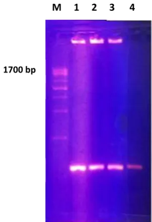
Figure 1. The Electrophoresis results of PCR BCSP-31
Brucella abortus
M= Marker; 1,2,3=Brucellosis abortus field isolates;4 = positive control
The sequencing result of gene Surface Protein-31 (BCSP-31) Brucella abortus field isolate shows the nucleotide sequence as follows:
TCTTGCACATCACTTCGGGCGATACAGGACTCCGGCCTTTACGCAGTCAGACGTTGCCTATTGGGC CTATAACGGCACCGGCCTTTATGATGGCAAGGGCAAGGTGGAAGATTTGCGCCTTCTGGCGACGCT TTACCCGGAAACGATCCATATCGTTGCGCGTAAGGATGCAAACATCAAATCGGTCGCAGACCTGA AAGGCAA
The homology analysis then be done by using the data in Gene Bank using the BLAST methods, the result is that the gene Surface Protein-31 Brucella abortus field isolate shows the homology for about 97% with some other strain Brucella abortus provided in the Gene Bank, including the Brucella abortus S19 ( Table 1 ).
Table 1:
Brucella abortus
sequences that produce higest significant alignments with the BCSP-31
gene of the
B
.
abortus
field isolate using nucleotide BLAST.
No.
Accession No.
Description
Query Coverage
Max.
Identify
1.
CP 003176.1
Brucella abortus
A13334 chromosome 1
93%
97%
2.
HO 132292.1
Brucella abortus
KOL-79 BCSP31
93%
97%
3.
O 132291.1
Brucella abortus
AHM-900 BCSP31
97%
97%
1700 bp
1200 bp
1000 bp
500 bp
224bp
227 | P a g e
4.
GO 167387.1
Brucella abortus
BMA
2008 BCSP31
97%
97%
5.
CP 000887.1
Brucella abortus
S19
chromosome1
97%
97%
IV.
CONCLUSION
The Gene Surface Protein-31 (BCSP-31) Brucella abortus field isolate can be characterized has nucleotide lenght of 224 bp with level of homology 97%.
REFERENCES
[1] Office de Intenational Epizooties (OIE), 2009, Mannual of Standards for Diagnostic Testand Vaccine for Terrestial Animals, References Laboratories for Bovine Brucellosis, www.unau.es/microbial/Brucellosis 2003 proceeding.pdf.
[2] D.H Kim, B.G Son; J.J. Lim; J.J. Lee; D.G. Kim; H.J. Lee; W. Min; M.H. Rhee; K.D. Kim; H.H Chang and S. Kim, 2013, The Role of Brucella abortus Lipoprotein in intracellular Replication and Pathogenecity in Experimentally Infected Mice,Microbial Pathogenesis 54(2013) 34-39
[3] Annonymous,2009, Penyakit Keluron Menular (Brucellosis), Dinas Peternakan Provinsi Jatim, Surabaya( write in Bahasa Indonesia)
[4] J. S Shamira, L. Hongseok Tae, Sarmead., D. Preston, L. Meluer, C. Franck, A. Dickerman, L. G. Adams and H.R. Garrier. 2012. Comparism of Genome Diversity of Brucella spp. Field isolates using Universat Bio-Signature Detection Array based Whole Genome Sequencing reveals limitations of Current Diagnostic Methods. ELSEVIER Gene 509 pp. 142 – 147.
[5] E Jawetz, J.L.Melnick and E.A. Adelberg, 2002. Mikrobiologi untuk Profesi Kedokteran.ECG. Penerbit Buku Kedokteran. Jakarta.
[6] G.G Alton, L. M. Jones, R.D. Angus and J.M. Verger, 1988. Techniques for the Brucellosis Laboratory. INRA., Paris, France.
[7] P.J.Quinn, B.K.Markey, M.E Carter, W.J.Donnelly and F.C.Leonar. 2002. Veterinary Microbiology and Microbial Disease. Blackwell Publishing. Great Britain.
[8] I.A. Merchant, and R. A. Packer. 1971. Veterinary Bacteriology and Virology. 7th Ed 3rd printing. The Iowa State University Press, Ames, Iowa. USA
[9] M.Asif, A.R. Awan, M.E. Babar, Ahmad Ali, S. Firyal and Q. M. Khan. 2009. Development of Genetic Marker for Molecular detection of Brucella abortus. Pakistan J. Zool. Suppl. Ser. 9 : 267-271 [10] Al-Attas, R.A., M. Al-Khalifa and A.R. Al-Qurashi. 2000. Evaluation of PCR, culture and serology for