Available Online atwww.ijcsmc.com
International Journal of Computer Science and Mobile Computing
A Monthly Journal of Computer Science and Information Technology
ISSN 2320–088X
IMPACT FACTOR: 6.199
IJCSMC, Vol. 8, Issue. 5, May 2019, pg.126 – 131
DIABETIC RETINOPATHY
DETECTION USING DEEP
NEURAL NETWORK
Akhila T
1; Ambarish A
2; Unnikrishnan S Kumar
3 ¹M.Tech Scholar, MCET, Thrissur, Kerala²Asst. Prof (CSE Department), MCET, Kerala
3
Asst. Prof (CSE Department), MCET, Kerala
1
[email protected]; 2 [email protected]; 3 [email protected]
Abstract– Diabetic Retinopathy (die-uh-BET-ik ret-ih-Nop-uh-thee) is a diabetic complication that effects eyes. It is caused by damage to the blood vessels of the light – sensitive tissues at the back of eye (retina). The condition can developed in anyone who has type 1 or type 2 diabetes. This paper focus on a desktop application that will help you to the identification of diabetic retinopathy. The screening occur in real time. The application can be developed using a tensor flow deep neural network architecture. Here it is trained and tested more than thousands of images. During the creation of deep neural network we will create five layers, 2 pool layer and 2 convolution layer and one fc layer. Fc layers are used to detect specific global configurations of the features detected by the lower layers in the net. In this model there are two options for screening that are one for image screening and one for real time screening. For this desktop application there is no need of internet connection for its working and it can be used as an easy manner.
Keywords: Diabetic Retinopathy, tensor flow, pool layer, convolution layer, FC layer
I. INTRODUCTION
Diabetic Retinopathy (DR) is the disease affecting blood vessels in the retina where capillary vessels in
particular are vulnerable to high glucose levels caused by diabetes. DR is the most common complication of diabetes
and the leading cause of blindness. The most significant predictor of the prevalence of DR is the duration of the
diabetes [8]. Patients with diabetes are at risk of developing retinal micro vascular complications that can cause vision
loss, and indeed, diabetes is the leading cause of incident blindness among the working age population. Early detection
of Diabetic Retinopathy will help to stop the leading to blindness [9]. Annual screening of the retina is recommended
but presents a huge challenge, given that the global prevalence of diabetes was estimated to be 9% among adults in
2015. If detect early enough, effective treatment of Diabetic Retinopathy is a strong area of research [7].
Convolutional Neural Networks (CNNs), a branch of deep learning, have a large record for applications in image
data were routinely built already before many years with many helpful applications and surpassed other approaches to
many highly challenging tasks like handwritten character recognition [3]. However, it is not until several breakthroughs
in neural networks such as the implementation, rectified linear units of them and the increase in computing power
through graphical processor units that they become viable for more complex image recognition problems.
In this application we categorize the images as the images which effected with Diabetic Retinopathy and images
that doesn‟t effected with Diabetic Retinopathy. Here we carried out to operations that are training and testing of
images [4]. Once we did it we can identify the effecting of Diabetic Retinopathy in the given image. Here we can do
two operations with the desktop applications that are check the occurrence of Diabetic Retinopathy in a given image
and also the real time screening through the camera.
II. METHODOLOGY
The methodology for this concept will consist of mainly five steps. The first step is the Database collection then
preprocessing the third step is Feature Extraction after that Training and final step is Testing. These steps can be
described as below:
DATASET COLLECTION
It is necessary to collect the required image for training. Diabetic Retinopathy dataset with annotation and images are
collected during this step.
PREPROCESSING
Preprocessing will help to filter the images in the dataset and avoid noises. This organization of image data helps in
training the neural network. Once the data is organized it is ready for preprocessing which involves two steps [6]:
Filtering: To make the images more smooth and reliable a convolution filter is used. The images in the dataset will
consist of some noises such as color variations, shadows etc. the step of filtering will help to avoid these type of noises.
Conversion: The step of conversion will included that the conversion of size. That is in a single word „resizing‟. Here
it is resizes all the images in the dataset into 256X256 pixels [10].
FEATURE EXTRACTION
Each class will contain certain features, so the feature of each class must be extracted and separated. The images will be
divided into five layers in this step. That is 2 pool layers, 2 convolution layer and one FC layer.
TRAINING
In the training phase we train out machine learning model with the features and the system will generate a knowledge
base.
TESTING
It is the final step of the methodology. Here the system classifies the new input with the knowledge bases.
Implementing convolutional neural network has become a popular method in the biomedical field. Furthermore,
the neural network can be used in detecting brain tumors and analyzing x-ray images. The application is powered by a
tensor flow deep neural network architecture that is trained and tested on 16,798 fundus images. These images are
preprocessed to remove noise and prepare them to be fed into neural network [5][11]. Preprocessing steps involve
averaging all the images using a 5x5 filter to improve the quality of images and then these images are resized to
The developed desktop application was tested in real time on test dataset images. Since the test dataset contains
images of both categories of Diabetic Retinopathy and no Diabetic Retinopathy, so it is used as source for real time
image analysis as one would be capturing image of an actual subject [12]. Once the process is done, for all the images
the same process will repeated.
We can also calculate some factors such as specificity, accuracy and sensitivity for all the images that we want
to be tested and the images that will capture at the time of real time screening.
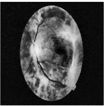
Figure 1: fundus image database set
Figure 2: no Diabetic Retinopathy Figure 3: Diabetic Retinopathy
III. DEEP NURAL NETWORK
Deep learning is a part of boarder family or machine learning methods based on artificial neural networks.
Learning can be supervised, semi-supervised or unsupervised. Deep neural network is mode of Deep learning architecture.
It is an artificial network [2]. These artificial network may be used for predictive modeling, adaptive control and
The utilized neural network architecture is based on Mobile Nets. This network is built on depth wise
convolution layers which are further divided into depth wise and point wise convolution, except for the first layer which is
a fully connected layer [1]. Depth wise convolution is used for applying a single filter on every input channel while point
wise convolution is used to form a linear combination of the output from the depth wise layer. There are two non-linearity
used: batch norm and ReLU after each layer.
Neural network process information in a similar way that the human brain does the network is composed of a large
number of highly interconnected processing elements working in parallel to solve a specific problem. It cannot be
programmed to perform specific tasks.
IV. RESULT
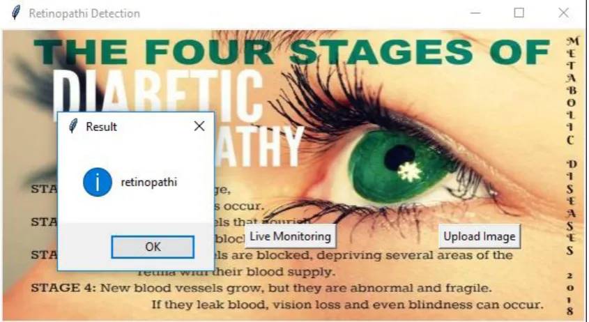
The result of the proposed system is that a message box that contains the alert that Retinopathy and no
Retinopathy. And there are two options for checking, one is for checking the presence of retinopathy in image that is
uploaded by the user, and another option is for real time screening by capture the image through the camera.
Figure 5: Result while uploading image
V. CONCLUSION
The described neural network model based Desktop application works well for identification of Diabetic
Retinopathy. The application makes use of the deep neural network architecture that is trained on thousands of images on
the collected dataset of images. Now the application only work for give an alert that the image of eye is effected with
Diabetic Retinopathy or not. We can develop a system with spotting the effected part of the given image. The system can
be developed easily using the python language. Rather than the desktop application, we can developed it also as an
REFERENCES
[1]Wang, D., Khosla, A., Gargeya, R., Irshad, H., & Beck, A.(2016). Deep Learning for Identifying Metastatic Breast
Cancer. ArXiv: 1606.05718[q-bio.QM].
[2]Abadi, Agarwal, Barham, Brevdo, Chen, Citro, Zheng. (2016). TensorFlow: Large-Scale Machine Learning on
Heterogeneous Distributed Systems.arXiv:1603.04467 [cs.DC].
[3]M. Abadi, A. Agarwal, P. Barham, E. Brevdo, Z. Chen,C. Citro, G. S. Corrado, A. Davis, J. Dean, M. Devin, et
al.Tensorflow: Large-scale machine learning on heterogeneous systems, 2015. Software available from tensorflow.
Org, 1, 2015. arXiv:1603.04467 [cs.DC].
[4]T. Tieleman and G. Hinton. Lecture 6.5-rmsprop: Divide the gradient by a running average of its recent magnitude.
COURSERA: Neural Networks for Machine Learning, 4(2),2012.
[5]K. Goatman, A. Charnley, L. Webster, S. Nussey, “Assessment of automated disease detection in diabetic
retinopathy screening using twofield photography,” PLoS One, vol. 6, no. 12, e27524, 2011.
[6]Philip S, Fleming AD, Goatman KA, Fonseca S, McNamee P, “The efficacy of automated disease/no disease
grading for diabetic retinopathy in a systematic screening programme”, Br. J. Ophthalmol., vol. 91, no. 11, pp.
1512–1517, 2007.
[7]Fleming AD, Goatman KA, Philip S, Williams GJ, Prescott GJ, “The role of hemorrhage and exudate detection in
automated grading of diabetic retinopathy”, Br. J. Ophthalmol., vol. 94, no. 6, pp. 706-711, 2010.
[8]Fleming AD, Goatman KA, Philip S, Prescott GJ, Sharp PF, “Automated grading for diabetic retinopathy: a
large-scale audit using arbitration by clinical experts”, Br. J. Ophthalmol., vol. 94, no. 12, pp. 1606-1610,2010.
[9] M. Lalonde, M. Beaulieu and L. Gagnon, "Fast and robust optic disc detection using pyramidal decomposition and
hausdorff-based template matching", IEEE Trans. Medical imaging, vol. 20, no. 11, pp.1193-1200, 2001.
[10] 17. S. G. Mallat, "A theory for multiresolution signal decomposition: The wavelet representation", IEEE Trans.
Pattern Anal. Machine Intell., vol. 11, pp.674-693, 1989.
[11] 18. Sharath Kumar P N, Rajesh Kumar R, Anuja Sathar, Sahasranamam V, “Automatic Detection of Exudates in
Retinal Images Using Histogram Analysis,” in IEEE International Conference on Recent Advances in Intelligent
Computational Systems (RAICS), pp. 277-281, December 2013.
[12] 19. Y. Hatanaka, T. Nakagawa, Y. Hayash, A. Fujita, Y. Mizukusa, M. Kakogawa, K. Kawase, T. Hara, and H.
Fujita, “CAD scheme for detection of hemorrhages and exudates in ocular fundus images,” in Proc. SPIE Medical