Alan D. Irvine, B.A., B.A.I.
Supervisor
Prof. Daniel J. Kelly
External Examiner Internal Examiner
Prof. Jan Herman-Kuiper Prof. Rocco Lupoi
Monday 19th June 2017
I declare that this thesis has not been submitted as as an exercise for a degree at this or any other university and it is entirely my own work.
I agree to deposit this thesis in the University’s open access institutional repository or allow the library to do so on my behalf, subject to Irish Copyright Legislation and Trinity College Library conditions of use and acknowledgement.
Alan Irvine
Signature
T
ISSUE engineering is a potential alternative to conventional surgical techniques forregenerating damaged tissues that lack the ability to spontaneously repair. MSCs are a promising cell source for tissue engineering strategies intended to treat a range of injuries and diseases. Within the synovial joint environment, a complex array of biochemical, environmental and biomechanical stimuli arise. If MSCs are to be successfully usedin vivo, understanding the effects on their differentiation and function in response to these stimuli is vital. A central hypothesis of this thesis is that oxygen availability and mechanical stimulation can direct chondrogenic differentiation and hypertrophy of MSCs. This thesis examines the response of MSC-laden hydrogels to oxygen availability, substrate stiffness and dynamic compressive strain. The overall objective is to develop the precursor to an osteochondral graft by mechanically modulating chondrogenesis and hypertrophy of MSCs within a 3-dimensional hydrogel environment. To enable this, this thesis first systemically explored the response of MSCs undergoing chondrogenesis to altered levels of substrate stiffness, dynamic compression and oxygen availability. The application of dynamic compression after 21 days at different magnitudes in 20 % O2to dynamic compression to MSC-laden agarose hydrogels
was found to alter calcification in a magnitude dependent manner. High magnitudes (20 % strain) tended to reduce the increases in calcification produced at moderate strains.
strains (10 %).
The next stage of the thesis explored tailoring substrate stiffness, oxygen availability and local dynamic compressive strain magnitude in a gradient fashion, with the hypothesis that a precursor to an osteochondral-like tissue could be produced. Varying substrate stiffness throughout the depth in these alginate hydrogels and applying dynamic compression lead to spatial changes in MSC differentiation, but failed to produce an effective osteochondral precursor. The final stage of this thesis involved impregnating a MSC-laden alginate hydrogel into a 3-D printed polymeric scaffolds with a gradient profile. This created an environment that modulated local strain magnitude alone. Spatial differences in MSC chondrogenesis in gradient mesh constructs alone were observed. GAG production was enhanced in regions of low strain and hypertrophy was reduced in areas of high strain.
M
Ysincerest gratitude goes to Prof. Daniel Kelly for his guidance over the past four years.It has been privileged to work with someone who trusts your time as your own, yet also has great expectations and aspirations for each individual’s research. Thank you for providing me with this opportunity to undertake such a challenging experience.
Many thanks to Prof. Conor Buckley, whose support and mentorship from the onset has both encouraged me to grow and learn, as well as mould my technical abilities in the lab.
I have enjoyed working with everyone in the Trinity Centre of Bioengineering. Thanks especially to Grainne for her constant help in performing general lab procedures and protocols as well as other post-doctoral research fellows throughout my time including Olivier, Raja and particularly Binu, whose guidance in rt-PCR has been invaluable. Another thank you goes to Simon for instructing me through much of my technical engineering work from the onset of this project. Further thanks go to the rest of the lab.
Thanks to everyone in the workshop, Michael Reilly and Paul Normoyle for technical assistance in manufacturing and computation respectively.
Finally, I could not overlook the huge support from my very patient parents whose unwavering support helped make this possible.
Declaration i
Abstract iii
Acknowledegments v
List of Figures xv
List of Tables xix
Nomenclature xx
1 Introduction 1
1.1 Joint damage and degeneration . . . 1
1.2 MSC based osteochondral tissue engineering . . . 3
1.3 Project aims and objectives . . . 5
2 Literature Review 13 2.1 Composition, structure and mechanical properties of cartilage . . . 13
2.1.1 Composition and structure of articular cartilage . . . 13
2.1.2 Biomechanical properties of articular cartilage . . . 17
2.1.3 In Vivo deformation of articular cartilage . . . 18
2.2 Articular cartilage injury . . . 19
2.3.4 Hydrogels supporting MSC chondrogenesis . . . 28
2.3.4.1 Agarose hydrogel . . . 29
2.3.4.2 Alginate hydrogel . . . 30
2.4 Hypertrophy in cartilage tissue engineering . . . 32
2.4.1 Characterisation of chondrogenic hypertrophy . . . 33
2.4.2 Factors affecting chondrogenic hypertrophy . . . 34
2.4.3 Trophic effects of MSCs . . . 35
2.5 Oxygen tension as a regulator of MSC chondrogenesis . . . 38
2.5.1 Oxygen tension affecting MSC hypertrophy and endochondral ossification . . . 39
2.6 Cell shape . . . 41
2.6.1 Regulating myogenic and chondrogenic differentiation . . . 41
2.6.2 Regulating osteogenesis and adipogenesis differentiation . . . 44
2.6.3 Summary . . . 46
2.7 Substrate stiffness . . . 47
2.7.1 2-D culture . . . 47
2.7.2 3-D culture . . . 48
2.7.3 Summary . . . 53
2.8 Dynamic compression as regulator of chondrogenesis . . . 54
2.8.1 Chondrocyte response . . . 55
2.8.2 Initiation and progression MSC chondrogenesis . . . 55
2.8.3 MSC hypertrophy and endochondral ossification . . . 58
2.8.4 Summary . . . 61
2.9 Cellular mechanics as a result of mechanical cues . . . 62
2.10 Summary . . . 62
3.2 Introduction . . . 68
3.3 Materials and Methods . . . 70
3.3.1 Cell isolation, expansion and hydrogel encapsulation . . . 70
3.3.2 Dynamic compression application . . . 71
3.3.3 Mechanical testing . . . 72
3.3.4 Biochemical content . . . 74
3.3.5 Histology and immunohistochemistry . . . 74
3.3.6 Statistical analysis . . . 75
3.4 Results . . . 76
3.4.1 Agitation of the culture media within a bioreactor system may suppress chondrogenesis of MSCs . . . 76
3.4.2 Dynamic compression enhances the functional development of cartilage grafts engineered using BMSCs . . . 78
3.4.3 Intermediate levels of DC promote hypertrophy and calcification of engineering cartilage . . . 80
3.4.4 Increases in stiffness correlate with enhanced calcium accumulation in mechanically stimulated engineered tissues . . . 82
3.5 Discussion . . . 83
3.6 Conclusion . . . 85
4 Modulating both oxygen levels and substrate stiffness to regulate chondrogenesis and hypertrophy of MSCs 87 4.1 Abstract . . . 87
4.2 Introduction . . . 88
4.3 Materials and Methods . . . 90
reaction (rt-PCR) . . . 92
4.3.5 Biochemical content . . . 93
4.3.6 Histology and immunohistochemistry . . . 93
4.3.7 Statistical analysis . . . 95
4.4 Results . . . 96
4.4.1 Altering substrate stiffness in alginate hydrogels by varying ionic crosslinking concentration . . . 96
4.4.2 Soft hydrogels provide enhanced biological cues for chondrogenesis 97 4.4.3 Stiff hydrogels promote the development of a more mechanically functional cartilage tissue . . . 98
4.4.4 Both external oxygen levels and hydrogel stiffness influence spatial tissue deposition . . . 101
4.5 Discussion . . . 103
4.6 Conclusion . . . 105
5 The effect dynamic compression has on MSC chondrogenesis is dependent on substrate stiffness 107 5.1 Abstract . . . 107
5.2 Introduction . . . 108
5.3 Materials and Methods . . . 109
5.3.1 RGD incorporation and alginate preparation . . . 109
5.3.2 Cell isolation, expansion and hydrogel encapsulation . . . 110
5.3.3 Biochemical content . . . 111
5.3.4 Histology and immunohistochemistry . . . 112
5.4 Results . . . 116
5.4.1 Soft hydrogels enhance early chondrogenesis . . . 116
5.4.2 Dynamic compression does not enhance chondrogenesis of MSCs encapsulated in soft hydrogels . . . 119
5.4.3 Dynamic compression enhances chondrogenesis in stiff hydrogels while regulating hypertrophy in a strain dependent manner . . . . 121
5.5 Discussion . . . 123
5.6 Conclusion . . . 124
6 The effect of modulating mechanical cues and oxygen availability throughout a single hydrogel construct on MSC chondrogenesis and hypertrophy 127 6.1 Abstract . . . 127
6.2 Introduction . . . 128
6.3 Materials and Methods . . . 129
6.3.1 RGD incorporation and alginate preparation . . . 129
6.3.2 Cell isolation, expansion and hydrogel encapsulation . . . 130
6.3.3 Biochemical content . . . 132
6.3.4 Histology and immunohistochemistry . . . 132
6.3.5 Strain mapping using cell-tracking and digital image correlation (DIC)133 6.3.6 Dynamic compression application . . . 134
6.3.7 RNA isolation and real-time reverse transcriptase polymerase chain reaction (rt-PCR) . . . 134
6.3.8 Statistical analysis . . . 135
6.4 Results . . . 137
6.4.3 Late MSC chondrogenesis is enhanced in stiff hydrogels . . . 140
6.4.4 Dynamic compression enhances late MSC chondrogenesis in stiff hydrogels . . . 142
6.5 Discussion . . . 146
6.6 Conclusion . . . 148
7 Integrating a bilayered 3-D printed mesh into MSC-laden hydrogels to spatially modulate mechanical cues, chondrogenesis and hypertrophy 149 7.1 Abstract . . . 149
7.2 Introduction . . . 150
7.3 Materials and Methods . . . 151
7.3.1 RGD incorporation and alginate preparation . . . 151
7.3.2 Scaffold mesh 3-d bioprinting . . . 152
7.3.3 Cell isolation, expansion and hydrogel encapsulation . . . 153
7.3.4 Dynamic compression application . . . 154
7.3.5 Biochemical content . . . 156
7.3.6 Histology and immunohistochemistry . . . 156
7.3.7 Statistical analysis . . . 157
7.4 Results . . . 158
7.4.1 Scaffold design influences mechanical properties and the local strain environment within composite alginate-PCL constructs . . . 158
7.4.2 Dynamic compression enhances MSC chondrogenesis in constructs containing a mesh gradient . . . 161
7.6 Conclusion . . . 168
8 Discussion 169 8.1 Overview . . . 169
8.2 Modulators of MSC chondrogenesis . . . 171
8.2.1 Substrate stiffness . . . 171
8.2.2 Magnitude of dynamic compression . . . 173
8.2.3 Oxygen availability . . . 173
8.3 Modulators of MSC hypertrophy . . . 174
8.3.1 Magnitude of dynamic compression . . . 174
8.4 Experimental considerations . . . 174
8.4.1 Magnitude of dynamic compression . . . 174
8.4.2 Substrate Stiffness . . . 175
8.4.3 Oxygen availability . . . 175
8.4.4 Biomaterial choice . . . 176
8.4.5 Time points . . . 176
9 Conclusion 179 9.1 Main Results . . . 179
9.2 Future Directions . . . 180
Bibliography 183 Appendix A: Bioreactor Design 213 A.1 Dynamic Compression Bioreactor . . . 213
A.2 Design . . . 215
A.2.1 Rig . . . 215
A.2.3 Bioreactor Dish . . . 221
A.2.3.1 Base . . . 222
A.2.3.2 Walls . . . 223
A.2.3.3 Lid . . . 224
A.2.3.4 Platen . . . 225
A.2.3.5 Guides . . . 226
A.3 Sterilisation . . . 227
A.4 Viability Study Methods . . . 228
A.4.1 Cell isolation, expansion and culture . . . 228
A.4.2 Tri-potentiality . . . 229
A.4.3 Dynamic compression application . . . 229
A.4.4 Biochemical content . . . 230
A.5 Viability Study Results . . . 231
A.5.1 Tri-potentiality . . . 231
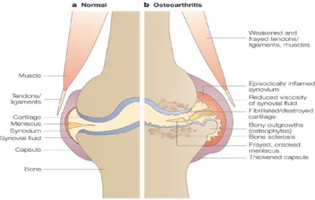
1.1 Knee anatomy in normal and osteoarthritic states. . . 3
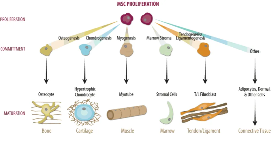
1.2 Differentiation pathways of stem cells derived from the mesenchyme. . 5
1.3 Full schematic of all thesis sub-objectives. . . 7
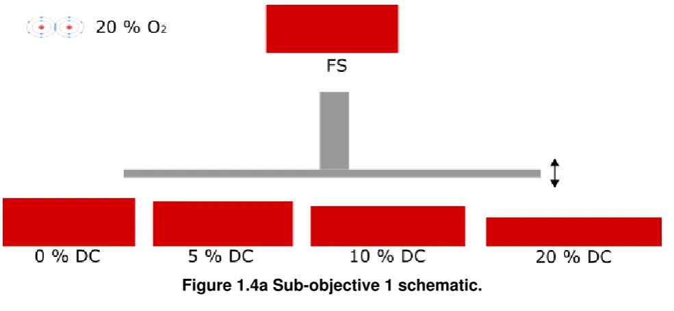
1.4a Sub-objective 1 schematic. . . 8
1.4b Sub-objective 2 schematic. . . 9
1.4c Sub-objective 3 schematic. . . 10
1.4d Sub-objective 4 schematic. . . 11
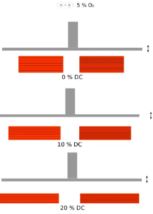
1.5 Modulating environmental cues to engineer MSCs with an osteochondral fate. . . 12
2.1 3-D Representation of the ultrastructural arrangement of the collagen network throughout the depth of articular cartilage. . . 16
2.2 The traditional triad approach of tissue engineering. . . 23
2.3 Defined events in chondrogenic differentiation of mesenchymal stem cells. 25 2.4 Schematic of the successive steps of the chondrocyte differentiation pathway. . . 28
2.5 Cell shape mediated changes in differentiation using micro-patterned islands. . . 42
3.3 The influence conventional 10 % strain DC magnitude has on chondrogenesis in comparison to 0 % agitated controls. . . 79 3.4 Effect different magnitudes of loading have on the progression of MSC
chondrogenesis in cartilage grafts. . . 81 3.5 Correlating increases in equilibrium modulus with calcium deposition. . 82
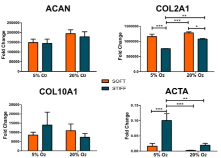
4.1 Altering substrate stiffness of alginate hydrogels by varying molarity of calcium chloride. . . 96 4.2 Soft hydrogels promote enhanced chondrogenic gene expression after
14 days of culture in chondrogenic conditions. . . 98 4.3 Biochemical content of MSCs in soft and stiff alginate hydrogels cultured
at 5 % or 20 % O2 after 28 days in TGF-β3 culture conditions. . . 100
4.4 Histological view of tissue produced in soft and stiff alginate hydrogels cultured at 5 % or 20 % O2after 28 days in TGF-β3. . . 102
5.1 Soft hydrogels are more chondrogenic in short term culture than stiff MSC laden hydrogels . . . 117 5.2 Soft hydrogels promote enhanced chondrogenic gene expression after a
further 5 days of culture in FS-mixed bioreactor conditions. . . 118 5.3 MSCs fail to respond to dynamic compression in soft hydrogels . . . 120 5.4 MSC chondrogenesis is enhanced by dynamic compression in stiff
hydrogels while hypertrophy is strain dependent . . . 122
6.1 Schematic of experimental design . . . 131 6.2 Altering substrate stiffness in MSC laden hydrogels . . . 138 6.3 Digital image correlation analysis (DIC) to determine effect of substrate
throughout the depth in all hydrogels . . . 145
7.1 Ideal scaffold design and geometry. . . 153
7.2 Schematic of study design. . . 155
7.3 Stereomacroscopic characterisation of both mesh designs . . . 159
7.4 Digital image correlation analysis (DIC) to determine effect of different meshing designs on local strain magnitudes . . . 160
7.5 Gradient meshed constructs have a greater chondrogenic response to dynamic compression . . . 162
7.6 Visual representation of tissue content within MSC-laden hydrogel-mesh constructs subjected to 15 % dynamic compression . . . 163
7.7 Calcium content of MSC-laden MSC-laden, gradient meshed or homogenous meshed, RGD-alginate hydrogels subjected to dynamic compression . . . 165
7.8 Differences in tissue composition of loaded alginate-PCL constructs . . 166
A.1 Original bioreactor system developed in 2012 during an undergraduate final year project. . . 213
A.2 Solidwork 3-D model of compression bioreactor. . . 214
A.3 Solidwork 3-D model of compression bioreactor rig assembly used to place and hold parts. . . 215
A.4 Technical drawings for the back of the rig frame. . . 216
A.5 Technical drawings for the frame base. . . 217
A.6 Technical drawings for the actuator holding section of the rig frame. . . 218
A.7 Zaber NA08A30 linear, motor-driven actuator. . . 219
A.8 Matlab controller and script section. . . 220
A.13 Technical drawings for the compressing platen that was magnetically coupled to the actuator-load cell assembly. . . 225 A.14 Technical drawings for the guide posts to direct motion of the platen by
preventing possible rotation. . . 226 A.15 Bioreactor sterilised and within incubator duringin vitroculture. . . 227 A.16 Histological content of MSC cultured in pellet and monolayer form with
or without certain growth factor incorporation to assess stemness. . . . 232 A.17 Biochemical content of alginate hydrogels within the bioreactor culture
2.1 Summary of literature findings on MSC differentiation in response to cell
shape. . . 46
2.2 Summary of literature findings on MSC differentiation in response to substrate stiffness. . . 53
2.3 Summary of literature findings on MSC differentiation in response to dynamic compression. . . 61
4.1 Primer sequences used for rt-PCR. . . 94
5.1 Primer sequences used for rt-PCR. . . 115
g 1 gram is a unit of mass. It is equivalent to mass that 1 one-thousandth cubic metre of water holds.
kP a 1 Pascal is a unit of pressure that refers to the weight of 1 Newton acting over a 1 square metre area. 1 kPa refers to 1000 Pascals (Pa) of pressure.
Introduction
1.1
Joint damage and degeneration
A
RTICULAR cartilage is a complex tissue that lines the edges of bones in diarthrodialjoints (fig. 1.1). Defects in cartilage can arise either through traumatic overloading of the joint (e.g. sporting injuries) or pathogenically through a degenerative disease known as osteoarthritis (OA) [1,2]. The lesions that arise from such defects have adverse structural and biological effects that cause thinning of the tissue and a loss of mechanical integrity. To compound the issue, cartilage is avascular in nature as well as having a highly organised structure that make both natural and artificial repair of these defects difficult [3,4]. The low grade synovial inflammation (synovitis) that also occurs in both the early and late phases of OA also contributes to the progression of the disease. This induces a vicious circle that promotes further joint degradation.
may be coupled with micro-fracturing of the subchondral bone layer in areas of partial or total cartilage loss. These micro punctures attempt to invoke a natural repair response by allowing blood to inflitrate from the underlying bone tissue and marrow. A more invasive method of repairing chondral and osteochondral defects is through the use of allografts and autografts. Mosaicplasty involves taking a number of osteochondral autografts from a donor site that are then re-implanted in the affected areas. Allografts from other patients can also be used [7]. Despite short-term success, all the aforementioned techniques have efficacy concerns relating to undesirable outcomes such as periosteal hypertrophy, delamination of the implant, arthofibrosis, disease transmission, immune rejection and ultimately implant or tissue regeneration failure [8]. In the case of microfracture, the repair tissue also tends to consist of fibrous cartilage, which does not have the biomechanical competence of the hyaline type that lines articulating joints.
The concern surrounding previous joint repair and regeneration procedures has lead to extensive research into alternative methods that either attempt to engineer or regenerate the tissue using novel approaches. Currently approved tissue engineering approaches include autologous cartilage implantation (ACI) and matrix-assisted ACI (MACI) [9]. In these procedures cartilage cells (extracted and expanded from the patient), termed chondrocytes, are surgically placed in the defect site; either alone, or within a biocompatible matrix material. Despite success in regenerating largely hyaline like cartilage, these approaches have associated limitations; an additional defect site is created in the patient on a non-load bearing cartilage region so that chondrocytes can be isolated from this tissue. Equally, there is an inability to obtain sufficient numbers of differentiated chondrocytes from this cartilage tissue, particularly in elderly and osteoarthritic patients.
osteochondral tissue [12]. The ability to control the differentiation of MSCs into specialised cell types will be central to unlocking their regenerative potential.
Figure 1.1 Knee anatomy in normal and osteoarthritic states. Taken from https://sciencebasedmedicine.org/.
1.2
MSC based osteochondral tissue engineering
MSCs have an affinity to differentiate into cell types specific to certain tissues such as cartilage and bone, which make them a promising cell source for osteochondral tissue engineering. Transforming Growth Factor-β3has been shown to promote chondrogenesis
of MSCs[10,11,13–15]. TGF-β3 primed MSCs are therefore widely used for cartilage
therapies is the prevention of terminal differentiation in which chondrocytes, differentiated from MSCs, become hypertrophic, increase in size and progress along the endochondral pathway before terminally differentiating into bone forming cells [16,17]. This apparent obstacle in MSC-based articular cartilage tissue engineering has recently been realised as a potential advantage in both endochondral bone tissue engineering and osteochondral tissue engineering [18–20]. A means by which chondrogenesis and endochondral ossification could be modulated would potentially allow the formation of both stable cartilage, or endochondral bone; through either inhibiting, or promoting the endochondral pathway (EC) respectively. This opens the possibility of spatially directing MSCs towards either a stable hyaline phenotype or along an EC pathway to engineer osteochondral tissues by managing the potential shift from cartilage to endochondral bone (fig. 1.2).
Having established endochondral ossification as a promising approach for engineering osteochondral grafts, the challenge becomes spatially modulating the chondrogenic differentiation of MSCs. Current approaches to altering chondrogenic hypertrophy include the use of different oxygen culturing conditions [21] and biomechanical stimuli, such as substrate stiffness [22] and dynamic compression [23]; among other more biochemically focused approaches that will not be considered as part of this thesis.
shown that osteogenic genes were upregulated when osteoblasts were subjected to 10 % dynamic compressive strain [33]. This suggests DC may have the potential to either enhance or suppress hypertrophy, possibly depending on the magnitude of the applied stimulus mimicking the differences in dynamic compressive strain within the respective tissues during articulation. Therefore the question remains how can oxygen tension, substrate stiffness and dynamic compression be combined and spatially regulated to modulate MSC chondrogenesis and hypertrophy in order to develop a strategy for engineering a viable osteochondral grafts.
Figure 1.2 Differentiation pathways of stem cells derived from the mesenchyme. Taken from https://www.rndsystems.com/resources/.
1.3
Project aims and objectives
initiation of chondrogenesis, and more recently have been implicated in the progression of MSC chondrogenesis towards either stable, hyaline-like cartilage or terminally-differentiated, hypertrophic cartilage that leads to bone formation through endochondral ossification. Mechanical stimuli are clearly a determining factor in both processes so the correct combination of these may alone provide a means of engineering an osteochondral graft from an engineered cartilage template.
Figure 1.3 Full schematic of all thesis sub-objectives.
1. Investigate the role that the magnitude of dynamic compression has on MSC chondrogenesis and progression along the endochondral pathway.
2. Determine the role of substrate stiffness and oxygen availability on the initiation and progression of chondrogenesis in MSCs.
3. Explore the interaction between substrate stiffness and dynamic compression on the initiation and progression of MSC chondrogenesis.
4. Biofabricate MSC-laden constructs with spatially defined mechanical properties to regulate MSC fate and promote the development of tissues destined for an osteochondral fate.
Figure 1.4d Sub-objective 4 schematic.
The overall aim being to spatially modulate the environmental cues developed within MSC-laden hydrogels in order to achieve the thesis global objective. Using a configuration such as the following:
Figure 1.5 Modulating environmental cues to engineer MSCs with an osteochondral fate.
Literature Review
2.1
Composition, structure and mechanical properties of
cartilage
A
RTICULAR cartilage is a complex tissue consisting of an intricate anatomical structure. This structure provides the means by which cartilage can withstand its challenging biomechanical environment. Therefore there is a need to understand the composition and anatomical structure of articular cartilage as well as the biomechanical properties of the tissue.2.1.1
Composition and structure of articular cartilage
The sparsely distributed cells in articular cartilage; named chondrocytes, account for less than 10 % of the tissue volume. This equates to a cell density of the order of five million per cm cubed in comparison to the typical density of a few hundred million per cm cubed found in other tissues throughout the body [34].
content from 3 to 10 %, both by wet weight; the remaining 60 to 87 % comprises water, inorganic salts, and other small quantities of different matrix proteins, glycoproteins, and lipids [4]. Collagen fibrils and PGs are the structural components supporting the mechanical forces developed from applied loads [37,38]. It is the combination of these components with the interstitial fluid pressure produced by the containment of water in a tight organic matrix that provide the biomechanical properties of articular cartilage.
Collagen, the most abundant protein in the body, has a high level of structural organisation in articular cartilage that provides a fibrous ultrastructure for the tissue [4,35,39]. In general, this ultrastructure is heterogeneous in collagen fibril distribution, but layers within the tissue have been identified with a relatively homogenous distribution of these fibrils. The variation in collagen fibre orientation is mirrored by zonal variations of collagen content (fig. 2.1), which is greatest at the surface and remains relatively constant throughout the deeper zones [40]. This ultrastructure serves greatly in enhancing the biomechanical properties of articular cartilage by maximising interstitial fluid pressure support and minimising solid matrix stress within the tissue; the former being very important as collagen fibrils alone exhibits little resistance to compression due to their high slenderness ratio [41,42]. The fibrils can, however, resist high tensile loads, which again aid in the biomechanical behaviour of characteristics [43].
Cartilage PGs are large protein-polysaccharide molecules consisting of a protein core to which one or more glycosaminoglycans (GAGs) are attached [36]. Aggrecan consists of an approximately 200mm long protein core to with about 150 GAG chains are covalently attached to [36]. Keratan sulphate and chondroitin sulphate, the two sulphated GAGs found in articular cartilage, are polymer sequences of specific repeating disaccharide units.
promotes immobilisation of the PGs within the collagen network, adding structural rigidity to the extracellular matrix [36,44,45].
Figure 2.1 3-D Representation of the ultrastructural arrangement of the collagen network throughout the depth of articular cartilage. In the superficial tangential zone (STZ), collagen fibres are tightly woven into sheets arranged parallel to the articular surface. In the middle zone (MZ), randomly arranged fibrils are less densely packed to accommodate the high density of proteoglycans and water. The collagen fibrils of deep zone (DZ) form larger, radially orientated fibre bundles which cross the tidemark, enter the calcified zone (CZ), and anchor the tissue to the underlying bone (SB).
When a stress is applied to the cartilage surface, deformation occurs primarily due to a change in the PG molecular domain. A sufficient external stress can cause an internal matrix pressure to exceed the swelling pressure, and liquid begins to exude from the tissue. As the fluid flows out the PG concentrations increases; this results in an increase in osmotic pressure, charge-charge repulsive force and compressive stress until equilibrium is achieved with the external stress. In addition, a change in a solution’s pH or ion concentration will alter the PG intermolecular charge repulsive forces causing a change in the size of the aggregate domain. Therefore the osmotic swelling pressure associated with fixed charge density due to the ionic groups in the GAGs, as well as the bulk compressive stiffness of the PG aggregates trapped in the collagen network, are the two physiochemical properties of the PG that allow it to resist compression and complement the role in tension played by the collagen network [43,58].
2.1.2
Biomechanical properties of articular cartilage
Cartilage is defined as a viscoelastic material meaning that the mechanical response of the tissue to a constant load or deformation varies with time. Therefore a modelling the response of articular cartilage to mechanical loading must account for the combination of the viscous fluid and the elastic solid that encompass cartilage.
The two fundamental responses of a viscoelastic material under mechanical loading are creep and stress relaxation. The action of either a constant load or deformation determines creep or stress relaxation respectively. Generally, a rapid initial deformation followed by a slow (time-dependent) and progressively increasing deformation, known as creep, is the response of a viscoelastic solid until equilibrium is reached. In the instance of applying a constant deformation, a viscoelastic solid responds with a high initial stress followed by a slow and progressively decreasing stress required to maintain the deformation.
behaviour of cartilage in response to interstitial fluid flow. The component due to macro-molecular motion is known as flow-independent (or intrinsic) and viscoelastic behaviour of the collagen-PG matrix [58].
Creep in articular cartilage is caused by exudation of the interstitial fluid. Shown in Figure is the biphasic creep response of articular cartilage in a one-dimensional confined compression test. The graph clearly highlights that following a constant force initiated at time t0, articular cartilage creeps until it reaches its equilibrium value; at which point the force is removed. Creep creases once the compressive stress within the solid matrix is enough to support the applied stress alone. Since the rate of creep is governed primarily by the rate of fluid exudation, it can be used to determine the permeability coefficient of the tissue. At equilibrium, no fluid flow occurs, indicating that the collagen-PG matrix intrinsic compressive modulus can be isolated from the equilibrium deformation. This has been found to range from 0.1 to 2.0 MPa [58].
Stress relaxation occurs when the stress begins to fall following an increase during a constant displacement rate of the tissue until a deformation d0 is reached. During this time, the stress continuously decreases until equilibrium is reached. The stress rise in the compression phase is related to fluid exudation, while stress relaxation is related to fluid redistribution within the porous matrix. This stress relaxation ceases when stress within the solid matrix reaches the intrinsic compressive modulus as described above [58].
2.1.3
In Vivo deformation of articular cartilage
equivalent to body weight, e.g. 0.9 x body weight in the glen humeral joint during abduction [60].
In general, the loading regime in a di-arthrodial joint is cyclical and/or intermittent. The mean contact stresses produced by these loads during non-strenuous daily activities are in the region of 2 MPa, whilst strenuous activity can result in stresses approaching 6 MPa [62,65,66]. It is estimated that the largest contact stress in non-traumatic conditions is 12.6 MPa [67]; although in vivo measurements of femoral prostheses have measured contact stresses as high as 18 MPa [61]. These loads highlight the importance of the interstitial fluid in balancing these extremely high contact stresses. The high water content, low permeability and composition of articular allow this pressurization of the interstitial fluid in order to resist these large contact stresses.
Changes in cartilage thickness from 6 - 20 % have been reported for physiological loading levels of 1 to 5 x body weight [68]. This highlights the large intermittent mechanical environment that this tissue is subjected to and therefore must be of some consideration before either implanting inferior biomechanical constructs or cells that do not respond well to such stimulus.
2.2
Articular cartilage injury
The major need for cartilage tissue engineering, as outlined previously, is the inability of cartilage tissue to self-repair following injury. Defects in cartilage can arise either through overloading of the joint (e.g. sporting injuries) or pathogenically through a degenerative disease known as osteoarthritis (OA). Defects caused by such exist in three types; matrix disruption, partial thickness defects and full thickness defects.
Partial thickness defects (e.g. fissures, etc.) are disruption to the cartilage surface that do not penetrate the subchondral bone. For some unknown reason, nearby residing cells begin to proliferate immediately following the incident in a cellular attempt to fill the defect but terminate this activity before fully repairing the tissue.
Full thickness defects penetrate through the cartilage layer into the underlying subchondral bone. The normal wound healing response ensues as a fibrin clot fills this defect following damage. This injury is unique in the fact that it has access to bone marrow stem/progenitor cells, which can migrate to fill the defect [69,70]. The resulting repair tissue formed is generally an intermediary between fibro- and hyaline cartilage. The eventual degradation of this inferior tissue is thought to be due to its less stiff and more permeable characteristics [70].
Collectively it can be seen that each of these three defect types has an inability to fully repair in order to restore tissue function. Insufficient mechanical function of cartilage only leads to enhanced degeneration cartilage tissue and surrounding areas, such as the subchondral bone, during daily motion. This vicious circle of injury and degeneration is a major problem especially considering the lack of viable regenerative medicine strategies that exist to date.
This degenerative state and lack of tissue function causes severe pain during articulation of the joint and can seriously debilitate a person’s life. According to estimates from the National Institute of Arthritis and Musculoskeletal and Skin Diseases more than 20 million Americans currently suffer from OA [7]. Conservative estimates suggest that 35-40 million Europeans have OA. Statistical data from epidemiological studies suggest that arthritis is the number one condition associated with functional limitation and physical disability among US population aged 65 and older, in addition to affecting 30 % of the population [7]. It is expected that by 2030, 20 % of adults will have developed OA in Western Europe and North America. OA is an important cause of disability-adjusted-life years in both the developed and developing world [5]. Therefore, OA is expected to be a heavy economic burden on healthcare systems and community services in Europe, North America and the rest of the world as the population expands and the number of older people increases. 3 An effect means of solving this growing problem is therefore extremely important [7].
2.3
Cartilage tissue engineering
The development of a successful repair strategy for both chondral and osteochondral defects require the establishment of certain design criteria. Firstly, viable cells are needed to engineer the proteins necessary to recapitulate the structure and composition of articular cartilage or subchondral bone. Secondly, the proteins produced by these cells must be tissue and site specific in order to restore the structure of the required area. Thirdly, the ability to integrate firmly with the host tissue through either natural or artificial means is important. Finally, the strength to withstand the highly loaded, biomechanical environment of the joint is also a key factor.
In order to meet these design criteria, tissue engineering traditionally uses a triad combination of approaches:
• A 3-D biomaterial that provides a structural support and housing for the cells in which to form the desired tissue.
• Biochemical (growth factor) and/or mechanical (e.g. deformation) stimulus to encourage differentiation of the cells and the enhanced production of extracellular matrix.
Figure 2.2 The traditional triad approach of tissue engineering. Taken from http://www.mdpi.com/.
2.3.1
Mesenchymal Stem Cells (MSCs)
The discovery that non-haematopoeitic MSCs of the bone marrow selectively adhered to plastic allowed for their isolation from the non-adherent haematopoeitic cells present in the bone marrow. These cells, when expanded on plastic, exhibit a homogenous population of cells with a spread, fibrotic morphology, which can form colonies during 2-D monolayer expansion. This gave rise to the original term of marrow stromal fibroblasts: known today as either mesenchymal stem cells (MSCs), mesenchymal stromal cells (MSCs) or mesenchymal progenitor cells (MPCs). Debate still remains as to whether these cells can be appropriately called stem cells [80–82]. Regardless they still exhibit some potency and can be differentiated down the adipogenic (fat), osteogenic (bone), chondrogenic (cartilage) and myogenic (muscle) lineages.
Bone-marrow derived MSCs have therefore good proliferative potential and possess the ability to differentiate into a range of tissues. It has been stated that only 0.001 - 0.01 % of cells resident in adult bone marrow comprise MSCs. Identification of a range of surface markers allows the characterisation of these cells [11]. Various techniques, such as flow cytometry and magnetic bead isolation, allow for the isolation of cells from the marrow population with the relevant surface markers pertaining to MSCs. Many researchers, however, continue to use the plastic adherence technique to isolate MSCs from the other cell populations within bone marrow aspirates.
2.3.2
Chondrogenic growth factors
Chondrogenic differentiation can be directed by different growth factors or cytokines. These include bone morphogenic proteins (BMPs) [83], members of the transforming growth factor-β(TGF-β) superfamily [14,84], fibroblast growth factors (FGFs) that can be also be used to increase proliferative potential during expansion, insulin like growth factor (IGF-1), Indian hedhehog and others [85]. The most widely used is TGF-β1 or TGF-β3. A timescale
and 8 - 21 days, which relates to gene expression, protein synthesis, and both the synthesis and accumulation of glycosaminoglycan (GAG) [86]. While the use of growth factors to direct MSC chondrogenesis generally results in a reasonable cartilage template, it fails to meet the standards of chondrocyte laden biomaterials in terms of tissue deposition and mechanical properties [87]. Therefore further avenues, such as incorporating mechanical stimuli, need to be sought in order to enhance and improve the cartilage template produced.
Figure 2.3 Defined events in chondrogenic differentiation of mesenchymal stem cells determined by the sequential expression of matrix components during pellet culture. [86]
2.3.3
Chondrocyte fate and differentiation
this process, the chondrogenically committed MSCs express extracellular matrix and cell adhesion molecules such as N-cadherin, N-CAM (Ncam1), tenascin C (Tnc), versican, and thrombospondin-4. Expression of mesenchymal and condensation markers are turned off by prechondrocytes that emerge in the centre of these condensations and start to express Col2a1 and other early cartilage markers.
A strong increase in cell proliferation and deposition of cartilage matrix are key indicators that prechondrocytes have overtly differentiated into fully committed and active chondrogenic cells. Col2a1 expression is upregulated and a different splice variant of the gene is produced (type IIB instead of type IIA [91]. The cells also start to express Agc1, Cartl1, the genes for the collagen types IX and XI, and other cartilage extracellular matrix components at high levels. These cells are referred to as chondroblasts rather than chondrocytes as they rapidly proliferate and build new tissue, rather than limiting their activity to maintaining the functional integrity of mature cartilage tissue [92].
During development of long bones, early chondroblasts in the metaphyses start to form columnar zones once the cells in the diaphyses have undergone prehypertrophy and proceed toward terminal maturation. Initially small and round, they become flattened and organized into parallel, longitudinal columns. They proliferate at the highest rate at the top of the columns (away from the primary ossification center) and progressively decrease their proliferation rate as they move down the columns [93]. They undergo irreversible growth arrest as they convert into prehypertrophic chondrocytes, one layer at a time, at the bottom of the columnar zone. The cells in the diaphyses, however, at the prehypertrophic stage, they contain higher levels of RNA for Col2a1, Agc1, and most other early cartilage matrix genes than chondroblasts, and sequentially activate the genes for the parathyroid hormone and parathyroid hormone-related peptide receptor (Pthr1), Ihh, and Col10a1. At the hypertrophic stage, they cease to express early cartilage matrix genes and also terminate expression of Pthr1 and Ihh. They upregulate expression of Col10a1 and activate the gene for the vascular endothelial growth factor (Vegf). This large shift in cell fate, and the mechanical cues regulating this, will be discussed further in subsequent sections [92,93].
Figure 2.4 Schematic of the successive steps of the chondrocyte differentiation pathway as they occur during development of endochondral bones, highlighting major histological features, extracellular matrix markers, and regulatory markers expressed at each step.
2.3.4
Hydrogels supporting MSC chondrogenesis
biogradeable, non-toxic both initially and following enzymatic degradation, non-immunogenic and easily manufactured [94]. Scaffolds must have both high porosity and permeability to permit the transport of nutrients and waste to and from cells located at the scaffold core. Scaffolds must be able to withstand biomechanical loading.
Hydrogels are a commonly used class of scaffold in many tissue engineering disciplines including that for cartilage. Hydrogel materials include agarose, alginate, chitosan, collagen, fibrin, poly(ethylene glycol), poly(vinyl alcohol) (PVA) and pluronics as examples [95]. They are beneficial in the fact that many have the potential to be formed in situ. The swelling nature of hydrogels also produces an aqueous environment for encapsulated cells that is comparable to soft tissues, such as cartilage, and the exchange of nutrients is facilitated by their high water content. They can also be combined with plastic, biocompatible scaffolding platforms. These platforms can be easily bioprinted and have the advantage of providing biomechanical integrity while also allowing the hydrogel within to provide the necessary cellular stimuli to direct differentiation.
2.3.4.1 Agarose hydrogel
Agarose, a linear polysaccharide extracted from marine red algae, is a thermosetting hydrogel that undergoes gelation in response to reduction in temperature due to hydrogen bonding of entangled chains that form the gel. Agarose has been extensively used for studies involving the addition of biomechanical stimuli in which gels are deformed. It is an attractive gel for these purposes due to its ability to withstand sustained mechanical loading as well as its strong chondrogenic potential.
2.3.4.2 Alginate hydrogel
Agarose is classically used in mechanobiology studies for the reasons above but lacks the required degradation to permit vascularisation and bone formation. Therefore while a good hydrogel candidate for mechanobiology studies involving chondrogeneis, it is not suitable for the bone layer in osteochondral tissue engineering approaches. Alginate has been extensively used for both chondrogenesis, endochondral ossification and osteogenesis. Therefore it would suit initial stages of osteochondral graft development with the long-term goal of using a biphasic hydrogel approach to recapitulate both the cartilage and bone layer. Alginate is a naturally occurring anionic polymer typically obtained from brown seaweed. Its biocompatibility, low toxicity, relatively low cost, and mild gelation by addition of divalent cations, such as Ca2+, make it suitable for use in many biomedical applications. In addition,
there are several cross-linking methods for gelating alginate making it a versatile biomaterial; and also the structural similarity of alginate to extracellular matrices of living tissues allows its application in wound healing, delivery of bioactive agents such as small chemical drugs and proteins, and cell transplantation [101].
Alginate varies in molecular structure and as such it is important to understand the chemical constituents that encompass the biomaterial. D-mannuronate and the more recently discovered L-guluronate residue [102] are regarded as the major components of alginate. The use of magnesium and calcium salts in fractional precipitation showed that alginate is composed of copolymer blocks, which has a ratio of guluronate to mannuronate that varies depending on the natural source [103]. Alginate is now known to be a whole family of linear copolymers containing blocks of (1,4)-linkedβ-D-mannuronate (M) andα-L-guluronate (G)
residues. The blocks are composed of consecutive G residues (GGGG), consecutive M residues (MMMM), and alternating G and M residues (GMGM). Different alginate extract sources have been shown to change the M and G contents as well as the length of each respective block, and more than 200 different alginates are currently undergoing manufacturer [104]. Divalent cations (e.g., Ca2+) are only believed to cross-link the G-blocks of alginate
G-block length, and molecular weight are thus critical factors affecting the physical properties of alginate and its resultant hydrogels [105].
There are various ways to cross-link alginate chains to prepare gels. Thermal gelation and covalent crosslinking chemistry are among two of the three ways in which alginate gels can be formed. Thermal gelation can be problematic to work with in vitro despite its in vivo potential as an in situ forming gel. Covalent crosslinking involves the use of potentially toxic chemicals that can be hazardous to cell viability. Therefore, the last of the three options, ionic crosslinking has proven the most popular means of gelating alginate chains. Combining the viscous alginate solution with exogenous, ionic-crosslinking agents, such as divalent Ca2+cations, solely allows binding of the guluronate blocks of the alginate chains enabling
alginate gelation. Gelation using ionic agents binds the guluronate blocks of one polymer with adjacent guluronate polymer chains in of aformation of cross-linking known as the egg-box model [106]. Calcium chloride (CaCl2) is one of the most frequently used agents
to ionically cross-link alginate. The advantage of rapid gelation using calcium chloride is counteracted, in part, by poorly controlled gelation caused by high solubility of calcium chloride in aqueous solutions. Other solutions can be mixed with alginate in a syringe system to allow slow, controlled gelation. This, however, can lead to bubbles forming within the solution and therefore substantial loss of cells containing material during gel fabrication. Higher mechanical integrity and greater uniformity can, however, be achieved.
Chemically-modified alginates, which typically incorporate cell-binding peptides to enable greater cell-substrate interactions, provide a means to mimic native cell-matrix interactions in biomaterials that lack natural cell-matrix binding sites. Carbodiimide chemistry can be used to couple various cell-binding peptides via the carboxylic groups of the sugar residues in alginate chains. This attaches a cell-binding peptide as a side chain to the alginate polymer structure. Peptides including the sequence arginine-glycine-aspartic acid (RGD) have been extensively used as model adhesion ligands, due to the wide-spread presence of integrin receptors (e.g.,αvβ3,α5β1) for this ligand on various cell types. Water-soluble carbodiimide
cells. The affinity of the RGD peptides is also important as it can increase the potency of the peptide; thus lowering the effective concentration of peptide needed to change cellular adhesion. Cyclic peptides tend to have a greater potency than linear RGD peptides [101]
Alginate gels have been used across many tissue engineering disciplines. MSCs within alginate hydrogels have the ability to undergo chondrogenesis, myogenesis, adipogenesis and osteogenesis. The in situ gelation of alginate motivates its use as a delivery vehicle for endothelium cells to damaged areas of myocardium tissue. Alginate gels are also being to be used to assess how biomechanical stimuli affect stem cell fate due to the ease with which the stiffness of these hydrogels can be varied [29]. Both crosslinking density and polymeric concentration can be used to effect bulk stiffness of ionically crosslinked alginate gels.
There is a small area of debate surrounding the use of RGD-modified alginate gels in the context of cartilage tissue engineering. Some papers suggest peptide introduction alone to alginate chains is inhibitory to chondrogenesis [107]. The addition of higher RGD concentrations results in an enhanced inhibitory effect. Others suggest the incorporation of RGD-peptides in microcavity forming alginate gels to be beneficial [108]. Regardless of its effect on chondrogenesis, it is still a useful tool to possibly enhance the mechanosensing of matrix deformations through integrin binding complexes. These integrin complexes have been shown to be involved in multiple mechanotransduction mechanisms [109,110]. This suggests a benefit of RGD-peptide incorportation in tissue engineering experiments involving different biomechanical stimuli.
2.4
Hypertrophy in cartilage tissue engineering
endochondral bone, however, leverages this mechanism by mimicking the process of long bone growth. An ability to modulate the process in different conditions and recapitulating these in a single construct could lead to the development of an osteochondral graft of both stable and hypertrophic cartilage respectively.
2.4.1
Characterisation of chondrogenic hypertrophy
Chondrogenic hypertrophy is characterised by a greater than 10-fold increase in cell volume, as well as ECM structural remodelling [113]. Cell function is affected by this dramatic expansion in cell volume [114]. Changes in intracellular and extracellular osmolarity; peripheral ECM degradation; and increasing number of organelles around the cell occur as a result of the explosive change in cell volume [115]. Images taken steroeologically show osmotic swelling to be responsible for most of the cell volume increase. An increase in cytoplasmic concentration or a decrease in extracellular osmolarity can result in swelling that is followed by aquaporin-mediated movement of water to re-establish iso-osmotic conditions [116]. The abundance of proteoglycans coupled with their high negative fixed charge mean that these ECM molecules are the prime contributors to generation of osmotic pressure within cartilage. It remains unclear whether the expression of terminal markers results in increased cell volume or vice versa.
[121]; COLX serves as a framework for subsequent calcification through matrix vesicles (MV); ALP hydrolyses pyrophosphate (PPi) to inorganic phosphate (Pi) which, in the presence of calcium, forms hydroxyapatite [122]; and IHH induces the proliferation of non-hypertrophic chondrocytes [123].
Calcification of cartilage ECM originates at MV [124]. ECM mineralization to endochondral bone formation consists of three steps: (1) Hydroxyapatite crystals are formed inside the MV; (2) Hydroxyapatite crystals penetrate MV into the ECM; and (3) Endochondral ossification. The final stages of endochondral ossification include the degradation of the calcified matrix, VEGF-mediated vascular invasion of the calcified zone, and deposition of osteoid on the calcified trabeculae by osteoblasts, are all under the control of MMPs [125]. Development of MV depends on MMPs that can calcify the growth plate. Finally, calcification is substituted by endochondral bone. MMP13 binding to the MV membrane and cooperating with MMP9 could promote the release of VEGF in apoptotic chondrocytes, further accelerating the formation of vascularity in the growth plate [126].
2.4.2
Factors affecting chondrogenic hypertrophy
Bian et al. found that dynamic compressive loading increased the mechanical properties, as well as the glycosaminoglycan (GAG) and collagen contents of human BMSC-seeded hyaluronic acid hydrogel constructs in a seeding density dependent manner. Hypertrophic markers were shown to be significantly reduced and calcification was suppressed within these constructs [22]. This is one example of several papers that have linked dynamic compression with a suppression of MSC hypertrophy following TGF-βinduction chondrogenesis. The authors further reported that hydrogel properties such as polymeric density and crosslinking time that correlate with stiffness both affect the extent of chondrogenic hypertrophy. A low stiffness gel acted to accelerate hypertrophy based on greater calcium and collagen type X deposition [22]. In addition to these factors, normoxia has been shown to enhance late stage MSC hypertrophy. Hypoxia alternatively has been shown to delay hypertrophic development within chondrogenically primed MSCs [21].
Taken together this highlights the need to optimize environmental and biomechanical conditions in order to control MSC hypertrophy and endochondral ossification. All in all biomechanical stimuli clearly play a role in MSC hypertrophy. This is further evidence for investigating their use in engineering osteochondral grafts using MSCs.
2.4.3
Trophic effects of MSCs
MSCs in models of infarct (injured heart), stroke (brain), or meniscus regeneration models are reviewed within the context of MSC-mediated trophic effects in tissue repair [127].
MSCs do more than respond to stimuli and differentiate. It has been documented that newly committed progenitors synthesize a broad spectrum of growth factors and cytokines that have effects on cells in their vicinity [128]. The function (paracrine and autocrine) of the secreted bioactive factors can be either direct or indirect or even both. Direct by causing intracellular signalling or indirect by causing another cell in the vicinity to secrete the functionally active agent. This indirect action is referred to as trophic. Therefore MSCs can have two functions: MSCs can provide replacement units for expired cells in mesenchymal tissue, and MSCs can have trophic effects on cells in their vicinity without generating newly differentiated mesenchymal phenotypes. There are three distinct experimental tissue repair models in which the introduction of MSCs have dramatic effects without extensive differentiation of the therapeutic MSCsMyocardial Infarct, Strike and Meniscus Regeneration. The brain is a complex, multi-cellular, multi- compartmented organ with its various layers and compartments nurtured by the vascular system. An interruption in vascular flow or blockade results in an injury response associated with the resulting ischemia. The cumulative effect of a vascular blockage, ischemia, and injury is summarily referred to as stroke. MSCs are capable of differentiating into neural elements and as such if introduced into an ischemic sector of the brain, may enable the restoration of pre-existing pathways or development of circumnavigating routes to re-establish neural components. Chopp and his collaborators, as well as others, have shown that MSCs can re-establish parts of coordinated function when introduced directly or systemically into the affected brain. MSC-mediated restoration of coordinated function in old rats suggested that MSCs do not differentiate into neurons or neuronal support cells and instead use an alternative mechanism [129,130]. It is proposed that the MSCs supply bioactive agents that inhibit scar formation, inhibit apoptosis, increase angiogenesis, and stimulate the action of intrinsic neural progenitor cells to regenerate functional neurological pathways (synaptogenesis, neurogenesis) with the resulting gain of coordinated function.
cardaic markers with implanted MSCs. However, the therapeutic contribution of MSCs to increased heart function can be caused by multiple factors including: neo-vascularization, inhibition of scarring, decreased cardiomyocyte apoptosis, increased nerve sprouting, and direct differentiation into cardiomyocytes [131]. Despite most studies highlighting that he contribution of donor MSCs is through their differentiation into cardiomyocytes, most authors also acknowledge the extent to which heart function is restored cannot be solely attributed to this mechanism. Recent studies document the effects of transplanted MSCs expressing the pro-survival gene Akt1 [132]. The trophic effects of MSCs have been documented by Tang et al., who showed that MSCs implanted into ischemic myocardium simulated an increased production of vascular endothelial growth factor (VEGF), increased vascular density and blood flow, and decreased apoptosis, all of which were likely influenced by the secretion of bioactive molecules; the authors also present evidence that some MSCs differentiated directly into endothelial cells [133].
punitive observations in the stroke model and a strong undocumented case in meniscus repair.
2.5
Oxygen tension as a regulator of MSC chondrogenesis
Cartilage tissue has a poor intrinsic repair capability due, in part, to its avascular nature. In vivo, cartilage resides in a low oxygen microenvironment and is exposed to hypoxic conditions of as low as 1 % oxygen [135]. Most in vitro tissue engineering strategies are performed within a 20 % oxygen environment, in conditions known as normoxia. Hypoxia is generally associated with oxygen levels below 2 % oxygen; other conditions below normoxic conditions of 20 % oxygen are generally termed low oxygen.
The ability of cells to sense and respond to changes in oxygen tension is vital for many physiological events [136]. Hypoxia-inducible factor (HIF-1) is a key transcriptional regulator of cellular responses to low ambient oxygen levels. HIF-1 is an oxygen-sensitive, dimeric complex composed of HIF-1α and HIF-1βsubunits. During normoxia HIF-1α resides in the
cytosol but is rapidly degraded by the ubiquitin-proteosome pathway. A reduced oxygen environment is one means of stabilising HIF-1αallowing HIF-1αto subsequently translocate
to the nucleus and, in concert with HIF-1β, initiate the transcription of hypoxia-related genes
such as vascular endothelial growth factor [137].
Expansion under hypoxia was reported to enhance the preservation of stemness and the subsequent differentiation potential of bone marrow-derived MSCs in vitro [138]. Another study highlighted the positive influence hypoxia (2 % O2) has on chondrogenesis of rat
bone marrow MSCs with increased Safarin-O staining and collagen type II deposition. It also demonstrated a link with HIF-1α nuclear translocation. Taken together hypoxia is a
2.5.1
Oxygen tension affecting MSC hypertrophy and endochondral
ossification
During embryonic development, the permanent articular cartilage and the transient hypertrophic cartilage both arise from the same cartilaginous anlage. However, specific sets of stimuli drive these two hyaline cartilages into distinct differentiation programs. Attempts to identify the required stimuli to drive the formation of permanent articular cartilage have been largely unsuccessful to date. Recent studies suggested that oxygen levels might play a role in driving hypertrophic differentiation [139,140]. Interestingly, in the cartilage anlage, permanent articular cartilage is formed under hypoxic conditions, whereas hypertrophic differentiation of cartilage and subsequent endochondral ossification are associated with vascular invasion and, consequently, much higher levels of oxygen [141].
Various oxygen levels have linked low oxygen to a suppression of MSC hypertrophy such as Sheehy at el. in which differentiation at low oxygen (5 % O2) of bone marrow-derived
MSCs in both pellets and hydrogels was revealed to suppress markers of hypertrophy such as collagen X and alkaline phosphatase [21]. Similarly, Gawlitta et al. showed that the deposition of collagen type X in MSC aggregates was evidenced in both chondrogenically and hypertrophically stimulated cultures. However, mineralization was exclusively observed in hypertrophically stimulated, normoxic cultures [142]. Overall, the progression of hypertrophy was delayed in hypoxic compared with normoxic groups.
In a separate study conducted by Leijten et al., alternate 2.5 % O2 levels enhanced
the chondrogenesis of hMSCs micromass pellets in which GAG staining was more abundant throughout the construct in these low oxygen levels compared with lower staining concentrated predominantly in the core of the construct under normoxic conditions [141]. 2.5 % O2levels created a more hyaline-like, articular cartilage over the 35 day culture period as
upregulation of hypertrophic markers of COLX, MMP13 and PANX3 in normoxia was severely suppressed in the 2.5 % O2levels. Switching of oxygen levels from low to high at the 3 week
including GREM, FRZB, DKK1 [141]. Furthermore, the study highlighted that with low oxygen pre-conditioning MSC-laden alginate implants in a 5 week, nude mouse subcutaneous model prevented the hypertrophic development of collagen type II; in contrast to the normoxia pre-implantation conditions that promoted both hypertrophy and vascular invasion [141].
2.6
Cell shape
There are several studies presenting evidence that cell shape plays a role in MSC lineage commitment as well as the degree to which that commitment occurs. Several researchers have investigated the effect of spreading area and shape of stem cells cultured on both micro- and nano-patterned surfaces on differentiation lineage commitment while others have used hydrogels to present a more physiologically relevant 3-D environment. The basic motivation being connective tissue cells differ greatly in phenotype. Although they descend from a common mesenchymal stem cell (MSC) precursor, differentiated adipocytes are round and fat-laden [143,144], while osteoblasts vary from elongated to cuboidal, depending on their matrix deposition activity [145,146]. The shapes of these cells serve their specialized functions, while simultaneously driving their multicellular organization. A round, spherical shape allows for maximal lipid storage in adipose tissue, while cell spreading facilitates osteoblast matrix deposition during bone remodeling [147].
2.6.1
Regulating myogenic and chondrogenic differentiation
TGF-β3has been linked with both myogenesis and chondrogenesis. Therefore when culturing
in TGF-β3supplemented media, there is the potential for a myogenic or chondrogenic lineage
commitment switch [148]. This potential has been clearly demonstrated by Gao et al. when culturing MSCs in these conditions on micro-patterned surfaces of varying sizes. The contact surface area enabled or inhibited cellular spreading. MSCs that took on a flattened and spread cell shape were found to differentiate towards a myogenic lineage and exhibited higher Rac1 activity. In contrast, when cell spreading is prevented TGF-β3fails to activate
found to be strongly correlated with myogenesis, with exposure to TGF-β3 increasing the
[image:66.595.55.538.207.468.2]expression of N-cadherin only in the spread cells [148].
Figure 2.5 Cell shape mediated changes in differentiation using micro-patterned islands [148]. Cells were placed on islands of area 1,024 or 10,000 µm2 in size and compared to cells allowed to spread in an unconstrained environment.
Tay et al. investigated the differentiation of hMSCs toward the myogenic lineage when these cells were cultured on thin PLGA films with micro-patterned fibronectin islands. Human MSCs cultured on micro-patterned surfaces in expansion medium were found to be highly elongated with small adhesive areas of approximately 2000 µm2, whereas hMSCs cultured
on unpatterned surfaces had flat morphologies with large adhesive areas of approximately 10,000 µm2 [149]. Several hallmark neurogenesis (NeuroD1, nestin, GFAP, and MAP2)
Myogenic lineage proteins, such as cardiac myosin heavy chain (MHC), predominantly existed in hMSCs cultured on micropatterned surfaces. The enforced cell shape distortion resulted in the rearrangement of the cytoskeletal network and altered the shape of the nucleus, indicating the mechanical deformation of hMSCs translated into a biochemical response and ultimately contributed to specific differentiation toward a specific lineage, such as the myocardial lineage [149].
Even in the absence of soluble factors MSCs can begin to differentiate along specific lineages in response to topographical cues alone. Seeding hMSCs onto thin films of polylactic- co-glycolic acid (PLGA) printed with fibronectin lead to an up-regulation of mainly myogenic markers, but expression of neurogenic markers were also observed, due to a highly elongated shape over a small area [149]. Controls on non-patterned substrata adhered to the surface causing them to be thin and spread, which produced no increase in myogenic or neurogenic markers. This biochemical response was perceived to be due to large-scale rearrangements in cytoskeletal components and altered nucleus shape, which are enforced by modulation of cell morphology [149].
As well as directing stem cell fate in the absence of specific growth factors, cell shape also modulates biochemical mediation of differentiation in 3-D environments [150]. Free swelling culture of adipose stem cells (ASCs) within a collagen type I hydrogel in chondrogenic media initially induces chondrogenic gene expression, but this is suppressed by day 14. After blocking β-1 integrin in order to hinder cellular spreading, chondrogenic markers were promoted [150]. Blocking β-1 integrin, a key trans-membrane receptor that controls attachment to the cell and its ECM, causes a down-regulation in Rock-1 and -2 genes, producing a rounded cell shape, both of which are key factors in modulating chondrogenic differentiation. Therefore integrin binding is important in late stages of chondrogenic growth and can be regarded as a putative mechano-sensor in chondrogenically primed SCs [150].
Thorpe et al. found that in agarose hydrogels, cell adopted a round morphology and underwent chondrogenesis, while in fibrin hydrogel MSCs spread and expressed markers of myogenesis [151].
have also been used to systematically investigate the influence of cell shape and cell-matrix interactions on SC fate. It was found that interactions with the RGD-modified hydrogels promoted BMSC spreading in a density-dependent manner and involved in receptors. In the presence of media containing chondrogenic supplements, the RGD modified agarose gels inhibited the stimulation of sGAG formation, but disrupting the F-actin cytoskeleton with cytochalasin D could prevent this reduction in sGAG formation. In the presence of serum-supplemented media, however, osteogenic gene markers were enhanced. These results show that the cell shape effects stem cell differentiation and this depends on the biochemical composition of the microenvironment [107]. Other studies contract these findings by showing that culture in RGD-modified hydrogels produced significantly greater cartilage-specific gene up-regulation and ECM production than in pellet culture or unmodified poly(ethylene glycol) gels [152].
To add to all this, chondrocyte growth in 2D culture that stimulates a flattened cell shape leads to “de-differentiation” and a shift from a chondrogenic phenotype to a more fibroblastic phenotype [153]. The native shape of chondrocytes is retained in a 3D shape using pellet culture [154] or by encapsulation in a gel such as agarose or alginate [155]. Interestingly, chemical alteration of the actin cytoskeleton can partially restore some of the phenotypic changes [156,157] . In this light, a number of studies have shown that the differentiation of adult or embryonic stem cells into a chondrogenic phenotype requires a rounded cell shape, either through pellet (e.g., micromass) culture, or through the use of gel-based artificial encapsulation systems [14,158]. Indeed, chondrogenic markers were enhanced in round morphologies when directly comparing the cell and nuclear shape of bone-marrow derived MSCs [159].
2.6.2
Regulating osteogenesis and adipogenesis differentiation
Similarly to the use of TGF-β3, a mixed osteo-adipo media can enable commitment of MSCs

![Figure 2.5 Cell shape mediated changes in differentiation using micro-patternedislands [148]](https://thumb-us.123doks.com/thumbv2/123dok_us/1411097.676596/66.595.55.538.207.468/figure-cell-shape-mediated-changes-differentiation-using-patternedislands.webp)