University of South Carolina
Scholar Commons
Theses and Dissertations
2017
Functional Role of the Homeobox Transcription
Factor Six1 in Neoplastic Transformation of
Human Keratinocytes
Maria Hosseinipour
University of South Carolina
Follow this and additional works at:https://scholarcommons.sc.edu/etd Part of theBiomedical and Dental Materials Commons
This Open Access Dissertation is brought to you by Scholar Commons. It has been accepted for inclusion in Theses and Dissertations by an authorized administrator of Scholar Commons. For more information, please [email protected].
Recommended Citation
F
UNCTIONALR
OLE OF THEH
OMEOBOXT
RANSCRIPTIONF
ACTORSIX1
IN NEOPLASTIC TRANSFORMATION OF HUMAN KERATINOCYTESBy
Maria Hosseinipour
Bachelor of Science University of Florida, 2009
Submitted in Partial Fulfillment of the Requirements
For the Degree of Doctor of Philosophy in
Biomedical Science
School of Medicine
University of South Carolina
2017
Accepted by:
Lucia A. Pirisi, Major Professor
Udai Singh, Chair, Examining Committee
Kim E. Creek, Committee Member
Wayne Carver, Committee Member
Maria Marjorette Peña, Committee Member
DEDICATION
To my Mom and Dad:
Yesterday I was clever, so I wanted to change the world. Today I am wise, so I am
changing myself… I ran from what was comfortable, and forgot safety. I lived where I
feared to live, and destroyed my reputation. In the end, I became notorious.
ACKNOWLEDGEMENTS
The process of earning a doctorate and writing a dissertation is lengthy and
arduous- and it is without a doubt not done singlehandedly. First and foremost, I would
like to thank my mentors Dr. Lucia Pirisi and Dr. Kim Creek. They have become my
non-biological parents during the course of my studies at USC. Without their continuous
support, I would not have survived this path towards the completion of my Ph.D. in
Columbia, SC. I will eternally be grateful for their kindness and generosity.
I would like to thank my committee members Dr. Wayne Carver, Dr. Udai Singh
and Dr. Marj Peña. Thank you for always believing in me and not allowing me to fail
during the toughest moments I had experienced at the School of Medicine.
I would also like to extend a special thank you to Dr. Carver and Dr. Edie
Goldsmith. I was a Master’s student at the beginning of 2011 and they believed that I
belonged in the Biomedical Sciences PhD program. Without their encouragement and full
faith in me, I would have never entered into this doctoral program in 2012.
I would like to further thank Dr. Shekhar Patel, Dr. Swapan Ray and Dr. Peter
Botrous. Dr. Patel always had my back and wanted me to strive and achieve great things.
Dr. Ray offered me advice and explained to me, in detail, how to sustain a great track
record with my studies during my time at USC. Then there were the chance encounters I
stopping in the hallways and talking to me. I enjoyed each and every conversation and
will deeply treasure the memories and laughter.
I would also like to extend a thank you to Dr. Jeff Twiss and Dr. Seung Joon Lee.
Without their efforts, training and guidance, I would not have been able to work with the
ddPCR system, gain knowledge from this novel technology and present my data in a
proper manner.
To all my lab members who have come and gone, thank you. Elena, Susannah and
Carolyn- I will miss our lunches. Susannah- thank you for teaching me a lot of the skill
sets that I have now developed. Dr. Phillip Buckhaults and Dr. Carolyn Banister- thank
you for all the fun moments, allowing me to use the laboratory and training me whenever
possible. Fadi, Justin, Geraldine, Erica and Rupa- thank you for all the help that you have
provided me with over the years. Kiona- thank you for believing in me and making sure
my head was always held up high. Diego- you provided me with guidance on our
microarray studies and my western blot technique. Thank you for being kind and patient
with me. I always felt comfortable in your presence and you were the tough, but caring,
critic that I needed during my PhD studies. Swati- you taught me everything I know as a
doctoral student in Lucia’s lab. Hanwen- you taught me everything I know about SIX1.
Yvon- Thank you for the deep conversations and the always insightful advice. I will
never forget what you told me when we were both down in our luck as PhD students,
“Never forget that we are truly blessed to be in Lucia’s lab, because no other mentor
could have possibly been more understanding, caring or encouraging as her. We both
need to complete our projects, grow as scientists and nurture us as if we were her own
children and not just students in her lab.”
Last but never the least, I would like to thank my family and close friends.
Without their unconditional love and support, I would not have been able to achieve this
great moment in my life. To my mom, dad, sister, Aunt Helen and grandmother Shahin-
without them I would not be chasing my dreams and striving to excel on a daily basis. I
feel very blessed to have them all in my life.
To the people I cherish and look up to. I dedicate this piece of work to them. To
my role models whose eternal spirit is in a far better place than here on Earth-
ABSTRACT
The homeobox transcription factor SIX1 contributes to both tumor development
and progression. Numerous studies have determined that the inappropriate expression of
embryonic genes, in particular transcription factors, contributes to carcinogenesis. SIX1
is essential for the development of numerous organs including the auditory and olfactory
system as well as the kidney, by promoting proliferation, survival and migration of
progenitor cells during embryogenesis. SIX1 has also been shown to increase cancer cell
proliferation, survival and invasion. The aberrant expression of SIX1 occurs in numerous
adult and pediatric cancers. We have previously determined that our in-vitro model
system for HPV16-mediated tumorigenesis shares many important features with cervical
cancer and enables us to study the molecular mechanisms of transformation and
immortalization in our cell human keratinocyte (HKc) lines. SIX1 mRNA and protein
levels are overexpressed in our HPV16-transformed HKc lines at the
differentiation-resistant stage (HKc/DR) compared with early passage, HPV16-immortalized HKc
(HKc/HPV16) and in HKc/HPV16 compared to normal HKc. Furthermore, we have
recently determined that SIX1 overexpression in HKc/HPV16 induces the
differentiation-resistant phenotype characteristic of HKc/DR, and that SIX1 overexpression in HKc/DR
induces tumorigenicity. In this study, we explored the role of SIX1 as a regulator of
a transformed phenotype in HKc/HPV16 and HKc/DR. We determined that loss of SIX1
is not tolerated by HKc/DR, which appear to be “addicted” to this oncogene. Decreased
SIX1 expression results in slower proliferation and decreased HPV16 E6/E7 mRNA
levels. We utilized Affymetrix GeneChip Arrays to explore the gene expression changes
associated with decreased SIX1 expression in HKc/DR. Ingenuity Pathway Analysis,
real-time PCR and functional cell-based assays determined that SIX1 is vital for cell
survival; the decline in SIX1 causes a transition from the mesenchymal phenotype
characteristic of HKc/DR towards the standard epithelial phenotype
(mesenchymal-epithelial transition, MET). MET is accompanied by a switch in TGF-β signaling from an
EMT-inducing tumor promoter to a tumor suppressor in HKc/DR cells. Additionally, we
observed that SIX1 overexpression in normal HKc extends their lifespan and induces
epithelial-mesenchymal transition, EMT. In summary, our studies suggest that SIX1 is
necessary for cell survival in HPV-16 –transformed cells and may potentially become a
T
ABLE OFC
ONTENTSDEDICATION ... iii
ACKNOWLEDGEMENTS ... iv
ABSTRACT ... vii
LIST OF TABLES ... xi
LIST OF FIGURES ... xi
LIST OF SYMBOLS ... xiv
LIST OF ABBREVIATIONS ...xv
CHAPTER1INTRODUCTION ...1
1.1SIX1INNORMALANDHPV-16TRANSFORMEDHUMAN KERATINOCYTES ... 1
1.2EPITHELIAL-MESENCHYMALTRANSITION(EMT)...4
1.3TRANSFORMINGGROWTHFACTOR-BETA(TGF-Β) ...7
1.4HUMANPAPILLOMAVIRUS(HPV)ANDCERVICALCANCER ...7
1.5IN-VITROMODELSYSTEMOFHPV16-MEDIATED TRANSFORMATIONOFNORMALHUMAN KERATINOCYTES(HKC) ... ...10
1.6SUMMARYANDGOALOFRESEARCH ...12
CHAPTER 2INHIBITIONOFSIX1EXPRESSIONPRODUCES MESENCHYMAL-EPITHELIALTRANSITIONANDDECREASES E6/E7EXPRESSIONINHPV16-TRANSFORMEDHUMAN KERATINOCYTES ...13
2.1INTRODUCTION ...13
2.3RESULTS ...24
2.4DISCUSSION ...41
CHAPTER3SIX1OVEREXPRESSIONEXTENDSTHELIFESPANOF NORMALHUMANKERATINOCYTESANDPROMOTES EPITHELIAL-MESENCHYMALTRANSITION ...47
3.1INTRODUCTION ...47
3.2MATERIALSANDMETHODS ...49
3.3RESULTS ...55
3.4DISCUSSION ...77
LIST OF TABLES
TABLE 2.1PRIMERSEQUENCESUSEDINREAL-TIMEPCR ...21
TABLE 2.2EXPRESSION OF EMT-ASSOCIATED GENES TARGETED BY SIX1 AND
TGF-Β IN HKC/DR ...35
TABLE 2.3EXPRESSION OF MET-ASSOCIATED GENES TARGETED BY SIX1 AND
TGF-Β IN HKC/DR ...35
TABLE 3.1PERCENTAGE OF WOUND CLOSURE BETWEEN NHKC,HKC/SIX1,
HKC/ALL3,HKC/HPV16 AND HKC/DR ...68
TABLE 3.2CLONAL GROWTH OF NHKC,HKC/SIX1 AND HKC/ALL310 DAYS IN
CULTURE ...70
TABLE 3.3CUMULATIVE POPULATION DOUBLINGS NHKC,HKC/SIX1 AND
HKC/ALL3 ...70
TABLE 3.4ESTIMATED DOUBLING TIMES OF NHKC,HKC/SIX1 AND
HKC/ALL3 ...70
TABLE 3.5TOTAL CELL OUTPUT BETWEEN NHKC,HKC/SIX1 AND HKC/ALL3
OVER THE COURSE OF 40 DAYS ...70
TABLE 3.6EXPRESSION OF EMT AND MET-ASSOCIATED GENES TARGETED BY
SIX1 AND TGF-Β IN NORMAL HKC ...74
TABLE 3.7EXPRESSION OF EMT AND MET-ASSOCIATED GENES TARGETED BY
LIST OF FIGURES
FIGURE 1.1TRANSITION BETWEEN EPITHELIAL AND MESENCHYMAL
STATE OF CELLS ...6
FIGURE 1.2THE METASTATIC SPREAD OF MALIGNANT CELLS FACILITATED
THROUGH EMT ...6
FIGURE 1.3HIGH-RISK HPV INFECTION AND PROGRESSION TO INVASIVE CERVICAL
CARCINOMA ...9
FIGURE 1.4IN-VITRO MODEL SYSTEM OF HPV16-MEDIATED TRANSFORMATION OF
NORMAL HKC ...11
FIGURE 2.1LOSS OF SIX1 IN HKC/DR AFFECTS PROLIFERATION AND
HPV16-E6/E7 MRNA EXPRESSION LEVELS ...25
FIGURE 2.2HKC/DR INFECTED WITH FOUR DIFFERENT SHRNA CONSTRUCTS
TARGETING HUMAN SIX1 GENE ...26
FIGURE 2.3LOSS OF SIX1 WITH PRE-FORMED ANTI-SIX1 SIRNAS CAUSES
DECREASES OF HPV16-E6/E7 MRNA LEVELS IN HKC/DR...28
FIGURE 2.4INCREASED SIX1RNA EXPRESSION IN NORMAL HKC AND HKC
/E7-EXPRESSING CELLS ...31
FIGURE 2.5INCREASED SIX1 AND HPV16-E6 MRNA EXPRESSION IN
HKC/DR-E7-EXPRESSING CELLS ...32
FIGURE 2.6KNOCKDOWN OF HPV16-E7 WITH PRE-FORMED ANTI-E7 SIRNAS
RESULTS IN DECREASED MRNA LEVELS OF HPV16-E6 AND SIX1 ...33
FIGURE 2.7GENE EXPRESSION PROFILES OF SIX1 KNOCKDOWN IN HKC/DR
INVOLVE TGF-Β SIGNALING,MET AND APOPTOSIS ...36
FIGURE 2.8SIX1 KNOCKDOWN INDUCES MARKERS OF MET IN HKC/DR ...39
FIGURE 2.9SIX1 KNOCKDOWN IN HKC/DR RESETS THE LEVELS OF TGF-Β
FIGURE 3.1THE EXPRESSION OF SIX1 SIGNIFICANTLY INCREASES IN
NORMAL HKC ...56
FIGURE 3.2THE EXPRESSION LEVELS OF HRAS-V12 IN HKC/SIX1
AND HKC/ALL3 ...57
FIGURE 3.3THE EXPRESSION LEVELS OF P53 AND RB IN HKC/SIX1
AND HKC/ALL3 ...59
FIGURE 3.4THE EXPRESSION OF P53 AND RB DECREASE IN NORMAL HKC
TRANSFECTED WITH PRE-FORMED ANTI-SIX1 SIRNAS ...60
FIGURE 3.5SIX1 OVEREXPRESSION IN NORMAL HKC AND HFB ...62
FIGURE 3.6SIX1-EXPRESSING CELLS ARE CULTURED FROM KERATINOCYTES AND EXHIBIT EPITHELIAL FEATURES, DESPITE THEIR
FIBROBLASTIC MORPHOLOGY ...64
FIGURE 3.7SIX1 OVEREXPRESSION INDUCES ALTERATIONS IN GROWTH,
INVASION AND MIGRATION IN NORMAL HKC ...67
FIGURE 3.8HKC/SIX1 AND HKC/ALL3 DISPLAY HIGH COLONY DENSITY
AND INCREASED REPLICATIVE CAPABILITY ...69
FIGURE 3.9HKC/SIX1 AND HKC/ALL3 SURVIVE IN SUSPENSION CULTURE ...72
FIGURE 3.10SIX1 OVEREXPRESSION IN NORMAL HKC PROMOTES EMT ...75
FIGURE 3.11INCREASE IN SIX1 EXPRESSION MODULATES TGF-Β RECEPTOR
LIST OF SYMBOLS
* p values ≤ 0.05
** p values ≤ 0.01
LIST OF ABBREVIATIONS
BME ... Basement Membrane Extract
BOR ... Branchio-oto-renal Syndrome
BPE ... Bovine Pituitary Extract
CCK8 ...Cell Counting Kit-8
CCN2 ... Connective Tissue Growth Factor
CIN ...Cervical Intraepithelial Neoplasia
COX-2 ... Cyclooxygenase-2
CRC...Colorectal Cancer
CSC ... Cancer Stem Cell
CTGF ... Connective Tissue Growth Factor
CTNNβ1 ... β-catenin
DACH ... Drosophila Dachshund (dac) Gene
ddPCR ... Droplet Digital PCR
DMEM ... Dulbecco’s Modified Eagle’s Medium
DR ... Differentiation Resistant
EGF ... Epidermal Growth Factor
EGFR ... Epidermal Growth Factor Receptor
ELISA ... Enzyme-linked Immunosorbent Assay
EMT ... Epithelial-Mesenchymal Transition
ETS1 ... ETS Proto-Oncogene 1, Transcription Factor
FBS ...Fetal Bovine Serum
GAPDH ...Glyceraldehyde 3-Phosphate Dehydrogenase
gDNA ... Genomic DNA
GFI ... Growth Factor Independent
GFP ...Green Fluorescent Protein
GUSB ... Glucuronidase-beta
HFb ...Human Fibroblast
HFb/ALL3...HFb transfected with human SIX1 gene, Ras-V12 plasmid, p53i shRNA
HFb/SIX1 ... HFb transfected with human SIX1 gene
HKc ... Human Keratinocyte
HKc/ALL3 ... HKc transfected with human SIX1 gene, Ras-V12 plasmid, p53i shRNA
HKc/SIX1 ... HKc transfected with human SIX1 gene
HPV... Human Papilloma Virus
HPV16... Human Papilloma Virus Type 16
HRas ... Harvey Rat Sarcoma Viral Oncogene Homolog
HRas-V12 ... HRas mutation replacing glycine with valine at position 12
HSV2... Herpes Simplex Virus 2
IPA ... Ingenuity Pathway Analysis
K14 ...Keratin 14
K15 ...Keratin 15
K19 ...Keratin 19
KSFM ... Keratinocyte Serum-free Medium
MET ... Mesenchymal-Epithelial Transition
OCLN ... Occludin
p53i ... p53 shRNA-based vector
PANC-1... human Pancreatic Adenocarcinoma Cell Line 1
Pap... Papanicolaou smear test
PPARγ ... Peroxisome Proliferator Activated Receptor Gamma
PTGS2 ... Prostaglandin-endoperoxide Synthase 2
RDGN ... Retinal Determination Gene Network
RPL26 ... Ribosomal Protein L26
shRNA... short-hairpin RNA
siRNA ...small (or short) interfering RNA
SIX ... Sine Oculis Homeobox Homolog Family Proteins
SIX1 ... Sine Oculis Homeobox Homolog 1
STD ... Sexually Transmitted Disease
TAC... Transcriptome Analysis Console
TβRI ... Transforming Growth Factor-beta Receptor Type I
TβRII ... Transforming Growth Factor-beta Receptor Type II
TβRIII ... Transforming Growth Factor-beta Receptor Type III
TGF-α... Transforming Growth Factor-alpha
TGF-β ... Transforming Growth Factor-beta
CHAPTER
1
INTRODUCTION
1.1 SIX1 IN NORMAL AND HPV-16 TRANSFORMED HUMAN KERATINOCYTES
The retinal determination gene network (RDGN) was first discovered in
Drosophilia eye formation and, over the years, has become a model system for analyzing
the genetic and molecular mechanisms of cell fate determination. RDGN is composed of
dachshund (dac/ Dach), tyrosine phosphatase eyes absent (eya/ Eya) and the SIX family
sine oculis (so/ SIX) (Liu et. al., 2016). Genes from this network are highly conserved
during evolution and encode nuclear transcription factors and cofactors that regulate
morphogenesis in mammals. This network is aberrantly expressed in several pediatric and
adult cancers: Eya and SIX genes are known to be upregulated and Dach genes are shown
to be downregulated (Liu et. al., 2016).
SIX1 belongs to the SIX superfamily of homeobox genes, which encode
transcription factors. These proteins have an essential role in organogenesis and
tumorigenesis through the activation and inhibition of numerous downstream targets. The
SIX superfamily are homologous to Drosophilia sine oculis (so),optix and DSix4 family
of genes. The SIX family members in flies have homologs in many organisms within the
shown that each of the SIX1 gene has two members of each subclass: Six 1/2 (sine oculis,
so),Six 3/6 (optix) and Six 4/5 (DSix4) (Kumar et. al., 2009).
The SIX family members are DNA-specific transcription factors that consist of
two evolutionarily conserved domains. DNA binding is elicited through the homeobox
nucleic acid recognition domain (HD), which are 60 amino acids in length. The SIX
domain (SD) is directly adjacent to HD, 146 amino acids in length and contributes to
protein-protein interactions (Kumar et. al., 2009). SIX1 is ubiquitously expressed in
humans and plays a critical role in the development of major organ systems including the
muscle, kidney, brain, auditory and olfactory. SIX1 utilizes cofactors, coactivators and
corepressors to regulate the transcription of downstream targets. In the absence of Eya,
Dach regulates SIX1 to suppress gene expression. On the other hand, Eya phosphatase
recruits coactivators to initiate transcription of target genes (Kumar et. al., 2009).
Numerous studies have highlighted the importance of SIX1/EYA in development
and disease, including cancer. For example, mutation of the SIX1 gene causes the human
disease brancchio-oto-renal (BOR) syndrome. Classified as a developmental disorder,
BOR syndrome is clinically characterized with hearing loss and malformations of the
kidney and/-or urinary tract. Specifically, researchers have observed one SD mutation as
well as two HD mutations that affect SIX1-EYA interaction and SIX1- DNA binding
(Ruf et. al., 2004) Overall, targeting this transcriptional complex will benefit patients in
the clinical setting.
SIX1 is a master regulator of development where it plays a role in normal
embryonic genes in cancer, in particular transcription factors, contribute to
carcinogenesis (Abate-Shen et. al., 2002). Elevated SIX1 levels in malignant cells is
sufficient to increase cellular proliferation, survival and invasion, similar to what is
observed during embryonic development (Christensen et. al., 2008). In breast cancer cell
lines, gene amplification is a type of mechanism shown to increase SIX1 expression
(Reichenberger et. al., 2005).The aberrant expression of SIX1 occurs in numerous adult
human cancers, including breast, ovarian, cervical and hepatocellular carcinomas, and
pediatric malignancies including rhabdomyosarcoma and Wilms’ tumor (Coletta et. al.,
2010).
SIX1 overexpression has been shown to correlate with increased malignancy,
lymph node metastasis and poor survival in cancer patients (Coletta et. al., 2008;
Micalizzi et. al., 2009). For example, studies have established that the overexpression of
SIX1 in immortalized, non-tumorigenic mammary epithelial cells induces malignant
transformation, leading to highly aggressive and invasive tumors in nude mice. Studies
have demonstrated that SIX1 is overexpressed in half of primary mammary carcinomas
and most metastatic lesions (Coletta et. al., 2008). Along the same lines, SIX1
overexpression in human breast cancer cell lines induces epithelial-mesenchymal
transition (EMT), enhancing metastasis in-vitro and in-vivo(Coletta et. al., 2008;
Micalizzi et. al., 2009).SIX1 has also been shown to be overexpressed in cervical cancer
cell lines and tissues, which are correlated with increased malignancy and metastasis
(Tan et. al., 2011; Zheng et. al., 2010). SIX1-induced EMT is associated with
of TGF-β receptor type I (TβRI), allowing SIX1 to induce lymphangiogenesis and
metastasis by up-regulating VEGF-C, a lymphangiogenic factor (Wang et. al., 2012).
1.2 EPITHELIAL-MESENCHYMAL TRANSITION (EMT)
SIX1 overexpression is associated with tumor initiation, progression and
metastasis. Whole genome sequencing of tumor tissues implicate SIX1 in its ability to
decrease epithelial-related genes and increase mesenchymal-related genes and that
SIX1-induced tumors undergo EMT, primarily depending on the activation of TGF-β and
p38-MAPK (Xu H et. al., 2014; Xu H et. al., 2015). Through the interaction of SIX1 with
Wnt and mitogen-activated protein kinase (MAPK)/ERK signaling, tumors are able to
attain EMT and stem cell-like features (Xu H et. al., 2014).
Epithelial and mesenchymal cell phenotypes are established early in normal
development. However, these two cell phenotypes are highly dynamic and constantly in
motion, depending on the cell type and environment. The conversion between these cell
phenotypes, mechanistically labelled as Epithelial-Mesenchymal Transition (EMT) and
the reverse Mesenchymal-Epithelial Transition (MET), provides elasticity during
embryogenesis, wound healing and regeneration of differentiated tissues (Micalizzi et.
al., 2010). During developmental EMT, there are highly organized and well-defined
multi-step events that allow the transition of immotile, polarized and cuboidal epithelial
cells to gain a motile, apolar and fibroblastic phenotype. During carcinogenesis, epithelial
cells undergoing EMT acquire resistance to suspension-induced cell death (anoikis) with
respond to extracellular signals, involving extracellular matrix (ECM) components and
soluble growth factors including transforming growth factor-beta (TGF-β) and epidermal
growth factor (EGF) (Lee et. al., 2012; Micalizzi et. al., 2010). The reverse holds true for
mesenchymal cells where they are able to begin MET (Figure 1.1) (Micalizzi et. al.,
2010).
Cancer-associated EMT has been observed in several malignancies including
breast, cervical and colon. EMT becomes exaggerated in malignant cells and is associated
with the downregulation of epithelial markers, such as E-cadherin, and the upregulation
of mesenchymal markers, such as N-cadherin and Vimentin- the major hallmarks in the
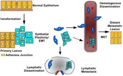
EMT cascade (Lee et. al., 2012; Micalizzi et. al., 2010). The metastatic spread of
malignant cells occurs in a multi-stepwise fashion, following EMT. Tumorigenic cells
leave their primary site, invade the basement membrane, intravasate into the local
lymphovascular, disseminate through the circulation, and extravasate into a secondary
site, thereby colonizing distant organs, depending on the cancer type, setting the stage for
metastasis (Figure 1.2) (Micalizzi et. al., 2010). Additionally, numerous regulators of
EMT have been misexpressed in cancers such as TGF-β, Wnt, Snail/Slug, Twist and
SIX1. Potentially, these and various other transcription factors appear to have a clinical
significance and have been recently identified to be prospective novel drug targets (Lee
Figure 1.1 Transition between epithelial and mesenchymal state of cells.
Figure 1.2 The metastatic spread of malignant cells facilitated through EMT.
1.3 TRANSFORMING GROWTH FACTOR-BETA (TGF-β)
Similar to many developmental signaling pathways misused in cancer, TGF-β
signaling is known to be connected to numerous pathologic processes and its function
during organogenesis parallels its role in neoplasia (Micalizzi et. al., 2009). TGF-β is an
ubiquitously expressed cytokine that contributes a dual role in tumor formation-
suppressing tumor growth in normal cells and early neoplastic lesions, while promoting
invasive migration and metastasis in the later stages of carcinogenesis (Micalizzi et. al.,
2009; Xu H et. al., 2014; Xu H et. al., 2015). This soluble growth factor is heavily
involved in EMT and is a regulator of tumorigenesis. SIX1 has been shown to increase
TGF-β signaling; this signaling pathway is a critical contributor to cancer progression
and metastasis by inducing oncogenic EMT. SIX1 expression selectively promotes the
pro-tumorigenic activity of TGF-β while halting its growth-inhibitory functions
(Micalizzi et. al., 2009). TGF-β contains three isoforms: TGFβ1, TGFβ2 and TGFβ3; and
mediates signaling by binding to three receptors: TGF-β receptor type I (TβRI), type II
(TβRII) and type III (TβRIII) (Meulmeester et. al., 2011; Xu H et. al., 2014; Xu H et. al.,
2015).
1.4 HUMAN PAPILLOMAVIRUS (HPV) AND CERVICAL CANCER
Cervical cancer is the leading cause of cancer morbidity and second leading cause
of cancer mortality in women worldwide. There are about 500,000 new cases and
300,000 deaths annually due to cervical cancer (Peralta-Zaragoza et. al., 2012). Virtually
all cervical cancer cases are due to persistent infection with a high-risk human papilloma
(STD). Currently, 79 million people in the USA have acquired an HPV infection. By the
age of 50, about 80% of women will have acquired an HPV infection (Society AC., 2013;
Peralta-Zaragoza et. al., 2012; Saslow et. al., 2012).
HPVs are small, non-enveloped viruses containing circular, double-stranded
DNA. HPV infects the epithelial cells of skin and mucosae. The HPV genome encodes
six to eight early and two late proteins. There are approximately 200 HPV types, 40 of
which can infect the genital mucosa (Faridi et. al., 2011). HPVs can be further classified
based on whether or not they lead to cervical cancer or benign genital warts: hence, we
distinguish high-risk (oncogenic) types- that cause virtually 99% of cervical cancer cases-
from low-risk (non-oncogenic) types that cause anogenital warts. Among the oncogenic
HPV types, HPV16 and 18 are the most common, causing about 70% of cervical cancers.
The non-oncogenic HPV types include HPV6 and 11, responsible for about 95% of
genital warts (Crow JM, 2012; Grainge et. al., 2005).
Most HPV infections are asymptomatic and spontaneously clear within 9-18
months, or (in some cases) the HPV remains latent at undetectable levels in the cervical
epithelium. However, approximately 10% of infected individuals cannot naturally clear a
high-risk HPV infection and are at risk for developing cervical cancer, which is known to
be a slow but progressive disease. Progression to cervical carcinogenesis first starts with
an infection to the genital mucosae of a high-risk HPV type. As the infection persists due
to a multitude of factors, this can progress through early and then late stages of dysplasia-
Cervical Intraepithelial Neoplasia I, II and III (CIN I, CIN II, CIN III) before developing
into invasive cervical carcinoma (Saslow et. al., 2012). Although new infections decrease
methods known to prevent or detect persistent, high-risk HPV infection. Primary
prevention with a prophylactic Gardasil-9 vaccine can prevent infections by nine HPV
types: 6, 11, 16, 18, 31, 33, 45, 52 and 58 (Harper DM et. al., 2017). Secondary
prevention with the Papanicolaou (Pap) smear test can detect abnormal cytology of
infected cervical cells (Figure 1. 3) (Crow JM, 2012).
Figure 1.3 High-risk HPV infection and progression to invasive cervical carcinoma. Cervical cells can clear an infection through the host immune system or potentially progress. Persistors can progress to CIN I, which can potentially regress or progress to CIN II/III. At the later stages of pre-malignancy, clinical regression is possible but progression is probable. The main methods to prevent and/or detect an HPV infection include a prophylactic Gardasil-9 vaccine or the Pap test.
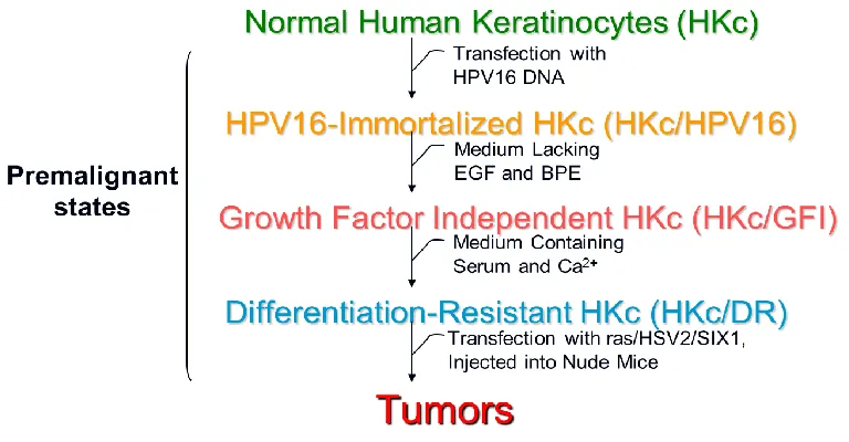
1.5 IN-VITRO MODEL SYSTEM OF HPV16-MEDIATED TRANSFORMATION OF
NORMAL HUMAN KERATINOCYTES (HKc)
Our laboratory has developed an in-vitro model system of HPV16-mediated
transformation of normal cells to explore the cellular and molecular events associated
with HPV infection. Primary or secondary cultures of human foreskin keratinocytes go
through a series of defined premalignant steps that represent the early and late stages of
cervical carcinogenesis. Normal human keratinocytes (HKc) were isolated from neonatal
foreskin tissue and transfected with pMHPV16d, which contains two copies of the
HPV16 DNA in a head-to-tail configuration, and selected with G418. This resulted in
immortalization of the transfected cells (HKc/HPV16), which represents the first
transformed stage in the in-vitro model. With continuous passaging of HKc/HPV16,
HPV16 sequences are amplified and integration into host chromosomes take place
(Pirisi et. al., 1987). Similar to normal HKc, these immortalized cells were cultured in
keratinocyte-serum free medium (KSFM), requiring both epidermal growth factor (EGF)
and bovine pituitary extract (BPE) for proliferation. In addition, normal HKc and
HKc/HPV16 cells are sensitive to differentiation triggered by serum and high calcium
levels. Nonetheless, HKc/HPV16 cells can be cultured in the absence of EGF and BPE.
This gives rise to growth factor independent HKc (HKc/GFI) cells. An additional
selection step was performed, where HKc/GFI were exposed to 5% fetal bovine serum
(FBS) and 1mM calcium chloride (Figure 1.4). In this step, cells that do not differentiate
in response to these two reagents are obtained and are consequently called differentiation
resistant cells (HKc/DR) (Pirisi et. al., 1987, 1988). This is the last stage of premalignant
into nude mice. In order to undergo malignant conversion, HKc/DR need to be
transfected with other transforming genes such as viral ras or herpes simplex virus type 2
(HSV2) DNA (DiPaolo et. al., 1989; DiPaolo et. al., 1990). We have repeatedly shown
that HKc/DR are completely resistant to the anti-proliferative effects of TGF-β, whereas
HKc/HPV16 and normal HKc are sensitive to the tumor inhibiting effects of TGF-β
(Creek et. al., 1995).
We have established this in-vitro system in order to investigate various stages of
malignant transformation. We utilized microarrays to identify genes whose altered
expression may lead to transformation in this multi-step, pre-malignant process of
in-vitro progression. Among the gene expression profiles that we had identified and that
needed to be further studied was SIX1 (Wan F et. al., 2008). This particular gene was
shown to be altered during the early and later stages of our model system in comparison
to normal cells.
1.6 SUMMARY AND GOAL OF RESEARCH
SIX1 overexpression has been shown to increase cancer cell proliferation,
survival and invasion. The overexpression of SIX1 is associated with tumorigenesis and
metastasis with poorer overall survival (Christensen et. al., 2008). We have previously
determined that SIX1 mRNA and protein levels are overexpressed in our
HPV16-transformed HKc lines at the differentiation-resistant stage (HKc/DR) compared with
early passage, HPV16-immortalized HKc (HKc/HPV16) and in HKc/HPV16 compared
to normal HKc. Furthermore, we have recently determined that SIX1 overexpression in
HKc/HPV16 induces the differentiation-resistant phenotype characteristic of HKc/DR,
and that SIX1 overexpression in HKc/DR induces tumorigenicity. These findings
strongly suggest that SIX1 has a role in regulating the growth and transformation of
normal HKc, and that it plays an important role in the maintenance of growth and a
transformed phenotype in HKc/HPV16 and HKc/DR. In summary, our studies suggest
that SIX1 is necessary for cell survival in HPV-16 –transformed cells and may potentially
become a suitable therapeutic target for HPV-driven cancers.
The main goal of this study is to explore the role of SIX1 in HPV16-transformed
human keratinocytes by following the cellular and molecular changes associated with the
CHAPTER 2
INHIBITION OF SIX1 EXPRESSION PRODUCES
MESENCHYMAL-EPITHELIAL TRANSITION AND DECREASES E6/E7 EXPRESSION IN
HPV16-TRANSFORMED HUMAN KERATINOCTYES
2.1 INTRODUCTION
Cervical cancer is the leading cause of cancer morbidity and second leading cause
of cancer mortality in women worldwide. There are about 500,000 new cases and
300,000 deaths annually due to cervical cancer. Virtually all cervical cancer cases are due
to persistent infection with a high-risk human papilloma virus (HPV). Infection with
HPV is the most common sexually transmitted viral disease (STD). Currently, 79 million
people in the USA have an HPV infection. By the age of 50, about 80% of women will
have acquired an HPV infection (American Cancer Society; Peralta-Zaragoza et. al.,
2012; Saslow et. al., 2012).
HPVs are small, non-enveloped viruses containing circular, double-stranded
DNA. HPV infects the epithelial cells of skin and mucosae. The HPV genome encodes
six to eight early and two late proteins. There are approximately 200 HPV types, 40 of
which can infect the genital mucosa. HPVs can be further classified based on whether or
not they lead to cervical cancer or benign genital warts: hence, we distinguish high-risk
(non-oncogenic) types that cause anogenital warts. Among the oncogenic HPV types, HPV16
and 18 are the most common, causing about 70% of cervical cancers. The non-oncogenic
HPV types include HPV6 and 11, responsible for about 95% of genital warts. Most HPV
infections are asymptomatic and are spontaneously cleared within 9-18 months, or (in
some cases) the HPV remains latent at undetectable levels in the cervical epithelium.
However, approximately 10% of infected individuals cannot naturally clear a high-risk
HPV infection and are at risk for developing cervical cancer. The Gardasil-9 vaccine can
prevent infections by nine HPV types: 6, 11, 16, 18, 31, 33, 45, 52 and 58 (Harper DM et.
al., 2017).
Numerous studies have determined that the inappropriate expression of
embryonic genes in cancer, in particular transcription factors, contribute to
carcinogenesis (Abate-Shen C et. al., 2002). Among the embryonic genes that also play a
role in cancer development, the sine oculis homeobox homolog 1 (SIX1) is essential for
the development of numerous organs including the auditory and olfactory system as well
as the kidney by promoting proliferation, survival and migration of progenitor cells
during embryogenesis. SIX1 has been shown to increase cancer cell proliferation,
survival and invasion, similar to what is observed during embryonic development
(Christensen et. al., 2008). In fact, many parallels exist between normal development and
tumorigenesis. The aberrant expression of SIX1 occurs in numerous adult human cancers,
including breast, ovarian, cervical and hepatocellular carcinomas as well as pediatric
SIX1 overexpression has been shown to correlate with increased malignancy,
lymph node metastasis and poor survival in patients afflicted with cancer (Coletta et. al.,
2008; Micalizzi et. al., 2009). For example, SIX1 is overexpressed in half of primary
mammary carcinomas and most metastatic lesions (Coletta et. al., 2008). SIX1 has also
been shown to be overexpressed in cervical cancer cell lines and tissues, which are
correlated with increased malignancy and metastasis (Tan et. al., 2011; Zheng et. al.,
2010). Studies have established that the overexpression of SIX1 in immortalized and
non-tumorigenic mammary epithelial cells induces malignant transformation, leading to
highly aggressive and invasive tumors in nude mice. Moreover, SIX1 overexpression in
human breast cancer cell lines induces epithelial-mesenchymal transition (EMT),
enhancing metastasis in-vitro and in-vivo (Coletta et. al., 2008; Micalizzi et. al., 2009).
Nonetheless, very few studies have elicited the effects of the loss of SIX1, especially in
cervical carcinogenesis. Determining the role of SIX1 in the tumorigenesis and
progression of cervical cancer could facilitate diagnosis and treatment. This work would
contribute in our understanding of the deteriorative functions of SIX1 in cancers as a
global regulator of malignant transformation.
In order to study the cellular and molecular mechanisms of HPV-mediated
transformation, Pirisi et. al. established an in-vitro model system to study keratinocytes
progression toward the premalignant stages of cervical cancer. In brief, primary human
keratinocytes (HKc) were immortalized by transfection with pMHPV16d, which contains
a head-to-tail HPV16 DNA dimer, and selection with G418. These immortalized human
A differentiation-resistant (HKc/DR) phenotype was then obtained by selection in serum
and calcium -supplemented medium. HKc/DR cells are not tumorigenic but can form
tumors in nude mice when these cells are transfected with activated Ras, Herpes Simplex
Virus 2 (HSV2) or the SIX1 oncogene (DiPaolo et. al., 1989; DiPaolo et. al., 1990).
Microarray studies conducted in our laboratory indicated that the in-vitro model
system reflects many cellular and molecular alterations characteristic of cervical cancer.
Therefore, our system for HPV16-mediated multi-step carcinogenesis shares important
features with cervical cancer and enables us to study the molecular mechanisms of
transformation and immortalization in our cell lines. It has been shown that SIX1 mRNA
and protein levels are overexpressed in all 4 cell lines (HKc/DR vs. HKc/HPV16), and
SIX1 mRNA and protein levels are consistently up-regulated during progression from
HKc/HPV16 to HKc/DR. SIX1 overexpression was also confirmed in about 25% of
cervical cancer tissue samples in a tissue array. Microarray results identified the
increased expression of SIX1 as a potential biomarker for cervical cancer (Wan F et. al.,
2008; Xu H et. al., 2014; Xu H et. al., 2015).
HPV16 is the most common among the high-risk oncogenic papillomaviruses that
contributes to cervical carcinogenesis (Zheng XH et. al., 2010; Zheng ZM et. al., 2011).
HPV16-mediated transformation requires the continuous expression of the viral
oncoproteins E6 and E7. These two oncogenes are necessary for malignant conversion
where E6 binds to and promotes the degradation of the p53 tumor suppressor protein; and
E7 binds to the Rb tumor suppressor protein thereby releasing the E2F transcription
factor. Consequently, this leads to tumor initiation, proliferation and genomic instability
SIX1 overexpression in HKc/DR is associated to the increasing expression of
HPV16 E7 that marks premalignant progression of these cells. SIX1 is known to be
primarily under the control of E2F (Young AP et. al., 2003). When E7 binds to
unphosphorylated Rb, this tumor suppressor protein becomes inactivated and releases
E2F. The displacement of E2F from Rb allows E2F to activate the cell cycle and
promotes the aberrant expression of the embryonic gene SIX1 (Young AP et. al., 2003).
We have determined that SIX1 overexpression in early-stage
HPV16-immortalized human keratinocytes (HKc/HPV16) induces a differentiation-resistant
phenotype, and that SIX1 overexpression in differentiation-resistant HPV16-transformed
human keratinocytes (HKc/DR) induces tumorigenicity (Xu H et. al., 2014; Xu H et. al.,
2015). Furthermore, we have also shown that SIX1 modulates TGF-beta signaling by
decreasing canonical (Smad-mediated) responses and increasing non-canonical TGF-beta
signaling pathways in both HKc/HPV16 and HKc/DR (Xu H et. al., 2014; Xu H et. al.,
2015). We have also demonstrated that TGF-beta signaling through the Smads decreases
E6 and E7 expression, and that in turn, E7 decreases cells’ sensitivity to growth inhibition
by TGF-beta (Baldwin et. al., 2004). Therefore, it is reasonable to expect that a decrease
in SIX1 levels may be accompanied by a decrease in E6 and E7 expression, which would
in turn be reflected in the increase of p53 and Rb levels.
With this knowledge in mind, we studied the role of SIX1 as a regulator of growth
and transformation in HPV16-transformed human keratinocytes. Specifically, we studied
the functional consequences of suppressing SIX1 at late stages of HPV16-mediated
SIX1 sh- or siRNAs resulted in widespread cell death. The knockdown of SIX1 resulted
in slower proliferation rates and was associated with, decreased E6 and E7 mRNA and
increased p53 and Rb protein levels. We conclude that SIX1 expression is vital for cell
survival, particularly in our HKc/DR cell lines. The results of these studies will give us
valuable insight into the complex pathways affected through SIX1 and the functional
significance of its inappropriate expression and activity in cancers, particularly those due
to HPV.
2.2 MATERIALS AND METHODS
Cell culture and treatment
The cell lines discussed in this work, HKc/HPV16-d1, HKc/DRd-1 and
HKc/DRd-2, have been described previously (Pirisi et. al., 1987, 1988) In short, normal
HKc are isolated from neonatal foreskins and maintained in keratinocyte serum-free
medium supplemented with epidermal growth factor (EGF) and bovine pituitary extract
(BPE) (KSFM, Invitrogen, Carlsbad, CA). This medium will be referred as complete
medium. Normal HKc were transfected with a recombinant plasmid containing two
copies of the HPV16 DNA in a head to tail configuration. This gave rise to
HPV16-immortalized human keratinocytes, which are also cultured in KSFM. The HPV16-immortalized
cells were then selected in medium devoid of EGF and BPE, giving rise to Growth Factor
Independent HKc (HKc/GFI). These cells were then further selected in basal KSFM
supplemented with 5% fetal bovine serum (FBS) and 1 mM calcium chloride to give rise
differentiation-resistant (DR) medium. HKc/DR were transfected with SIX1, giving rise
to HKc/DR-SIX1. These cells were also maintained in DR medium. All cells were
maintained in a humidified atmosphere of 5% CO2 at 37° C.
Plasmid constructs and stable transfection
Independently-derived HKc/DRd-1 and HKc/DRd-2 cell lines at 70% confluency
were transfected with a pSuper vector containing either a specific shRNA against human
SIX1, HuSH-29 shRNA based vector (Oligoengine, Seattle, WA) or a scrambled shRNA.
Additional plasmids utilized in this work include a pLXSN.neo vector expressing the
HPV16- E7 viral oncogene and the Harvey rat sarcoma virus (Hras)-V12 oncogene in the
pBABE.puro vector (Addgene, Cambridge, MA). Up to 5ug of the pLXSN.neo- E7 and
pBABe.puro- Hras-V12 vector were used for transfection. Moreover, three different
shRNA constructs against E7- that target three different positions within the HPV16 –E7
mRNA sequence, cloned in the backbone of the pSUPER.retro plasmid were used. 4ug or
5ug of anti-SIX1 shRNA construct and anti- E7 plasmid were used. Transfection
efficiency was monitored with the co-transfection of the pSUPER/puro plasmid (3:1
w/v), expressing GFP. As a vector control, the pSUPER/puro plasmid was used. Cells
were transfected with Lipofectamine 3000 (Invitrogen) following the manufacturer’s
instructions. Stable transfectants were selected with 3 ug/ml Puromycin (Toku-E,
Bellingham, WA).
RNA interference (RNAi)
(control) duplexes using Lipofectamine RNAiMAX (Invitrogen) following the
manufacturer’s instructions. The following sequences were utilized in this study for
transfection with siRNAs targeting SIX1 and E7. SIX1-siR1: Forward
5’-CCAGCUCAGAAGAGGAAUU, Reverse 5’-AAUUCCUCUUCUGAGCUGG;
SIX1-siR2: Forward 5’-CACGCCAGGAGCUCAAACU,
Reverse 5’-AGUUUGAGCUCCUGGCGUG; E7- siR1 (position 141):
Forward GGACAGAGCCCAUUACAAU, Reverse
5’-AUUGUAAUGGGCUCUGUCC; E7- siR2 (position 653): Forward 5’-
GCUCAGAGGAGGAGGAUGA, Reverse 5’-UCAUCCUCCUCCUCUGAGC.
90 pmol siRNA was used per well and cells were incubated for 24, 48 and 72 hours
before being harvested for RNA and protein extraction, for real-time PCR and
Enzyme-linked immunosorbent assay analysis (ELISA), respectively.
Retroviral Infection
PA317 cells were transfected with four different shRNA constructs (Origene,
Rockville, MD) against SIX1 that target four different positions within the human SIX1
mRNA sequence. A pSuper vector containing a scrambled shRNA was used as a control.
Stable transfectants were selected with 3 ug/ml puromycin. After approximately 24
hours, virus-producing PA317 were cultured until sub confluent, fed fresh complete
medium and the medium was collected after 24 h and filter-sterilized (0.22 u pore size).
Virus stocks were aliquoted and stored at -80 °C. High passage HKc/Drd-1 cells
at 70% confluency were infected with control virus stock or anti-SIX1 shRNA virus in
KSFM containing polybrene (6 ug/ml). HKc/DRd-1 cells were maintained in DR medium
removal of virus-containing medium, the infected cells were re-plated and selected with
3 ug/ml puromycin.
Real-time PCR
Total RNA was isolated from cells using the RNeasy Plus Micro Kit (Qiagen,
Hilden, Germany). Reverse transcription was carried out with 1 μg of total RNA using
iScript cDNA Synthesis Kit (Bio-Rad, Hercules, CA). Real-time PCR was performed
using iQ SYBR Green Supermix (Bio-Rad). All procedures followed the manufacturer’s
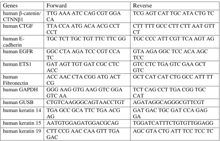
instructions. The sequences of the primers used for real-time PCR are listed in Table 2.1.
GAPDH was used as an internal control. All samples were assayed in duplicates.
Table 2.1 Primer sequences used in Real-time PCR. Primer sequence 5’-3’.
Genes Forward Reverse
human β-catenin/ CTNNβ1
TTG AAA ATC CAG CGT GGA CA
TCG AGT CAT TGC ATA CTG TC
human CTGF TTA CCA ATG ACA ACG CCT
CCT
CTT TTT GCC CTT CTT AAT GTT CT
human E-cadherin
TGC TCT TGC TGT TTC TTC GG TGC CCC ATT CGT TCA AGT AG
human EGFR GGC CTA AGA TCC CGT CCA
TC
GTA AGA GGC TCC ACA AGC TCC
human ETS1 GAT AGT TGT GAT CGC CTC
ACC
GTC CTC TGA GTC GAA GCT GTC
human Fibronectin
ACC AAC CTA CGG ATG ACT CG
GCT CAT CAT CTG GCC ATT TT
human GAPDH GGG AAG GTG AAG GTC GGA
GTC AA
TCT CAG CCT TGA CGG TGC CAT
human GUSB CTGTCAAGGGCAGTAACCTGT AGATAGGCAGGGCGTTCGT
human keratin 14 TGA GCC GCA TTC TGA ACG AG
GAT GAC TGC GAT CCA GAG GA
human keratin 15 AATGTGGAGATGGACGCAG TGGATCATTTCTGTGTTGGAGG
human ki-67 ACG CCT GGT TAC TAT CAA AAG G
CAG ACC CAT TTA CTT GTG TTG GA
human MMP1 AAA ATT ACA CGC CAG ATT
TGC C
GGT GTG ACA TTA CTC CAG AGT TG
human MMP9 AGT CCA CCC TTG TGC TCT
TCC C
TCT GCC ACC CGA GTG TAA CCA T
human N-cadherin
ACA GTG GCC ACC TAC AAA GG
CCG AGA TGG GGT TGA TAA TG
human Occludin ATG ACA AGC GGT TTT ATC
CA
CTC CAG CTC ATC ACA GGA CT
human p53 CGTGTGGAGTATTTGGATGAC AGTCTTCCAGTGTGATGATGG
human PPARγ GCC CTT TGG TGA CTT TAT
GGA
GCA GCA GGT TGT CTT GGA TG
human PTGS2 GCC CAG CAC TTC ACG CAT
CAG
AGA CCA GGC ACC AGA CCA AAG ACC
human Rb CAA ACT TGG AGT TCG CTT
GT
TTC AGA ATC CAT GGG AAA GA
human SIX1 ATT CTC ACC TCC CCA AAG TC ACT TAG GAC CCC AAG TCC AC
human SNAIL ACC CCA CAT CCT TCT CAC TG TAC AAA AAC CCA CGC AGA
CA
human TβRI CTT AAT TCC TCG AGA TAG
GC
GTG AGA TGC AGA CGA AGC
human TβRII GGT TCC TGT GTG CCC TTA TT TGC AAC CCA TGA AGG TAA
AA
human TWIST GTC CGC AGT CTT ACG AGG
AG
GCT TGA GGG TCT GAA TCT TGC T
human Vimentin GAG AAC TTT GCC GTT GAA
GC
TCC AGC AGC TTC CTG TAG GT
HPV16- E6 CAC AGT TAT GCA CAG AGC
TGC
CAT ATA TTC ATG CAA TGT AGG TGT A
HPV16- E7 CCG GAC AGA GCC CAT TAC
AAT
ACG TGT GTG CTT TGT ACG CAC
Enzyme-linked immunosorbent assay (ELISA)
Protein was isolated from cells using the Cell Lysis Buffer from the PathScan
Total p53 Sandwich ELISA kit (Cell Signaling, Danvers, MA). SigmaFAST Protease
Inhibitor (Sigma, Saint Louis, MO) was added to the resulting supernatant. PathScan
Total Rb Sandwich ELISA kit was then utilized according to the manufacturer’s
protein/sample was loaded into the Rb pre-coated wells. All samples were assayed in
replicates of two or three. Absorbance was read at 450 nm within 30 minutes using a
microplate reader.
Cell proliferation assay
To compare the growth rates of either HKc/DRd-1- SIX1sh or
HKc/Drd-2-SIX1sh and HKc/DRd-1-SCRAMsh or HKc/DRd-2-SCRAMsh, and HKc/HPV16d-1 the
WST-8 dye reagent from the Cell Counting Kit-8 (CCK8) sensitive colorimetric assay
was utilized (Dojindo, Rockville, MD). Proliferation was determined at 24-hour intervals.
5,000 cells were plated per well in 24-well plates in their respective media (1ml/well).
Cells were plated in triplicate wells and at the appropriate times, 50 ul of WST-8 dye
solution was added to each well and the plates were placed in a CO2 incubator for
2 hours. Absorbance was read at 450 nm with a microplate reader.
Gene-expression microarray analysis and validation by SYBR green real-time PCR
Total RNA from SIX1-siRNA1, SIX1-siRNA2 and
HKc/DR-control-siRNA was isolated using Qiagen’s RNeasy Plus Micro Kit according to the
manufacturer’s protocol. RNA quality was assessed using an Agilent 2100 Bioanalyzer
and RNA Integrity Numbers ranged from 9.5 to 10.0. Microarray experiments were
performed using the Affymetrix GeneChip Human Gene 2.0 ST Arrays as we have
Statistical Analysis
Results were expressed as the mean ± standard deviation (SD). Data were
interpreted using student’s t-test. P ≤ 0.05, P ≤ 0.01 or P ≤ 0.001 were considered
statistically significant and indicated in the figures by *, ** or ***, respectively.
2.3 RESULTS
2.3.1 THE KNOCKDOWN OF SIX1 AFFECTS CELL VIABILITY IN HKc/DR
We utilized shRNA constructs targeting the human SIX1 gene to explore the
effects of suppressing this oncogene in HPV16-transformed human keratinocytes.
Anti-SIX1-shRNA-encoding sequences and a control, scrambled shRNA were cloned in the
pSuper-retro plasmid backbone (Oligoengine) and the resulting plasmids transfected into
two HKc/DR cell lines, HKc/DRd-1 and HKc/DRd-2 (Pirisi et. al., 1987).Transfected
cells were briefly selected with puromycin until vector controls died (approx. 72 h). Cells
were then collected for RNA extraction and plated for a growth curve (Figure 2.1, A).
The shRNA decreased SIX1 mRNA levels to about 20% of control (maximum) (Figure
2.1, B) and cells transfected with the anti-SIX1 shRNA exhibited much slower growth
than their controls, although after 96h in culture, recovery could already be observed in
one of the transfected lines (Figure 2.1, A). Further cultivation of these cells resulted in
SIX1 mRNA levels returning to 100% of control and proliferation rates recovering
completely (data not shown). Upon visual inspection, phase contrast photographs taken
GFP, at various times after transfection, demonstrated that loss of SIX1 leads to cell
death in HKc/DR (Figure 2.1 D, E).
D
E
B
Figure 2.1 Loss of SIX1 in HKc/DR affects proliferation and HPV16- E6/E7 mRNA expression levels. (A) The rate of proliferation decreases in HKc/DRd-1 and HKc/DRd-2 transfected with SIX1-shRNA plasmid. Cell proliferation at 24-hour intervals was
determined utilizing the WST-8 dye reagent from the Cell Counting Kit-8 (CCK8) sensitive colorimetric assay. (B) The mRNA expression levels of SIX1, E6 and E7 decrease in HKc/HPV16 and HKc/DR transfected with a plasmid encoding anti-SIX1-shRNA. Real-time PCR analysis for SIX1, E6 and E7 mRNA in HKc/DRd-1 and
HKc/DRd-2 cell lines. Levels of expression were normalized to GAPDH. (C) The protein expression level of p53 increases while levels of Rb decrease in HKc/DRd-1 cell line transfected with SIX1-shRNA plasmid. ELISA for p53 and Rb in HKc/DRd-1 transfected with either control (anti-scram shRNA) or anti-SIX1 shRNA at indicated plasmid
concentrations. (D, E) Loss of SIX1 affects cell viability in HKc/DRd-1. Fluorescent and phase-contrast images depicting transfection efficiency and morphology, respectively, of HKc/DRd-1 cells 48 hours (top) and 4 days (bottom) post-transfection with anti-SIX1 shRNA plasmid. (E) Fluorescent and phase-contrast images depicting transfection efficiency and morphology, respectively, of HKc/DRd-1 cells 48 hours (top) and 4 days (bottom) post-transfection with control-shRNA and scrambled-shRNA plasmids. Images are shown at 200X magnification.
A
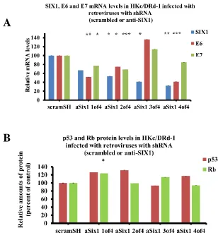
Figure 2.2 HKc/DR infected with four different shRNA constructs targeting human SIX1
gene. mRNA and protein expression levels of SIX1, E6 and E7 in HKc/DRd-1 cells infected with four different shRNA constructs targeting four different positions within the human SIX1 mRNA sequence. (A) Real-time PCR analysis of SIX1, E6 and E7 mRNA in HKc/DRd-1 cell lines. Levels of expression were normalized to GAPDH. (B) ELISA for p53 and Rb in HKc/DRd-1 infected with either control (anti-scram shRNA) or the indicated anti-SIX1 shRNA constructs.
To fully understand the necessity of SIX1 in these differentiation resistant cell
lines, HKc/DRd-1 cells were infected with anti-SIX1 shRNA PA317 conditioned
medium. SIX1 loss was observed in cells treated with all four different constructs with
concomitant E6 and E7 loss in three of the shRNA constructs (Figure 2.2, A). Similarly,
in regards to p53 and Rb levels, we observed slight increases in protein levels (Figure 2.2,
B). Overall, these results indicate that SIX1 expression is vital for cell survival in the
late-stage HPV16 transformed human keratinocytes.
2.3.2 LOSS OF SIX1 CAUSES DECREASES OF HPV16 E6/E7 mRNA LEVELS IN
HKc/DR
The experiments described above also provided a first hint that SIX1 expression
may affect HPV16 E6/E7 mRNA expression. We have demonstrated that SIX1
knockdown in HKc/DRd-1 results in the decrease of HPV16- E6/E7 mRNA levels with a
simultaneous increase in p53 and decrease in Rb protein expression levels (Figure 2.1 B,
C). Therefore, we set out to further investigate this possibility through transient
transfections with pre-formed anti-SIX1 siRNA. We utilized two different siRNA
mRNA levels decreased significantly, under these transient transfection conditions, at 24,
48 and 72 hours (Figure 2.3, A). Decreased SIX1 expression was associated with
decreases in HPV16-E6 and E7 mRNA levels (Figure 2.3 B, C). Consistent with a
decrease of active E6 levels, p53 protein levels increased significantly, with the greatest
rise observed 24 hours after SIX1 expression (Figure 2.3, D). Not surprisingly, p53
mRNA levels (which are high in HPV16-transformed cells) markedly decreased in
HKc/DR transfected cells with either of the two anti-SIX1 siRNA constructs (Figure 2.3,
E). Rb protein and mRNA levels followed similar trends, however to a lesser extent
(Figure 2.3 F, G).
A
C
Figure 2.3 Loss of SIX1 with pre-formed anti-SIX1 siRNAs causes decreases of
HPV16- E6/E7 mRNA levels in HKc/DR. (A) (B) (C) Real-time PCR analysis for SIX1, E6 and E7 mRNA in HKc/DRd-1 under transient transfection conditions at the indicated time points. Levels of expression were normalized to GAPDH. (D) (F) Relative protein expression of p53 and Rb, respectively, after transfection with pre-formed anti-SIX1 siRNAs in HKc/DRd-1 cells. p53 and Rb protein expression was detected by ELISA in HKc/DRd-1 at indicated 24-hour interval time points. (E) (G) Relative mRNA expression of p53 and Rb, respectively, in the HKc/DRd-1 cell line transfected with pre-formed anti-SIX1 siRNAs. p53 and Rb mRNA were quantified by RT/PCR in HKc/DRd-1 at the indicated 24-hour interval time points. Levels of expression were normalized to GAPDH.
Taken together, our data indicate that a loss of SIX1 expression in HKc/DR cells
is associated with decreased expression of the viral oncogenes E6 and E7. The
mechanisms in which SIX1 directly, or perhaps indirectly, affect E6 and E7 still need to
be elucidated.
D
E
2.3.3 KNOCKDOWN OF E7 RESULTS IN DECREASED mRNA LEVELS FOR SIX1
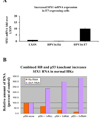
Previous unpublished work in our laboratory determined that SIX1 levels could
be increased dramatically in normal HKc by the expression of HPV-16 E7, and by
siRNA-mediated inhibition of Rb expression, provided that p53 levels were reduced as
well by shRNA (Figure 2.4 A, B). We have also overexpressed HPV16 -E7 in HKc/DR
cells. Increased E7 expression resulted in the subsequent increase of SIX1 and HPV16-
E6 mRNA (Figure 2.5, A). Concomitantly, Rb and p53 protein levels, respectively, were
downregulated (Figure 2.5 B-E).
To better pinpoint the functional consequence of E7 suppression in HKc/DR cells,
the HKc/DRd-1 cell line was transiently transfected with two different siRNA duplexes
targeting HPV16 -E7. Cells were then collected for RNA and protein extraction 48 hours
after transfection. Both siRNAs produced a significant decrease in both E6 and E7
mRNA levels, up to 70% inhibition (Figure 2.6, A) accompanied by approximately 50%
decrease in SIX1 mRNA levels (Figure 2.6, B). These changes in E6, E7 and SIX1
mRNA levels were also associated with the expected changes in p53 and Rb mRNA and
Figure 2.4 Increased SIX1 RNA expression in normal HKc and HKc/E7-expressing cells. (A) Real-time PCR analysis of SIX1 mRNA in normal HKc expressing HPV16E7. SIX1 mRNA expression was normalized to GAPDH; (B) Real-time PCR analysis for SIX1 and Rb mRNA in normal HKc co-transfected with a p53-shRNA plasmid and an anti-Rb siRNA duplex. Levels of expression were normalized to GUSB.
A
Figure 2.5 Increased SIX1 and HPV16- E6 mRNA expression in HKc/DR overexpressing HPV16 E7 from the pLXSN.neo- E7 vector. (A) Real-time PCR analysis for SIX1 and E6 mRNA in HKc/DR expressing HPV16 E7. Levels of expression were normalized to GAPDH. (B) (D) Real-time PCR analysis of Rb and p53 mRNA in HKc/DR
overexpressing HPV16- E7. Levels of expression were normalized to GAPDH. (C) (E) ELISA for Rb and p53 protein expression in HKc/DR overexpressing HPV16- E7 with the use of pLXSN.neo- E7 vector.
A
B
C
D
Figure 2.6 Knockdown of HPV16-E7 with pre-formed anti-E7 siRNAs results in
decreased mRNA levels of HPV16-E6 and SIX1. (A) (B) Real-time PCR analysis for E6, E7 and SIX1 mRNA in HKc/DRd-1 under transient transfection conditions at 48 hours. Levels of expression were normalized to GAPDH. (C) Real-time PCR analysis for p53 mRNA in HKc/DRd-1 48 hours after transient transfection. p53 expression was
normalized to GAPDH. (D) (F) ELISA for p53 and Rb protein expression in HKc/DRd-1 48 hours after transient transfection. (E) Real-time PCR analysis of p53 mRNA in
HKc/DRd-1 at 48 hours. Rb expression was normalized to GAPDH.
Collectively, these data suggest that a loss of E6/E7 expression is associated with
a decrease in the mRNA levels of SIX1. We tentatively explain these changes with the
interrelated effects of E7 and SIX1 on TGF-β signaling, as detailed in the Discussion.
A
B
D
C
2.3.4 SIX1 KNOCKDOWN IN HKc/DR PROMOTES MESENCHYMAL-EPITHELIAL
TRANSITION (MET) AND RESETS THE LEVELS OF TGF-Β RECEPTORS 1 AND 2
We set out to investigate global gene expression during transient SIX1
knockdown in HKc/DRd-1 cells on Affymetrix GeneChip Human Gene 2.0 ST Arrays.
Direct, paired comparisons of gene expression were made among four individual clones
of siRNA1and HKc/DR-control-siRNA, and between
HKc/DR-SIX1-siRNA2 and HKc/DR-control-siRNA. We used Transcriptome Analysis Console (TAC)
software from Affymetrix to analyze the microarray results. To determine the
differentially expressed genes in the pairwise comparisons, we used a fold change >1.4
(up and down) and a P value ˂ 0.01 as cutoff values. In addition, Ingenuity Pathway
Analysis (IPA) was performed on the differentially expressed genes. Pairwise
comparisons identified 2447 differentially expressed genes after treatment with
SIX1-siRNA1: 794 genes were upregulated and 1653 genes were downregulated.
Correspondingly, 2693 differentially expressed genes were identified after treatment with
SIX1-siRNA2: 1580 genes were upregulated and 1113 genes were downregulated. We
then manually identified common genes between the two siRNA duplexes that were
associated with a transition towards an epithelial phenotype. We were interested in nine
genes that were downregulated and four genes that were upregulated through siRNA
Table 2.2 Expression of EMT-associated genes targeted by SIX1 and TGF-β in HKc/DR. Decrease in target gene expression during transient SIX1 knockdown in HKc/DRd-1 cell lines.
Unsupervised cluster analysis clearly differentiated the SIX1 knockdown cells
from controls and allowed for the identification of a cluster of genes that best separate the
two (Figure 2.7 A, B). IPA pathway analysis showed that the pathways most heavily
affected by decreased SIX1 expression were consistent with MET (Figure 2.7, C).
A
B
Figure 2.7 Gene expression profiles of SIX1 knockdown in HKc/DR involve TGF-β signaling, MET and apoptosis. (A) Unsupervised hierarchical clustering of differentially expressed genes between HKc/DR control siR (left) and HKc/DR SIX1-siR2 (right). The color represents the expression level of a gene above (red), below (green), or at neutral (black) the mean expression level of that gene across all samples. Pairwise comparisons identified 2693 differentially expressed genes after treatment with SIX1-siRNA2: 1580 genes were upregulated and 1113 genes were downregulated. (B) Heatmap depicting select genes relevant to MET. (C) Illustration of downstream functional effects of gene transcripts that are upregulated and downregulated. Canonical pathways and diseases & functions that are differentially expressed as a result of SIX1 loss in HKc/DR (1.4 fold change up and down; p-value ˂ .01). Ingenuity Pathway Analysis (IPA) indicates deregulation of pathways related to invasion, proliferation, migration and apoptosis.
Among the many changes within these pathways, we observed a reduction in the
expression of the mesenchymal-related gene Vimentin (VIM) and an increase in the
expression of the epithelial-related genes Keratin 15 (K15) and Occludin (OCLN).
Furthermore, there was an increase in expression of the inflammatory response gene
prostaglandin-endoperoxide synthase 2, PTGS2 (cyclooxygenase-2, COX-2). There was
a decrease in the expression of the cell-cell adhesion complex molecule, β-catenin
(CTNNβ1) (Gumbiner et. al., 2005). We noted a decrease in the pro-survival factor
epidermal growth factor receptor (EGFR) and its associated ligand, transforming growth
factor alpha (TGF-α). There was a decrease in the angiogenic matricellular marker
connective tissue growth factor (CTGF/CCN2) and its downstream signaling effector
ETS proto-oncogene 1, transcription factor (ETS1) (Geisinger et. al., 2012; Ubink et. al.,
2016). mRNA levels for the markers for invasiveness, matrix metalloproteinase 1 and 9
(MMP1, MMP9), were also decreased. Lastly, there was a decrease in peroxisome
proliferator activated receptor gamma (PPARƴ), a ubiquitously expressed gene known to
suggest that mesenchymal to epithelial transition is a functional consequence of SIX1
loss in the late-stages of HPV16-transformed human keratinocytes.
To validate data from the array analysis we performed real-time PCR on
HKc/DRd-1 cells treated with SIX1-siRNA2 48 hours after transfection. The mRNA
levels of K15 and OCLN both increased approximately 2.2-fold while VIM mRNA
levels decreased approximately 20%. Additionally, there was a significant increase, up
to 40%, in PTGS2/COX-2. These cells might be experiencing cell stress as a result of
SIX1 loss; and therefore, they react by increasing levels of inflammatory-response
genes. While the array data showed a 1.54-fold decrease in β-catenin expression,
real-time PCR results revealed a 3-fold increase in mRNA levels. The shift away from a
mesenchymal phenotype can be associated with changes that follow a gain in cell-cell
adherens junctions typically seen in normal epithelial cells and β-catenin is a
well-known epithelial marker. There was a 40% decrease in EGFR mRNA levels following
transfection. Additionally, there was a 55% and 40% decrease in CTGF/CCN2 and
ETS1 mRNA levels, respectively. Tumorigenic cells are known to metastasize via blood
or lymphatic vessels through an increase in the mesenchymal-associated gene
CTGF/CCN2 (Ubink et. al., 2016).In addition, there was a 70% and 40% decrease in
MMP1 and MMP9 mRNA levels, respectively. Malignant cells can damage the
basement membrane thru the increase of proteolytic enzymes such as MMP1 and
MMP9.Lastly, there was a significant decrease in the proliferation and differentiation
marker PPARƴ. Therefore, a significant decrease of these EMT-inducing proteins is an
Figure 2.8 SIX1 knockdown induces markers of MET in HKc/DR. Levels of mRNA expression of CTGF, CTNNβ1, EGFR, ETS1, K15, MMP1 and 9, OCLN, PPARγ, PTGS2 and VIM were determined by real-time PCR analysis in HKc/DR control siR and HKc/DR SIX1-siR2. Data were normalized to GAPDH expression.
Last, but not least, microarray data revealed a 1.57-fold increase in TβRI and a
1.84-fold decrease in TβRII. Real-time PCR confirmed these changes. We analyzed our
array data to locate gene expression changes that are regulated by TGF-β signaling
(Figure 2.9). We also identified increases in K15 (1.72 fold) and OCLN (2.11 fold) and
decreases in Hic1 (1.4), VIM (2.05), CTGF (1.72) and PLAT (3) in response to loss of
Figure 2.9 SIX1 knockdown in HKc/DR resets the levels of TGF-β receptors 1 and 2. Levels of mRNA expression of TGFβRI and TGFβRII were determined by Real-time PCR analysis in HKc/DR control siR and HKc/DR SIX1-siR2. Data were normalized to GAPDH expression.
2.4 DISCUSSION
The results shown above, taken together with our prior observation on the role of
SIX1 in HPV16-mediated transformation, point to the fact that SIX1 expression is
necessary for growth and survival of differentiation-resistant, HPV16-transformed
human keratinocytes (HKc/DR) and that these cells do not tolerate a decrease in SIX1
levels. Given the fact that SIX1 is virtually not expressed in normal adult cells, this gene
may become a suitable therapeutic target for HPV-driven cancers.
Deregulation of homeobox genes in cancer results in an out of context activation
of their development-restricted functions. There have been countless studies identifying
the link between normal organogenesis and neoplasia. Indeed, they both share many
pathways and basic molecular processes between them, demonstrating undoubtedly that
tumorigenesis and progression is an aberrant form of morphogenesis. The oncoprotein
SIX1 has an important role in the expansion of progenitor cell populations during