Detection of Retinal Disease Using SLO
Technique
Rupali Sudamrao Wagh, Prof S.G Kole
Student, Department of Electronics & Telecommunication Engineering, JSPMNarhe Technical Campus Pune,
Savitribai Phule University Pune, India
Guide, Department of Electronics & Telecommunication Engineering, JSPM Narhe Technical Campus Pune, Savitribai
Phule University Pune, India
ABSTRACT: Taking a gander at laser ophthalmoscopes (SLOs) can be utilized for as a part of front of date-book unmistakable confirmation of retinal sicknesses. With the occurrence to most recent screening improvement, the upside of utilizing SLO is its wide field of perspective, which can picture an expansive part of the retina for better finding of the retinal sicknesses. Then again, amidst the imaging process, outdated rarities, for occasion, eyelashes and eyelids are in like way imaged close-by the retinal region. This brings an imperative test on the most proficient procedure to block these old-fashioned rarities. In this paper, I propose a novel way to deal with oversee really remove out veritable retinal zone from a SLO picture in context of picture dealing with and machine learning approaches. To lessen the multifaceted method for picture dealing with errands and give a strong primitive picture arrangement, we have aggregated pixels into various regions considering the territorial size and conservativeness, called super pixels. The system then registers picture based parts reflecting textural and vital data and solicitations between retinal area and old rarities. The trial assessment results have demonstrated fantastic execution with a general precision of 92%.
KEYWORDS-Preprocessing, Feature selection, retinal artefacts extraction, retinal image analysis, is scanning laser ophthalmoscope (SLO), Super Pixel Classification.
I. INTRODUCTION
the retina. In the event that these structures are emptied, this won't just backing the persuading examination regarding retinal zone, additionally empower to choose multi view pictures into a montage, understanding a completely recognizable retina for sickness examination. In this study, I have developed a novel structure for the extraction of retinal locale in SLO pictures. The three key strides for building our structure include:
1) Determination of parts that can be utilized to see the retinal district and the old rarities;
2) Selection of parts which are most key to the solicitation;
3) Construction of the classifier which can assemble out the retinal zone from SLO pictures.
For disconnecting between the retinal space and the relics, I have picked unmistakable picture based sections which reflect grayscale, textural, and central data at different resolutions. By then, I have picked the parts among the critical summary of limits, which are pertinent to the social occasion. The part determination process enhances the classifier execution to the degree computational time. At long last, I have developed the classifier for confining between the retinal zone and the old rarities. Our model has satisfied normal strategy precision of 92% on the dataset.
II. RELATED WORK
Our composed work study is started with the frameworks for zone and division of eyelids and eyelashes related on photographs of the front of the eye, which contains the understudy, eyelids, and eyelashes. On such a photograph, the eyelashes are when in doubt as lines or stack of lines gathered together. In this manner, the starting step of remembering them was the use of edge affirmation systems, for case, Sobel, Prewitt, Canny, Hough Transform [3], and Wavelet change [4]. The eyelashes on the iris were then cleared by applying nonlinear detaching on the suspected eyelash regions [5]. Coming about to eyelashes can be in either distinct structure or as different eyelashes gathered, Gaussian channel and Variance divert were related keeping in mind the end goal to see among both sorts of eyelashes [6]. The test demonstrated that detachable sorts of eyelashes were doubtlessly perceived by applying Gaussian channel; however Variance channels are more ideal for different eyelash division [7].
At to begin with, the eyelash hopefuls were compelled utilizing dynamic shape showing, and after that, eight-directional channel bank was related on the conceivable eyelash competitors. Kang and Park [8] utilized center score as a bit of sales to move the measure of convolution bits for eyelash affirmation. The size grouping of the convolution parts in like way confined between distinct eyelashes and different eyelashes. Min and Park [9] picked the parts in context of force and neighborhood standard grouping to pick eyelashes. They were edge utilizing Otsu's framework, which is an altered farthest point determination procedure considering specific suspicions about force spread. These frameworks have been related on CASIA database [10], which is an online database of Iris pictures. In a photograph picked up from SLO, the eyelashes show as either dull or astonishing locale showed up diversely in connection to retinal achieve subordinate upon how laser section is secured as it passes the eyelashes. The eyelids show as reflectance area with more essential reflectance reaction veered from retinal range. Subsequently, we have to discover parts, which can detach among genuine retinal zone and the obsolete rarities in SLO retinal compasses.
scatters quality [11]. For picking picture quality, the sections of area of vitality of anatomical structures, for case, optic nerve head (ONH) and Fovea have likewise been eviscerated [16].
The fragments included essential similarity once-over, zone, and visual descriptor and so on. A rate of the already expressed systems proposes the use of cross area examination, which can be a period productive technique to make segments of specific region rather than every pixel. Yet, matrix examination won't not be a positive approach to manage address sporadic locales in the photograph. Thusly, I picked the utilization of superpixels [17]–[20], which hide away pixels into various regions relying upon their territorial size and humbleness. Our technique depends on after isolating the SLO picture based parts, which are enlisted for a little range in the retinal picture called superpixels. The determination of highlight vector for each superpixel is computationally beneficial when showed up diversely in connection to highlight vector determination for every pixel. The superpixels from the photographs in the course of action set are doled out the class of either retinal space or relics relying upon the lion's offer of pixels in the superpixel having a spot with the specific class. The depiction is performed in the wake of arranging and determination of fragments comparably as suitability all together.
III. PROPOSED MECHANISM AND ARCHITECTURE
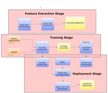
Fig. 1. Block diagram of retina detector framework Explanation-
around retinal extent. In the testing and evaluation masterminds, the modified remarks are performed on the "test set" of pictures and the classifier execution is surveyed against the manual clarifications for the determination of accuracy. Finally, the game plan stage performs the modified extraction of retinal district. The subtasks for get ready, testing, and game plan stages are immediately depicted as takes after Image Data Integration: It includes the coordination of picture information with their manual comments around genuine retinal zone. Image Preprocessing: Images are then preprocessed with a specific end goal to bring the power estimations of every picture into a specific reach. Generation of Superpixels: The preparation pictures subsequent to preprocessing are spoken to by little districts called superpixels. The era of the element vector for each superpixel makes the procedure computationally effective when contrasted with highlight vector era for every pixel. Feature Generation: I produce picture based components which are utilized to recognize the retinal region and the curios. The picture based elements reflect textural, grayscale, or territorial data and they were figured for each superpixel of the picture present in the preparation set. In testing stage, just those components will be produced which are chosen by highlight choice procedure. Feature Selection: Due to an extensive number of components, the element cluster should be diminished before classifier development. This includes highlights choice of the hugest elements for arrangement. Classifier Construction: In conjunction with manual comments, the chose elements are then used to develop the parallel classifier. The consequence of such a classifier is the superpixel speaking to either the "genuine retinal territory" or the "antiquities." Image Post handling: Image post preparing is performed by morphological sifting in order to decide the retinal range limit utilizing superpixels grouped by the arrangement model. The components of our location structure are examined as takes after.
A. Image Preprocessing
Images were normalized by applying a Gamma (γ) adjustment to bring the mean image intensity to a target value. γwas calculated .
B. Generation of Superpixels
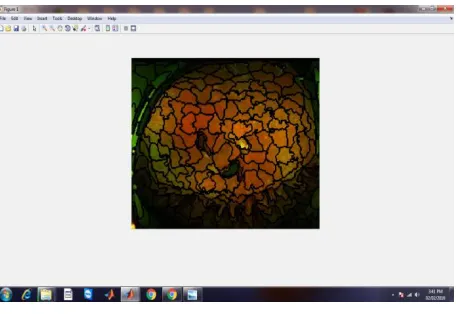
Fig 02 Generation of Superpixels
superpixels to be delivered. The value was set to 5000 as a tradeoff between computational constancy and desire precision. Highlight Generation
After the era of superpixels, the following step is to decide their components. I mean to separate between the retinal range and curios utilizing textural, grayscale slope, and provincial based elements. Textural and inclination based elements are figured from red and green channels on various Gaussian obscuring scales, otherwise called smoothing scales [23]. In SLO pictures, the blue channel is set to zero; accordingly, no component was figured for the blue channel.
IV. CONCLUSION
Perceiving bona fide retinal domain from old rarities in SLO pictures is a trying errand, which is similarly the essential basic step toward PC upheld affliction determination. In this study, I have proposed a novel framework for customized revelation of authentic retinal zone in SLO pictures. I have used super pixels to address different unusual regions minimalistically and reduce the preparing cost. Highlight decision enables the most basic components to be picked and, hence, diminishes handling cost too. A classifier has been developed considering picked components to independent out the retina zone. It has been appeared differently in relation to other two classifiers and was impeccable while saving the computational time. The trial evaluation result exhibits that our proposed framework can finish a precision of 92% in division of the honest to goodness retinal zone from a SLO picture.
REFERENCES
[1] M. S. Haleem, L. Han, J. van Hemert, and B. Li, “Automatic extraction of retinal features from colour retinal images for glaucoma diagnosis: A review,” Comput. Med. Imag. Graph., vol. 37, pp. 581–596, 2013.
[2] Optos. (2014). [Online]. Available: www.optos.com
[3] R. C. Gonzalez and R. E. Woods, Eds., Digital Image Processing, 3rd ed. Englewood Cliffs, NJ, USA: Prentice-Hall, 2006.
[4] M. J. Aligholizadeh, S. Javadi, R. S. Nadooshan, and K. Kangarloo, “Eyelidand eyelash segmentation based on wavelet transform for iris recognition,” in Proc. 4th Int. Congr. Image Signal Process., 2011, pp. 1231–1235.
[5] D. Zhang, D. Monro, and S. Rakshit, “Eyelash removal method for human iris recognition,” in Proc. IEEE Int. Conf. Image Process., 2006, pp. 285–288.
[6] A. V.Mire and B. L. Dhote, “Iris recognition system with accurate eyelash segmentation and improved FAR, FRR using textural and topological features,” Int. J. Comput. Appl., vol. 7, pp. 0975–8887, 2010.
[7] Y.-H. Li,M. Savvides, and T. Chen, “Investigating useful and distinguishing features around the eyelash region,” in Proc. 37th IEEE WorkshopAppl. Imag. Pattern Recog., 2008, pp. 1–6.
[8] B. J. Kang and K. R. Park, “A robust eyelash detection based on iris focus assessment,” Pattern Recog. Lett., vol. 28, pp. 1630–1639, 2007. [9] T. H. Min and R. H. Park, “Eyelid and eyelash detection method in the normalized iris image using the parabolic Hough model and Otsus thresholding method,” Pattern Recog. Lett., vol. 30, pp. 1138–1143, 2009. [10] Iris database. (2005). [Online]. Available: http://www.cbsr.ia.ac.cn/IrisDatabase.htm
[11] H. Davis, S. Russell, E. Barriga,M.Abramoff, and P. Soliz, “Vision-based, real-time retinal image quality assessment,” in Proc. 22nd IEEE Int. Symp.Comput.-Based Med. Syst., 2009, pp. 1–6.
[12] H. Yu, C. Agurto, S. Barriga, S. C. Nemeth, P. Soliz, and G. Zamora, “Automated image quality evaluation of retinal fundus photographs in diabetic retinopathy screening,” in Proc. IEEE Southwest Symp. ImageAnal. Interpretation, 2012, pp. 125–128.
[13] J. A. M. P. Dias, C. M. Oliveira, and L. A. d. S. Cruz, “Retinal image quality assessment using generic image quality indicators,” Inf. Fusion, vol. 13, pp. 1–18, 2012.
[14] M. Barker and W. Rayens, “Partial least squares for discrimination,” J. Chemom., vol. 17, pp. 166–173, 2003.