HIGHLIGHTED ARTICLE GENETICS | INVESTIGATION
Elevated Genome-Wide Instability in Yeast Mutants
Lacking RNase H Activity
Karen O’Connell, Sue Jinks-Robertson,1and Thomas D. Petes1 Department of Molecular Genetics and Microbiology, Duke University School of Medicine, Durham, North Carolina 27710
ABSTRACTTwo types of RNA:DNA associations can lead to genome instability: the formation of R-loops during transcription and the incorporation of ribonucleotide monophosphates (rNMPs) into DNA during replication. Both ribonuclease (RNase) H1 and RNase H2 degrade the RNA component of R-loops, whereas only RNase H2 can remove one or a few rNMPs from DNA. We performed high-resolution mapping of mitotic recombination events throughout the yeast genome in diploid strains of Saccharomyces cerevisiae lacking RNase H1 (rnh1D), RNase H2 (rnh201D), or both RNase H1 and RNase H2 (rnh1D rnh201D). We found little effect on recombination in the rnh1Dstrain, but elevated recombination in both the rnh201Dand the double-mutant strains; levels of re-combination in the double mutant were50% higher than in thernh201single-mutant strain. Anrnh201Dmutant that additionally contained a mutation that reduces rNMP incorporation by DNA polymerase e(pol2-M644L) had a level of instability similar to that observed in the presence of wild-type Pole. This result suggests that the elevated recombination observed in the absence of only RNase H2 is primarily a consequence of R-loops rather than misincorporated rNMPs.
KEYWORDSRNase H1; RNase H2; loss of heterozygosity; mitotic recombination; microarrays
R
NA transcripts can stably associate with the DNA template during transcription, giving rise to R-loop structures con-taining an RNA:DNA hybrid and an unpaired DNA strand. Such R-loops can stall DNA replication forks and are a major source of recombination events that are stimulated by high levels of transcription (reviewed by Aguilera and Garcia-Muse 2012). Transcription-associated recombination (TAR) is gen-erally strongest when the transcription machinery and a rep-lication fork approach each other head on (Prado and Aguilera 2005), but there is also an orientation-independent component to such conflicts (Azvolinskyet al.2009). Impor-tantly, TAR is highly elevated when RNA processing is per-turbed and R-loops accumulate, or when R-loop removal mechanisms are disabled. The RNA component of R-loops can be removed by the Sen1 RNA–DNA helicase (Mischoet al.2011) or degraded by ribonuclease (RNase) H1 or RNase
H2 (reviewed in Cerritelli and Crouch 2009), which are gen-erally considered to be functionally redundant.
R-loops in yeast have been quantified genome-wide using immunoprecipitation with an antibody specific to DNA:RNA hybrids, followed by hybridization to a microarray or by DNA sequencing. This type of analysis has been done in wild type (Chan et al.2014; El Hage et al.2014), mRNA processing defective (Chanet al.2014), RNase H-defective (Chanet al.
2014; El Hageet al.2014), and RNA–DNA helicase (Sen1 )-defective strains (Chan et al. 2014). In wild-type strains, Chanet al.(2014) observed RNA–DNA hybrid accumulation at Ty retrotransposons, near telomeres, within the ribosomal RNA (rRNA) gene cluster, and at highly transcribed GC-rich genes. El Hage et al. (2014) found hybrid accumulation in wild-type strains at highly transcribed genes, within the rRNA gene cluster, near tRNA genes, and near Ty retrotrans-posons. They also reported, however, that RNA:DNA accumu-lation in Ty retrotransposons was primarily a consequence of reverse transcription rather than R-loop formation. In strains lacking both RNases H1 and H2, both groups found increased R-loop accumulation, with the positions of the R-loops being similar to those observed in wild-type strains.
In addition to stable RNA:DNA association via R-loops, ribonucleotides (rNMPs) can be embedded into DNA during replication, either as remnants of Okazaki fragments or by
Copyright © 2015 by the Genetics Society of America doi: 10.1534/genetics.115.182725
Manuscript received September 9, 2015; accepted for publication September 16, 2015; published Early Online September 22, 2015.
Supporting information is available online at www.genetics.org/lookup/suppl/
doi:10.1534/genetics.115.182725/-/DC1.
1Corresponding authors: Department of Molecular Genetics and Microbiology, 213 Research Dr., CARL Bldg., Duke University Medical Center, Durham, NC 27710. E-mail: [email protected]; and [email protected]
direct incorporation (reviewed by Williams and Kunkel 2014). Despite the efficiency with which replicative DNA po-lymerases discriminate between rNTP and dNTP precursors, there are 15,000 rNMPs inserted into yeast DNA during each round of replication, which translates into 1 rNMP per 1000 nucleotides (Nick McElhinnyet al.2010b). rNMPs in budding yeast genomic DNA have recently been mapped to single-nucleotide resolution by several groups (Clausenet al.
2015; Koh et al.2015; Reijns et al.2015). In contrast to R-loops, which are removed by RNase H1 (encoded byRNH1) or RNase H2 (catalytic subunit encoded byRNH201), only RNase H2 is capable of removing of single rNMPs embedded in DNA (Cerritelli and Crouch 2009). In the absence of error-free removal by RNase H2, error-prone removal of single rNMPs can be initiated by Topoisomerase 1 (Top1), resulting in short deletions (Kim et al. 2011; Williams et al. 2013). Although genetic instability in the absence of RNase H1 and H2 is generally assumed to reflect persistent R-loops, path-ways that promote DNA-damage bypass during replication are essential in the complete absence of RNase H activity (Lazzaro et al. 2012). The requirement for bypass activity suggests that contiguous tracts of rNMPs in genomic DNA may also contribute to genetic instability.
In addition to the specific case of recombination associated with highly elevated transcription, yeast strains with reduced RNase H activity generally exhibit elevated genomic instability. Haploid strains lacking RNase H2, for example, have increased recombination between direct repeats (Iiet al.2011; Potenski
et al.2014).Top1is required for this increase in recombina-tion, suggesting that the initiating lesion is a Top1-mediated nick at an rNMP (Potenskiet al.2014). Contributions of RNase H activity to genome integrity have additionally been assessed by measuring artificial chromosome stability or loss of hetero-zygosity (LOH) on an endogenous yeast chromosome (Wahba
et al.2011). Mutations eliminating either RNase H1 or RNase H2 increased the rate of loss of an artificial chromosome, and lack of both enzymes had a synergistic effect. With regard to LOH, however, an increase was observed only in an rnh1D
rnh201Ddouble-mutant background.
It is clear that yeast strains lacking both RNase H1 and RNase H2 activity have elevated levels of recombination, but the relative contributions of R-loops and rNMPs have not been explored. In addition, most previous studies have used assays that are limited either to a single locus or a single chromosome arm without subsequent mapping of repair events. In the current study, we examine the location and distribution of LOH events that occur throughout the yeast genome or on the 1-Mb right arm of chromosome IV. Analyses were done in single-mutant rnh1D and rnh201D diploids, as well as in
double-mutant rnh1D rnh201D diploids. We found that
thernh1Dstrain had levels of recombination indistinguish-able from those in wild type, while LOH was elevated in an rnh201D strain. Experiments done under conditions of reduced rNMP incorporation into genomic DNA indicated that the elevated recombination in the rnh201D single-mutant background reflected primarily the accumulation
of R-loops. In all LOH assays, instability was further in-creased in the rnh1D rnh201D double-mutant relative to the rnh201D single-mutant strain. These findings have relevance to human disease, where hypomorphic muta-tions in RNase H2 lead to the neurodegenerative disor-der Aicardi-Goutiéres syndrome (Crow et al. 2006) and have been associated with systemic lupus erythematosus (Güntheret al.2015).
Materials and Methods
Strain construction
Haploid strains with various gene deletions were constructed by one-step transplacement in isogenic derivatives of W303-1A (genotype of derivative: MATaade2-1can1-100ura3-1 ho::hisGhis3-11,15leu2-3,112trp1-1) and YJM789 (geno-type of derivative:MATaade2-1ho::hisGgal2 ura3) genetic backgrounds using polymerase chain reaction (PCR)-generated DNA fragments containing a selectable drug-resistance marker. Deletions were confirmed using PCR. To introduce the pol2-M644Lmutant allele, two-step transplacement was done using theAgeI-digested plasmid p173-pol2-M644L (Nick McElhinny
et al.2010a).
LOH on chromosome IV was examined using hybrid diploid strains formed by mating W303-1A derivatives with YJM789 derivatives. Diploids had aURA3 gene fromKluyveromyces lactis(URA3-Kl) inserted near the right telomere of chromo-some IV (SaccharomycesGenome Database, SGD coordinate 14954320) on the W303-1A-derived homolog, and anADE2
gene inserted at the allelic position on the YJM789-derived homolog. For all experiments, at least two isolates of each diploid genotype were analyzed. In addition, for each dip-loid, at least 10 tetrads were dissected to confirm the correct genotypes.
A complete list of strains and the details of their construc-tion are provided inSupporting Information,Table S1and
File S1. Plasmids and primers used in the strain construction are listed in Table S2andTable S3, respectively. Table S4
summarizes which strains were used in different assays of LOH.
Accumulation of recombination events in subcultured cells
Mutant and wild-type strains were serially passaged on rich YPD medium (yeast, peptone, dextrose: 1% yeast extract, 2% bacto-peptone, 2% dextrose; 2% agar for plates) to allow accumulation of recombination events genome-wide. In each passage, a strain was grown from a single cell into a colony. This procedure was repeated 10 or 20 times, corresponding to
250 and 500 cell divisions, respectively. Genomic DNA from subcultured strains was examined using whole-genome single-nucleotide polymorphism (SNP) arrays.
Rates of LOH on chromosome IV
Single colonies grown on rich medium were diluted and plated onto solid synthetic dextrose medium supplemented
with all amino acids and bases except arginine. The concen-tration of adenine was reduced to 10mg/ml to allow the Ade2 sectors to develop their red color. Each plate contained
1000 colonies. The plates were screened for red/white sec-tored colonies using a dissecting microscope. Single colonies derived from the red and white sides of each sectored colony were isolated for subsequent phenotypic and physical analy-sis. To form a red/white sectored colony, the crossover must occur in thefirst division after the cells are plated. Thus, the frequency of red/white colonies divided by the total number of colonies is also the rate of sectored colony formation per cell division.
To produce a reciprocal crossover (RCO) using the sector-ing assay, both daughter cells containsector-ing the recombination products must be viable. For each strain, we examined the daughter-cell viability by following the division of 88 unbud-ded cells on solid medium. After60 min of growth at 30°, the mother and daughter cells were separated through mi-cromanipulation. After 2 days at 30°, the growth of individual cells into colonies was assessed. For all strains, at least 75% of the unbudded cells produced two viable daughter cells.
In separate experiments, we measured the rate of loss of theURA3-Kl marker by monitoring the rate of colonies re-sistant to 5-fluoro-orotate (5-FOA) as described in detail in
File S1. To measure rates, we determine the frequencies of 5-FOARderivatives in 10–20 individual cultures. These
fre-quencies are converted to rates of 5-FOA resistance (the num-ber of 5-FOARisolates generated per cell division) using the
method of the median (Lea and Coulson 1949). At least two independently derived diploid isolates were used for each estimate. Confidence intervals were obtained as described in Altman (1991).
Microarray hybridization and analysis
Diploids used to monitor LOH were heterozygous for55,000 SNPs. Chromosome IV-specific microarrays monitored1000 SNPs on the right arm of chromosome IV, and whole-genome microarrays monitored13,000 SNPs distributed through-out the genome (St Charles et al. 2012, 2013). On our custom Agilent microarrays, each SNP was represented by at least four 25-base oligonucleotides: probes identical to the Watson and Crick strands of the W303-1A-derived SNP and probes identical to the Watson and Crick strands of the YJM789-derived SNP. The sequences used for the oligo-nucleotides for these arrays are described in St Charleset al.
(2012, 2013).
DNA samples derived from the red or white sides of sectored colonies were labeled with a Cy5-taggedfluorescent nucleotide (St Charles et al.2012). DNA from a wild-type heterozygous control sample was labeled with a Cyanine3 (Cy3)-taggedfluorescent nucleotide. The labeled control and experimental DNAs were then mixed and hybridized to the microarrays. The hybridization levels for each Cy3- and Cy5-labeled sample were determined using a GenePix scan-ner. The ratio of Cy5 to Cy3 fluorescence (i.e., the ratio of medians) was normalized for each array (St Charles et al.
2012). If the normalized ratio of medians of a given sample for both W303-1A- and YJM789- derived oligonucleotides was 1, the locus is heterozygous. LOH events result in a nor-malized ratio of medians .1.5 for oligonucleotides repre-senting one homolog and a signal,0.5 for oligonucleotides representing the other homolog. We also required that at least two adjacent SNPs reflect LOH to be included in the dataset. In addition to detecting LOH events, the SNP micro-arrays revealed deletion and duplication events (reduced or elevated hybridization levels for SNPs from one homolog with no alteration in the level of hybridization of SNPs on the other homolog) and ploidy changes (trisomy).
For each SNP on the microarray, our analysis allows us to conclude whether the analyzed strain is homozygous for the W303-1A-derived SNP, the YJM789-derived SNP, or retains heterozygosity. The SGD coordinates for LOH events, dele-tions/duplications, and ploidy changes in the subcultured strains are shown in Table S5, Table S6, and Table S7, respectively. SGD coordinates for LOH events in sectored colonies are inTable S8. The patterns of LOH, deletions/ duplications, and aneuploidy for subcultured strains are shown schematically in Figure S1, Figure S2, and Figure S3; the location of LOH events on the chromosomes in sub-cultured strains is summarized inFigure S4,Figure S5, and
Figure S6. The patterns of LOH for sectored colonies are shown schematically inFigure S7.
Association of genome features with recombination events
The transition between heterozygous and homozygous SNPs should contain the site of the recombination-initiating lesion (St Charles and Petes 2013; Yin and Petes 2013). Breakpoint regions in subcultured mutant strains were examined to determine if specific chromosome elements (for example, autonomously replicating sequence, ARS elements) were overrepresented at the breakpoints. For interstitial LOH events (gene conversions), we used association windows that extended between the two heterozygousflanking SNPs that were closest to the homozygous SNPs at the termini of the conversion tracts. The association windows for terminal LOH events were defined by including the sequences located 10 kb centromere-proximal and 10 kb centromere-distal to thefirst homozygous SNP. For RCO events associated with sectored colonies, we used association windows that extended from the centromere-proximal heterozygous SNP of the event to the most centromere-distal homozygous SNP of the event. Other details of the association analysis are described inFile S1. The references for the locations of the chromosome ele-ments in the genome are in Table S9, and the number of elements per genomic microarray are inTable S10. After
de-fining the association windows for LOH events (as described above), we tallied the number of specific genomic elements within the association windows for each genotype for each type of experiment (whole-genome or chromosome IV-specific analyses). The total numbers of genomic elements within and outside of the association windows were compared
to the expected numbers byx2analysis. The expected
num-bers were based on the total numnum-bers of elements in the genome, and the amount of DNA inside and outside of the association windows for all strains analyzed (Song et al.
2014). Because multiple genome features were tested for association with recombination windows, we applied a cor-rection of theP-value (Hochberg and Benjamini 1990). The association analyses for the subcultured strains and the sec-tored colonies are inTable S11andTable S12, respectively.
Calculations of gene conversion tract lengths
Gene conversion tract lengths were calculated for RCO events in the sectoring assay and for interstitial LOH events in subcultured strains. Tract lengths were measured by calcu-lating the distances between the midpoints of heterozygous to homozygous transitions on the farthest edges of both sides of the tracts.
Data availability
Strains are available upon request. The contents of File S1,
Table S1,Table S2,Table S3,Table S4,Table S5,Table S6,
Table S7, Table S8, Table S9, Table S10, Table S11, and
Table S12, andFigure S1,Figure S2,Figure S3,Figure S4,
Figure S5,Figure S6, andFigure S7are described in the text. Microarray data are available at GEO with the accession num-ber GSE73334.
Results
To assess the contributions of incorporated ribonucleotides (rNMPs) and R-loops to genome instability and recombination in RNase H-defective strains, we compared genome instability in the following six diploid strains: wild type (KO198),rnh1D
(KO73/KO187), rnh201D (KO75/KO188/KO135),
pol2-M644L(KO234),rnh201Dpol2-M644L(KO244), andrnh1D
rnh201D(KO5/KO132/KO189); the complete genotypes of all strains are given in Table S1. Thernh1D and rnh201D mutants lack RNase H1 and the catalytic subunit of RNase H2, respectively (reviewed in Cerritelli and Crouch 2009). Thepol2-M644Lallele encodes a mutant form of the cata-lytic subunit of DNA polymerasee(Pole) that results in the insertion of 70% fewer rNMPs than the wild-type enzyme (Nick McElhinnyet al.2010a). Genome instability in mutant and wild-type diploid strains was monitored using two ap-proaches. Thefirst was to examine LOH throughout the ge-nome in cells that were serially passaged for500 divisions (20 cycles of growth from a single cell to a colony). The second approach involved identification of cells that had un-dergone a crossover on the right arm of chromosome IV. The locations of LOH events were determined by microarray anal-ysis. Details of these systems are described further below.
Genome-wide mapping of LOH events in subcultured RNase H-defective strains
Experiments utilized diploid strains that were derived by mating two sequence-diverged haploids (W303-1A and
YJM789) and were heterozygous for 55,000
single-nucleotide SNPs (St Charles et al. 2012). We developed oligonucleotide-based microarrays that detect LOH for
13,000 SNPs distributed throughout the genome, allowing mapping of events to1-kb resolution. Each heterozygous SNP on the microarray was represented by at least four 25-base oligonucleotides: two identical to the W303-1A form of the SNP and two identical to the YJM789 form. DNA from the serially passaged strains was labeled with Cy5-fluorescent dNTPs and mixed with DNA from a wild-type heterozy-gous control strain, which was labeled with Cy3-fluorescent dNTPs. The combined sample was then hybridized to a whole-genome microarray and the hybridization signal of DNA from the passaged strain was normalized to that from the heterozygous control strain (details in Materials and Meth-ods). A normalized ratio of about one for both the YJM789 and W303-1A SNP indicated that the subcultured sample had maintained heterozygosity at that position. LOH, as a conse-quence of a crossover, resulted in an elevation in the hybrid-ization signal for strain-specific SNPs derived from one homolog and a reduction in the signal of SNPs derived from the other homolog. Heterozygous deletions or duplications resulted in a loss or an increase, respectively, in the hybrid-ization of SNPs specific for one strain with no alteration in the hybridization level of the other strain-specific SNPs. Changes in ploidy were also readily detected.
The four common classes of events observed by this anal-ysis are shown in Figure 1. In this depiction, red and blue lines represent levels of hybridization to W303-1A- and YJM789-derived SNPs, respectively. An interstitial LOH event is shown in Figure 1A; from previous studies (St Charles et al.2012; St Charles and Petes 2013; Yin and Petes 2013), we know that these events reflect gene conversion, the nonreciprocal transfer of sequences from one homolog to the other. In this example, sequences of the YJM789 copy of chromosome II were copied into the W303-1A copy of chromosome II at two different locations. Figure 1B shows a duplication of se-quences derived from the W303-1A copy of chromosome XIII; such events are often generated by unequal recombination between two nonallelic Ty elements or other repeated genes (Songet al.2014). Figure 1C shows a terminal LOH event that includes most of the left arm of chromosome XI. This pattern of LOH can result from a crossover or break-induced replication (BIR) event between homologs. Lastly, Figure 1D shows the pattern of hybridization expected for chromosome V trisomy with a duplication of the YJM789-derived homo-log. No chromosome loss events were observed in our exper-iments. Schematic depictions of all of the LOH patterns observed in these strains are shown in Figure S1(terminal and interstitial LOH), Figure S2 (deletions/duplications), andFigure S3(aneuploidy).
We determined the numbers of events of various classes described above after 10–21 colonies of each genotype were subcultured 20 times, which corresponds to 500 genera-tions. These data are summarized in Figure 2. The average number of events (sum of all four classes) was about one per
strain in the wild-type,rnh1D, andpol2-M644Lstrains. There was no difference in the number of events for these three genotypes (Wilcoxon rank sum test,P.0.05). In contrast, the average number of events was elevated three- to fourfold in the rnh201D single- and rnh201D pol2-M644L double-mutant strains relative to wild type, and about sevenfold in thernh1Drnh201Ddouble mutant. Differences in the aver-age number of total events per strain primarily reflected dif-ferences in the number of terminal and interstitial LOH events. By the Wilcoxon rank sum test, all three of these strains had significantly (P,0.001) more genome alter-ations than in wild type, and the rnh1D rnh201D strain had significantly (P,0.001) more rearrangements than either thernh201Dor thernh201Dpol2-M644Lstrain.
The lack of an increase in recombination in the rnh1D strain, the greater instability in the rnh201D mutant, and the even greater instability in the rnh1D rnh201D double mutant could be explained in two ways. First, it is possible that the recombinogenic lesions primarily reflect persistent R-loops and RNase H2 is the primary enzyme involved in their removal, with RNase H1 playing a back-up role. The alternative possibility is that the primary recombinogenic le-sions are misincorporated rNMPs, with RNase H2 acting as the most important enzyme in their removal and RNase H1 having a relatively minor effect. Data obtained with the
rnh201D pol2-M644L double-mutant strain argue against the second possibility. As described previously, the pol2-M644Lallele encodes a mutant Polethat incorporates only one-third as many rNMPs as the wild-type enzyme (Nick McElhinnyet al.2010a). If rNMPs are the major recombino-genic lesion in thernh201Dsingle mutant, then the level of instability in thernh201Dpol2-M644Ldouble mutant should decrease to one-third of the level observed inrnh201Dsingle
mutant. No significant decrease in instability was observed (P= 0.37), however, indicating that rNMPs embedded into DNA by Pole are not the major source of instability in the
rnh201Dsingle-mutant strain. The most likely interpretation of the data in Figure 2 is that the hyperrecombination (hyper-rec) phenotype is driven primarily by persistent R-loops, which are present at inconsequential levels in thernh1D sin-gle mutant, high levels in thernh201D single mutant, and even higher levels in thernh1Drnh201Dmutant.
In addition to determining the number of LOH events and other genomic alterations, we used the microarray data to map the location of the transitions between heterozygous and homozygous SNPs. The coordinates of the SNPs adjacent to the transitions of LOH events are presented inTable S5,Table S6, andTable S7. These coordinates are based on the June 2008 (SGD/sacCer2) version of the SGD available on the University of California Santa Cruz Genome Browser (http:// genome.ucsc.edu/).
Several types of analyses were performed with these data. First, simple interstitial LOH events (class b1-2 fromFigure S1) were used to estimate the length of the gene conversion tract associated with each event. The conversion events that produce interstitial LOH are both conversion events that are unassociated with crossovers and conversion events in which the recombinant chromosomes cosegregate into the same cell. Tract size was estimated by averaging the distance be-tween the closest heterozygous sitesflanking the transition (the maximum tract size) and the sites of homozygous SNPs closest to the transitions (the minimum tract size). A sum-mary of conversion tract sizes is inTable S5. The median con-version tract lengths (in parentheses) for each strain were:
rnh201D(11.8 kb),rnh1Drnh201D(11.8 kb), andrnh201D
pol2-M644L (14.2 kb). The median length of conversion
Figure 1Classes of events found in subcultured strains. In each panel, the x-axis represents the chromosome coordinate, and the
y-axis represents the ratio of hy-bridization medians. The red and blue lines reflect the hybridization signal of the W303-1A-specific-and YJM789-specific oligonucleo-tides, respectively. The events in thisfigure are from the genome of a single passaged isolate of the
rnh1Drnh201Ddouble mutant.
events in the wild-type strain (based on only four events) was 12.7 kb, similar to the lengths observed in the mutant strains.
In the second type of analysis, we mapped the location of interstitial and terminal LOH events in yeast genome to search for potential hotspots. As shown inFigure S4,Figure S5, and
Figure S6, LOH events in the rnh201D,rnh1D rnh201D, andrnh201Dpol2-M644Lmutants were widely distributed throughout the genome with little evidence for strong hot-spots of recombination. For strains with large numbers of LOH events (rnh201D,rnh201Dpol2-M644L, andrnh1D
rnh201D), we also examined regions near the recombination breakpoints for enrichment of various chromosome elements. The rationale for this analysis is that the breakpoints are likely to be located at or near the site of the recombination-initiating DNA lesion. For each simple terminal and intersti-tial LOH event, we calculated an association window likely to contain the relevant lesion. For interstitial events, the asso-ciation windows were the DNA sequences between the het-erozygous SNPs that most closely flanked the LOH region (details are provided inMaterials and MethodsandFile S1). For terminal LOH events, we used a 20-kb window, extending 10 kb centromere-proximal and centromere-distal from the homozygous SNP located at the transition point. After calcu-lating the association window for each event, we determined the total number of each chromosome element/motif located within these windows. Based on the total number of an ele-ment in the genome and the fraction of genome located within and outside of the windows, we calculated an ex-pected number of elements within and outside of the win-dows. We then compared the observed numbers of events to the expected numbers byx2analysis, correcting theP-values
for multiple comparisons (Hochberg and Benjamini 1990). The list of the elements used in our analysis and the expected numbers of such elements are given in Table S9andTable
S10, respectively. The results of this analysis for the subcul-tured strains are inTable S11.
In our previous studies, breakpoints of spontaneous re-combination events in wild-type strains were enriched for replication fork-stalling motifs (St Charles and Petes 2013); a similar enrichment was observed for events associated with low levels of DNA Pola(Songet al.2014). In the current analysis, we first looked for overrepresented or underrep-resented genomic elements in the subcultured rnh201D,
rnh201D pol2-M644L, and rnh1D rnh201D strains. Based on results of several other groups, we expected overrepresen-tation of regions that accumulate R-loops and/or rNMPs among the recombination breakpoints. R-loop formation is promoted by high GC content, the ability to form G4 quadruplex structures on the nontranscribed strand, high levels of transcription, and unusually long genes (reviewed by Aguilera and Garcia-Muse 2012; Hamperl and Cimprich 2014). We found no significant enrichment for these factors (Table S11). Additionally, genomic sites of R-loop accumula-tion have been measured directly in wild-type and rnh1D
rnh201D strains (Chan et al. 2014; El Hage et al. 2014). Surprisingly, these sites also were not enriched at the LOH breakpoints in our study. An exception may be the ribosomal
RNA gene tandem array, where two independent rnh1D
rnh201Ddiploids had LOH events before subculturing began, indicating a high level of instability. We also found that one of 19 rnh201D and three of 17 rnh201D pol2-M644L subcul-tured colonies had LOH events in the rDNA; there were none among the 10 wild-type colonies analyzed. This difference, however, is not significant by the Fisher exact test (P= 0.56). We also looked for overrepresentation of sites of RNA polymerase II subunit Rbp3 accumulation during S phase (Fachinettiet al.2010) and binding sites for theRrm3 heli-case (Azvolinskyet al.2009) at LOH breakpoints. No signif-icant associations were observed for any of three mutant
Figure 2 Genome instability of subcultured wild type,
pol2-M644L, rnh1D, rnh201D, rnh201Dpol2-M644L, andrnh1Drnh201Dstrains. The total number of events per genome is indicated for each genotype as a black bar. For wild-type, pol2-M644L, rnh1D, rnh201D, rnh201D pol2-M644L, andrnh1Drnh201Dstrains, 10, 12, 10, 19, 17, and 22 genomes were analyzed by whole-genome microarrays, respectively. Since the wild-type strain was subcultured 10 times, and the mutant strains were sub-cultured 20 times (500 cell divisions), we doubled the number of events observed in the wild-type strain to make the comparisons among the strains valid.
strains (Table S11). Lastly, we calculated whether LOH events located within 25 kb of the telomere were overrepre-sented in the datasets. We found an overrepresentation of LOH events near the telomere in thernh201Dsingle mutant (x2analysis;P-value of 0.002), but not in thernh201D pol2-M644Lorrnh1Drnh201Ddouble mutant.
Frequency of recombination events on the right arm of chromosome IV
In addition to examining patterns of unselected LOH in sub-cultured mutant strains, we also employed a system that identifies cells that have undergone a RCO or BIR event on the1 Mb right arm of chromosome IV. As will be discussed below, this system allows inference of whether the recombi-nation-initiating lesion occurred in G1 or S/G2 of the cell
cycle. In the same hybrid genetic background used for the whole-genome analyses, we constructed diploids in which YJM789-derived copy of chromosome IV had an insertion of
ADE2 near the telomere of the right arm. The W303-1A-derived homolog had an insertion ofURA3at the same po-sition (Figure 3). The diploid was also homozygous for the
ade2-1allele at theADE2locus on chromosome XV. Strains without a wild-typeADE2gene on chromosome IV form red colonies.
An RCO event between the ADE2/URA3 insertions and
CEN4 can produce a red/white sectored colony in which the red side of the sector is Ura+and the white side is Ura2
(Figure 3A). In contrast, a BIR event initiated by a break on
the YJM789-derived homolog produces a red/white sectored colony in which both sides are Ura+(Figure 3B). It should be
noted that a red/white sectored colony in which both sectors are Ura+ also can result from loss of the YJM789-derived
chromosome, producing the red side of the sector. In this case, however, the red side of the sector would be Trp2. Of 173 red/white Ura+/Ura+sectors examined, the red sector
was Trp+in all but one, indicating that chromosome loss does
not significantly contribute to sector formation. It should be noted that if a BIR-initiating break occurs on the W303-1A-derived homolog, a red/white sectored colony is not formed (Figure 3C).
Using this system, we quantitated RCO and BIR events on the right arm of chromosome IV in two different ways. First, we counted red/white sectored colonies relative to total colonies in the following diploid strains: wild type, rnh1D,
pol2-M644L, rnh201D, rnh201D pol2-M644L, and rnh1D
rnh201D. Purified colonies from the red and white sectors were checked to determine whether they were Ura+or Ura2
to distinguish between BIR and RCO events. A summary of this analysis is shown in Figure 4. When normalized to the rate of sectored colonies observed in wild type, the rate of sectored colonies was 1.0 inrnh1D, 1.6 inpol2-M644L, 3.6 in
rnh201D, 4.0 in rnh201D pol2-M644L, and 5.7 in rnh1D
rnh201D. The sectoring results were in broad agreement with genome-wide analysis done using subcultured colonies:
rnh1D and pol2-M644L were indistinguishable from wild type (P= 0.92 andP= 0.16, respectively; contingencyx2),
sectoring was elevated inrnh201Drelative to wild type (P,
0.001), the presence of thepol2-M644Lallele did not reduce sectoring in thernh201Dbackground (P= 0.60), and sector-ing was elevated in thernh1Drnh201Ddouble mutant rela-tive to thernh201Dsingle mutant (P= 0.002). Relative to wild type, there were changes in the distribution between BIR and RCO events in thernh201Dpol2-M644Landrnh1D
rnh201Dstrains, where the BIR events were a larger fraction of LOH events. This difference was statistically significant only for thernh201Dpol2-M644Lstrain (P= 0.04 by Fisher exact test). In our analysis of subcultured colonies, it was not possible to distinguish between RCO and BIR events.
Our estimates of the rates of sectored colonies were based on a nonselective screening procedure and involved a rela-tively small number of sectored colonies, from 14 for the wild type to 126 for thernh201Dstrain. Since both BIR events that initiate on the W303-1A homolog and RCO events can result in derivatives that are Ura2 and, therefore, selectable on medium containing 5-FOA (Figure 3, A and C), we also de-termined the rates of 5-FOARcolonies in the same strains
used for the red/white sector analysis. These results (Figure 5) are also in good agreement with the LOH data from whole-genome analysis of the subcultured mutants (Figure 2). The wild-type,rnh1D, andpol2-M644Lstrains had similar rates of LOH on the right arm of chromosome IV (531025/cell
division). Thernh201Dandrnh201Dpol2-M644Lstrains had rates that were elevated 5-fold above wild type, and the
rnh1D rnh201Ddouble mutant had a rate10-fold higher
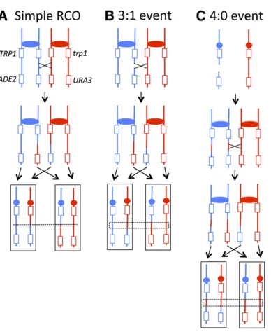
Figure 3 Mechanisms of repair leading to terminal LOH events in the red/ white sectoring system and in 5-FOA-resistant colonies. Blue and red lines represent the YJM789-derived and W303-derived chromosomes, tively. Rectangles and circles represent genes and centromeres, respec-tively. In two of the three types of repair in thisfigure, a red/white sectored colony is formed. (A) A double-strand break (DSB) leads to a reciprocal crossover (RCO) event. After thefirst mitotic division following the crossover, the sectored colony has reciprocal LOH products in the white, Ura2and the red Ura+ sides of the sector. (B) A DSB on the
YJM789-derived homolog repairs through break-induced replication (BIR) of the W303-1A-derived homolog, leading to a nonreciprocal terminal LOH event. Both sides of the red/white sector are Ura+. (C) A DSB on the
W303-1A-derived homolog repairs by BIR, using the YJM789-derived homolog as a template. Although no red/white sectored colony is formed, one half of the colony is Ura2and the other half is Ura+.
than wild type. Though the rates of LOH were similar for the
rnh201Dandrnh201Dpol2-M644Lstrains, it should be noted that there was a significant,20% decrease in the double mutant (P= 0.016 by Mann–Whitney test).
Mapping RCO events on the right arm of chromosome IV
The red and white sides of sectored colonies were analyzed using chromosome IV-specific microarrays, which allows the position of crossovers and their associated gene conversion tracts to be mapped (St. Charleset al.2012; St. Charles and Petes 2013). For RCO events unassociated with gene conver-sion, the transition between heterozygous and homozygous SNPs occurs at exactly the same position in both sectors, in-dicated by the dotted line in Figure 6A. Gene conversion events in which one sister chromatid is broken and subse-quently repaired using a nonsister chromatid result in an LOH pattern in which the breakpoints between heterozygous and homozygous SNPs are at different positions in the two sectors (Figure 6B). In the region boxed in Figure 6B, three of the chromatids have SNPs specific to W303-1A and one has SNPs specific to YJM789. This type of conversion is, there-fore, defined as a 3:1 conversion. We infer that the DNA lesion that initiated the crossover likely occurred in S or G2
of the cell cycle because only one sister chromatid received information from the homologous donor chromosome. An-other common LOH pattern observed for spontaneous cross-overs on chromosome IV is a 4:0 conversion (Figure 6C). This class of event reflects the repair of two sister chromatids broken at approximately the same position. The 4:0 pattern of conversion likely reflects double-strand break (DSB) for-mation in G1that is subsequently replicated to give two
bro-ken chromatids (Lee and Petes 2010). Microarray analyses of the red and white sectors with these patterns are in Figure 7. In the boxed region in Figure 7B, in the red sector, the hy-bridization signal is about one, indicating one copy of W303-1A-derived SNPs and one copy of YJM789-derived SNPs; in the white sector, the ratio of hybridization indicates that
there are two copies of YJM789-derived SNPs and no copies of W303-1A-derived SNPs within the boxed region. Thus, this region represents a 3:1 conversion tract. In Figure 7C, in the boxed regions of both the red and white sectors, the hybrid-ization signals indicate that there are two copies of YJM789-derived SNPs and no copies of W303-1A-YJM789-derived SNPs, as expected for a 4:0 conversion event. Among spontaneous crossovers, an additional pattern of conversion associated with crossovers is a hybrid 4:0/3:1 tract. This pattern is con-sistent with a G1-associated DSB in which the repair of the
two broken sister chromatids results in gene conversion tracts of different lengths (St Charles and Petes 2013).
In our previous analyses of spontaneous RCOs in a wild-type strain, most events were consistent with DSB formation in G1. The ratio of the numbers of 4:0 (or 4:0/3:1 hybrid) tracts
to 3:1 tracts to no detectable tracts was 90:28:20 (St Charles and Petes 2013). The corresponding ratios for thernh201D and rnh1D rnh201D strains in the current analysis were 7:2:12 and 2:8:4, respectively. These data are summarized in Figure 8. Though the numbers of sectored colonies ana-lyzed in the current study were relatively small, the distribu-tion of events in thernh201Dorrnh1Drnh201Ddiploid was significantly different from that in the wild-type strain (P,
0.001, Fisher exact test). For thernh201Dsingle mutant, the difference was driven by an increase in events with no de-tectable conversion tracts; when just 3:1 and 4:0 events were considered, there was no significant difference from wild type (P= 1). For thernh1Drnh201Ddouble mutant, how-ever, the difference reflected a strong shift from predomi-nantly G1events in wild type to predominantly S/G2events
in the double mutant. The distribution of events in the
rnh201Dsingle mutant also was significantly different from that in thernh1Drnh201Ddouble mutant (P= 0.016, Fisher exact test).
In the wild-type strain, only 20 of 138 (14%) RCOs had no detectable gene-conversion tract, whereas in the rnh201D strain 12 of 21 crossovers (57%) had no detectable tract (P,0.001 by Fisher exact test). The simplest explanation
Figure 4 Rate of red/white sector formation in RNase H-defective strains. The numbers of sectors among total colonies screened for wild type, pol2-M644L, rnh1D,
rnh201D, rnh201D pol2-M644L, and rnh1D rnh201D
strains were 14/134864, 44/266267, 22/218008, 126/337864, 59/143669, and 78/132302, respectively. Dark and light gray bars correspond to RCO and BIR events, respectively, among sectored colonies.
of this difference is that the conversion tracts in thernh201D strain are shorter than in the wild-type strain and, therefore, less likely to include a heterozygous SNP. Indeed, direct measurements of the conversion tract lengths confirmed this expectation, with tracts being significantly shorter in the
rnh201Dmutant than in wild type (P= 0.03, Wilcoxon rank sum test); conversion tract lengths in the rnh1D rnh201D strain were not significantly different from wild type (P= 0.63). The median tract lengths in the wild-type,rnh201D, andrnh1Drnh201D(95% confidence limits in parentheses) strains were 10.6 kb (8.2–13.6 kb; (St Charles and Petes 2013), 4.8 kb (1.7–17.1 kb), and 9.3 kb (2.2–19 kb), respec-tively. It should be emphasized that these conversion tracts are all associated with a crossover. The conversion tract lengths from the subcultured strains inTable S5represent a mixture of conversion events that are associated and unasso-ciated with crossovers.
Depictions of each class of sector identified in the current analysis are shown inFigure S7and the coordinates of the breakpoints in each event are listed inTable S8. We analyzed events on chromosome IV inrnh201Dand rnh1D rnh201D mutants for enrichment of various genetic elements using the same procedure as used for the subcultured strains. None of the genomic elements listed inTable S10were significantly over- or underrepresented at recombination breakpoints on the right arm of chromosome IV (Table S12).
Discussion
Previous work has shown that strains lacking RNase H1 and/ or RNase H2 have elevated levels of mitotic recombination and chromosome loss. RNase H2-defective haploids, for ex-ample, have elevated gene conversion between closely linked repeats (Potenskiet al.2014) and increased loss of markers
flanked by direct repeats (Iiet al. 2011). Although loss of either RNase H1 or RNase H2 promoted loss of an artificial chromosome, loss of both enzymes was required to elevate LOH on chromosome III in a diploid background (Wahba
et al. 2011). Our work extends analyses of instability to a genome-wide scale in diploid yeast strains that are partially
(rnh1Dandrnh201Dsingle mutants) or completely (rnh1D
rnh201Ddouble mutant) defective in RNase H activity and uses microarrays to provide a high-resolution map of recom-bination events resulting in LOH. Significantly, in each of three assays used, LOH was elevated in the rnh201D, but not the rnh1D single mutant, and was further elevated in thernh1Drnh201Ddouble mutant. We additionally used a mutant DNA polymerase that lowers the direct incorpora-tion of rNMPs into genomic DNA, allowing us to assess the relative contributions of R-loopsvs.rNMPs to LOH in the
rnh201Dbackground. The discussion below focuses on three related issues: (1) the nature of the recombination-initiating lesion in strains lacking RNase H activity, (2) the timing of formation of the recombinogenic lesion, and (3) factors that regulate the distribution of recombination events associated with loss of RNase H activity.
Nature of the recombinogenic lesions in strains lacking RNH1 and/or RNH201
The DNA alterations that lead to elevated recombination in strains lackingRNH1orRNH201are likely rNMPs embedded in DNA (expected to accumulate inrnh201Dstrains) and/or R-loops (expected to accumulate in rnh1D and rnh201D strains). In the case of loss of RNase H2, the elevated level of intrachromosomal recombination between repeats is de-pendent on Topoisomerase I (Top1) (Potenskiet al.2014). Further genetic and biochemical studies suggest that Top1-mediated cleavage at rNMPs is followed by the sequential action of Srs2 and Exo1, which produces a single-strand gap (Potenskiet al.2014). Subsequent replication of a gap-containing chromosome would be expected to produce a bro-ken, presumably recombinogenic, chromatid. That R-loop accumulation results in a hyperrec phenotype has been shown using mutants defective in transcript processing and/or in the removal of R-loops (Aguilera and Garcia-Muse 2012; Hamperl and Cimprich 2014). The corresponding recombinogenic DNA lesion could reflect either nicking of the unpaired DNA strand within an R-loop or conflicts be-tween the replication fork and an R-loop. Either of these mechanisms would likely result in single broken sister
Figure 5 Rate of terminal LOH on chromosome IV in RNase H mutant diploids. LOH was assessed by mea-suring the rate of 5-FOA resistance, which corresponds to loss of theURA3marker near the end of chromo-some IV. For wild-type,pol2-M644L,rnh1D,rnh201D,
rnh201D pol2M644L, and rnh1Drnh201D diploids, 17, 23, 16, 17, 25, and 19 independent cultures were used to derive the rates of instability, respectively.
chromatid in S phase. It is widely assumed that most, if not all, R-loops are redundantly processed by RNase H1 and RNase H2.
In our experiments, we did not detect a hyperrec pheno-type in rnh1D single mutants, but consistently observed a three- tofivefold increase in LOH inrnh201Dstrains. In all three assays, instability in the rnh1D rnh201D strains was elevated relative to that in thernh201Dsingle-mutant strains. One interpretation of the lack of a hyperrec phenotype in the
rnh1D mutant and the substantial hyperrec phenotype in the rnh201D mutant is that stimulation of recombination upon loss of RNase H2 is solely a consequence of misincor-porated rNMPs. Two arguments suggest that this extreme hypothesis is not correct. First, since the hyperrec phenotype is stronger in thernh1 rnh201 double mutant than in the
rnh1mutant, and RNase H1 has no activity on single ribonu-cleotides, the hyperrec phenotype of thernh201strain must reflect, at least in part, some other type of lesion than single
ribonucleotides. Second, if all of the recombination events in thernh201Dmutant reflect persistent rNMPs in DNA, then the hyperrec phenotype should be substantially reduced in an
rnh201Dpol2-M644Lstrain. Prior studies have demonstrated that the presence of thepol2-M644Lallele reduces the level of rNMPs in genomic DNA 70% relative to a strain with wild-type Pol 2 activity (Nick McElhinny et al.2010a). Al-though we did observe a small reduction in LOH in the
rnh201Dpol2-M644Lstrain relative to thernh201Dstrain in one of our LOH assays, the reduction was only20%. In a similar assay, the rate of LOH in anrnh201D strain was re-duced less than twofold by thepol2-M644Lmutation (Conover
et al.2015).
Based on subtle effect of the pol2-M644L mutation, we suggest that most of the LOH in thernh201Dmutant reflects persistent R-loops rather than persistent rNMPs. Our data indicate that RNase H2 can remove most, if not all, of the R-loops that accumulate in the absence of RNase H1, but that RNase H1 can remove only a relatively small fraction of the
Figure 6 Classes of red/white sectors resulting from RCO. As in Figure 3, blue and red lines indicate YJM789- and W303-1A-derived chromatids, respectively. Three common types of crossovers that result in a red Ura+
and a white Ura2sector can be distinguished by microarray analysis. (A) If a RCO is not associated with a gene conversion, the transition between heterozygous and homozygous SNPs occurs at the same position in the two sectors. Such crossovers provide no information about the likely timing of the recombinogenic DNA lesion. (B) A DSB formed in S or G2
on the YJM789-derived chromatid is associated with the nonreciprocal transfer of information from the W303-1A-derived chromatid, producing a 3:1 gene conversion event. In the dotted box, there are three chromo-somes with W303-1A-derived sequences and only one chromosome with YJM789-derived sequences. By microarray analysis, the red sector loses heterozygosity at a more centromere-proximal location than the white sector. (C) A DSB occurs on the YJM789-derived homolog in G1, and the
broken molecule is replicated to produce two sister chromatids that are broken at the same position. Repair of the two broken DNA molecules produces a 4:0 conversion tract as indicated by the dotted lines. Repair of one these breaks is associated with a crossover, producing the red/white sectored colony.
Figure 7 Examples of microarrays showing simple crossovers, crossovers with 3:1 conversion tracts, and crossovers with 4:0 conversion tracts. The red and white sectors of sectored colonies were examined by chromo-some IV-specific microarrays. The hybridization ratio of DNA derived from the sectors relative to heterozygous control DNA is shown on they-axis; the red and blue lines represent hybridization to W303-1A-specific or YJM789-specific oliognucleotides, respectively. Thex-axis shows the SGD coordinates on the right arm of chromosome IV. (A) In this sectored colony (KO135_5_2R and KO135_5_2W inTable S8), the red and white sectors have the same point of transition between heterozygous and homozygous markers (near SGD coordinate 530 kb), indicative of a simple crossover. (B) In this sectored colony (KO132_31_17R and KO132_31_17W in Table S8), the red sector has a transition point at about coordinate 500 kb, and the white sector has a transition near coordinate 555 kb. Thus, this sectored colony has a large (55 kb) 3:1 conversion tract associated with the crossover. In the boxed region, the red sector has one copy each of W303-1A- and YJM789-derived SNPs. In this region, the white sector has two copies of YJM789-derived SNPs and no copies of W303-1A-derived SNPs. (C) In this sectored colony (KO135_5_5R and KO135_5_5W in Table S8), there is a region of
20 kb that is homozygous for the YJM789-derived SNPs in both red and white sectors, consistent with a 4:0 conversion.
R-loops that accumulate in thernh201Dmutant. Why this par-ticular division of labor might not have been evident in prior studies could reflect the monitoring of instability in much smaller genetic intervals and/or the examination of RNase H activity only under conditions of pathological R-loop accumulation. The ability of RNase H2 to fully compensate for loss of RNase H1 activity may also be related to the induction of RNase H2 in strains that lack RNase H1 (Arudchandranet al.2000).
Is there any role of misincorporated rNMPs in stimulating LOH in diploids? Several types of data suggest that there is. First, as discussed above, Potenski et al. (2014) defined a pathway in whichTop1acts on rNMPs to produce recombi-nogenic lesions; a similar pathway produces mutagenic DNA lesions (Kimet al.2011). Second, we observed a small re-duction in LOH in thernh201Dpol2-M644Lstrain relative to the rnh201D strain; this reduction was statistically signifi -cant, however, only for the assay measuring the rate of LOH on chromosome IV (Figure 5). Altogether, our data sup-port the conclusion that recombination events in strains lack-ing RNase H1 and H2 are primarily a consequence of R-loop formation, with misincorporated rNMPs playing only a minor role. This conclusion, however, is based on the assumption that the reduced incorporation of ribonucleotides resulting from the pol2-M644L-encoded DNA polymerase is not af-fected by thernh201mutation or other aspects of the genetic background in thernh201 pol2-M644Ldiploid. In addition, we assume that thernh201 pol2-M644Lstrain does not have an alteration (for example, a significantly slower S phase) that affects the likelihood that the repair template is the sister chromatid rather than the homolog.
It should also be noted that triple mutant combination of
rnh1Drnh201Dpol2-M644Gis synthetically lethal, whereas the double mutantrnh201D pol2-M644Gis viable (Lazzaro
et al.2012). Since thepol2-M644Gallele encodes a form of Pol e that incorporates increased levels of ribonucleotides (Nick McElhinny et al. 2010a), this result was interpreted as indicating a possible role of RNase H1 in the removal of ribonucleotides (Lazzaroet al.2012), although other inter-pretations of the synthetic lethality are possible.
Cell-cycle timing of recombinogenic lesions in strains lacking RNase H1 and/or H2
Most of the proposed models for the recombinogenic effects of R-loops or misincorporated rNMPs predict a DNA lesion that leads to one broken chromatid in an S- or G2-phase cell. Such a
lesion could be repaired by sister-chromatid exchange (no observable LOH) or by an interaction with the homolog that is expected to result in an associated 3:1 conversion event. In a wild-type strain, only 30% of sectored colonies have the 3:1 pattern, indicating that most LOH is initiated in G1
(St Charles and Petes 2013). Most of the events in thernh1D
rnh201D double-mutant strain had the S/G2pattern,
how-ever, with 8 of the 10 sectored colonies having 3:1 conversion tracts. This result is consistent with the recombinogenic le-sion resulting from an interaction of the replication fork with an R-loop. In contrast, in thernh201D strain, seven of the nine events had 4:0 or 3:1/4:0 conversion tracts indicative of a G1-initiated event, and thus were similar to wild type. One
interpretation of this result is that RNase H2 might be spe-cialized to remove R-loops that give rise to recombinogenic lesions outside of S phase. Indeed, immunological data sug-gest that30% of pathological R-loops exert their recombi-nogenic effect outside of S phase (Wahbaet al.2011). The transcription of RNH201 is elevated prior to S phase and continues at a high level in S (Pramilaet al.2006); the tran-scription of RNH1 is not periodic (Pramilaet al. 2006). Alternatively, this result could reflect the production of a double-stranded DNA break reflecting the processing of rNMPs that are close (,10 bp) together on opposite strands of duplex DNA.
Distribution of recombination events associated with loss of RNase H activity
With the possible exception of the ribosomal RNA genes, no strong hotspots for LOH were observed in the subcultured
rnh201D,rnh201D pol2-M644L, or rnh1D rnh201D strains (Figure S4,Figure S5, andFigure S6). In particular, we found no correlation of LOH breakpoints with a number of chromo-some elements expected to be associated with R-loop forma-tion such as highly transcribed genes, intron-containing genes, or G4-forming motifs. We also observed no strong correlation between recombination breakpoints in subcul-tured mutant strains and regions of R-loop accumulation. Finally, we found no association between regions ofRpb3p
(a subunit of RNA polymerase II) accumulation and recom-bination breakpoints in thernh201D,rnh201Dpol2-M644L, orrnh1D rnh201Dstrains. These observations suggest that R-loop associated recombinogenic lesions are widely distrib-uted throughout the yeast genome and/or that a number of different factors each contribute to the hyperrec phenotype of strains lacking RNase H activity.
Although several studies reported that Ty elements accu-mulated RNA:DNA hybrids in strains lacking RNase H activity (Chanet al.2014; El Hageet al.2014), we found no enrich-ment of Ty eleenrich-ments among LOH events. We did, however,
Figure 8 Distribution of sector types by cell cycle stage in which the initiating lesion arose. Conversion tracts with a 4:0 region are indicative of a G1-associated DSB (gray bars), and 3:1 tracts indicate a G2-/S-phase
DSB (black bars). The stippled bars show simple CO events in which the timing of the recombinogenic lesion cannot be inferred.
find that deletion and duplication events frequently resulted from homologous recombination between nonallelic Ty ele-ments (Table S6). It is difficult to assess the significance of this observation since Ty elements are the primary type of large dispersed repeats in the yeast genome. In addition, non-allelic recombination between Ty elements is a common source of chromosome rearrangements in mutant yeast strains that do not accumulate R-loops (McCulley and Petes 2010; Songet al.2014). Several other relevant factors should be mentioned. First, our experiments were performed in dip-loid strains with bothMATaandMATainformation, and pre-vious studies showed that Ty transcription is repressed in such diploids (Erredeet al.1980). Second, RNA:DNA hybrids associated with Ty elements primarily involve cDNA copies rather than the genomic elements (El Hageet al.2014). Fi-nally, it should be pointed out that our genetic assays are fundamentally different than the physical analysis of R-loop formation. To be detected by our LOH assays, the recombi-nation event stimulated by R-loop formation must involve an interaction of the broken chromosome with the other homo-log; breaks that are repaired by equal sister-chromatid re-combination are genetically silent.
A significant enrichment of LOH events near the telomere was observed in thernh201Dstrain. Subtelomeric regions en-code a telomeric-repeat-containing RNA (TERRA) that accu-mulates in strains lacking RNase H2 (Yuet al.2014). Strains with increased levels of telomeric RNA:DNA hybrids have el-evated rates of telomere–telomere recombination (Yu et al.
2014). If elevated R-loops at the telomere cause a partial de-fect in telomere elongation by telomerase, there may be in-creased degradation of the ends of the chromosome, resulting in elevated levels of telomere-associated LOH. We note that Hackett and Greider (2003) previously showed an increase in terminal LOH in strains that had telomerase defects.
Summary
Our genome-wide analysis of instability associated with loss of RNase H in yeast shows that RNase H2 activity is much more important than RNase H1 activity in the maintenance of genome stability. However, strains that lack both RNase H1 and RNase H2 have qualitative and quantitative differences in genome stability relative to strains that lack only RNase H2. Our results suggest that R-loops contribute to most of the genetic instability of strains lacking RNase H activity, and that RNase H2 is uniquely able to process a subpopulation of R-loops. These results are relevant to human pathologies associated with defects in RNase H2 as well as the particular species of RNA:DNA hybrids that serve as the triggers for autoimmune disease. In the specific case of Aicardi-Goutiéres syndrome, recent work suggests that RNA:DNA hybrids are the likely immunogenic trigger of disease (Limet al.2015).
Acknowledgments
We thank J. L. Argueso and D. Koshland for communicating unpublished information and all members of the Petes and
Jinks-Robertson labs for useful suggestions. The research was supported by National Institutes of Health grants GM24110 and GM52319 to T.D.P., and GM038464 and GM101690 to S.J.-R. K.O. was supported by a National Science Foundation graduate research fellowship (1106401).
Note added in proof: See Conoveret al.2015 (pp.951–961) in this issue for a related work.
Literature Cited
Aguilera, A., and T. Garcia-Muse, 2012 R loops: from transcription byproducts to threats to genome stability. Mol. Cell 46: 115–124. Altman, D. G., 1991 Practical Statistics for Medical Research,
Chapman and Hall/CRC Press, Boca Raton, FL.
Arudchandran, A., S. Cerritelli, S. Narimatsu, M. Itaya, D. Y. Shin
et al., 2000 The absence of ribonuclease H1 or H2 alters the sensitivity ofSaccharomyces cerevisiaeto hydroxyurea, caffeine and ethyl methanesulphonate: implications for roles of RNases H in DNA replication and repair. Genes Cells 5: 789–802. Azvolinsky, A., P. G. Giresi, J. D. Lieb, and V. A. Zakian,
2009 Highly transcribed RNA polymerase II genes are imped-iments to replication fork progression inSaccharomyces cerevi-siae. Mol. Cell 34: 722–734.
Cerritelli, S. M., and R. J. Crouch, 2009 Ribonuclease H: the en-zymes in eukaryotes. FEBS J. 276: 1494–1505.
Chan, Y. A., M. J. Aristizabal, P. Y. Lu, Z. Luo, A. Hamza et al., 2014 Genome-wide profiling of yeast DNA:RNA hybrid prone sites with DRIP-chip. PLoS Genet. 10: e1004288.
Clausen, A. R., S. A. Lujan, A. B. Burkholder, C. D. Orebaugh, J. S. Williamset al., 2015 Tracking replication enzymology in vivo by genome-wide mapping of ribonucleotide incorpora-tion. Nat. Struct. Mol. Biol. 22: 185–191.
Conover, H., and S. Lujan, M. Chapman, D. Cornelio, R. Sharifet al.
2015 Stimulation of chromosomal rearrangements by ribonu-cleotides. Genetics 947–957.
Crow, Y. J., A. Leitch, B. E. Hayward, A. Garner, R. Parmaret al., 2006 Mutations in genes encoding ribonuclease H2 subunits cause Aicardi-Goutiéres syndrome and mimic congenital viral brain infection. Nat. Genet. 38: 910–916.
El Hage, A., S. Webb, A. Kerr, and D. Tollervey, 2014 Genome-wide distribution of RNA-DNA hybrids identifies RNase H tar-gets in tRNA genes, retrotransposons and mitochondria. PLoS Genet. 10: e1004716.
Errede, B., T. S. Cardillo, F. Sherman, E. Dubois, J. Deschamps
et al., 1980 Mating signals control expression of mutations resulting from insertion of a transposable repetitive element adjacent to diverse yeast genes. Cell 22: 427–436.
Fachinetti, D., R. Bermejo, A. Cocito, S. Minardi, Y. Katou et al., 2010 Replication termination at eukaryotic chromosomes is mediated by Top2 and occurs at genomic loci containing paus-ing elements. Mol. Cell 39: 595–605.
Günther, C., B. Kind, M. A. Reijns, N. Berndt, M. Martinez-Bueno
et al., 2015 Defective removal of ribonucleotides from DNA promotes systemic autoimmunity. J. Clin. Invest. 125: 413–424. Hackett, J. A., and C. W. Greider, 2003 End resection initiates genomic instability in the absence of telomerase. Mol. Cell. Biol. 23: 8450–8461.
Hamperl, S., and K. A. Cimprich, 2014 The contribution of co-transcriptional RNA:DNA hybrid structures to DNA damage and genome instability. DNA Repair (Amst.) 19: 84–94. Hochberg, Y., and Y. Benjamini, 1990 More powerful procedures
for multiple significance testing. Stat. Med. 9: 811–818. Ii, M., T. Ii, L. I. Mironova, and S. J. Brill, 2011 Epistasis analysis
between homologous recombination genes in Saccharomyces
cerevisiaeidentifies multiple repair pathways for Sgs1, Mus81-Mms4 and RNase H2. Mutat. Res. 714: 33–43.
Kim, N., S. Y. Huang, J. S. Williams, Y. C. Li, A. B. Clark et al., 2011 Mutagenic processing of ribonucleotides in DNA by yeast topoisomerase I. Science 332: 1561–1564.
Koh, K. D., S. Balachander, J. R. Hesselberth, and F. Storici, 2015 Ribose-seq: global mapping of ribonucleotides embed-ded in genomic DNA. Nat. Methods 12: 251–257.
Lazzaro, F., D. Novarina, F. Amara, D. L. Watt, J. E. Stoneet al., 2012 RNase H and postreplication repair protect cells from ribonucleotides incorporated in DNA. Mol. Cell 45: 99–110. Lea, D. E., and C. A. Coulson, 1949 The distribution of the
num-bers of mutants in bacterial populations. J. Genet. 49: 264–285. Lee, P. S., and T. D. Petes, 2010 Mitotic gene conversion events induced in G1-synchronized yeast cells by gamma rays are sim-ilar to spontaneous conversion events. Proc. Natl. Acad. Sci. USA 107: 7383–7388.
Lim, Y. W., L. A. Sanz, X. Xu, S. R. Hartono, and F. Chédin, 2015 Genome-wide DNA hypomethylation and RNA:DNA hy-brid accumulation in Aicardi-Goutières syndrome. eLife 4: doi: 10.7554/eLife.08007.
McCulley, J. L., and T. D. Petes, 2010 Chromosome rearrange-ments and aneuploidy in yeast strains lacking both Tel1p and Mec1p reflect deficiencies in two different mechanisms. Proc. Natl. Acad. Sci. USA 107: 11465–11470.
Mischo, H. E., B. Gomez-Gonzalez, P. Grzechnik, A. G. Rondon, W. Weiet al., 2011 Yeast Sen1 helicase protects the genome from transcription-associated instability. Mol. Cell 41: 21–32. Nick McElhinny, S. A., D. Kumar, A. B. Clark, D. L. Watt, B. E. Watts
et al., 2010a Genome instability due to ribonucleotide incor-poration into DNA. Nat. Chem. Biol. 6: 774–781.
Nick McElhinny, S. A., B. E. Watts, D. Kumar, D. L. Watt, E. B. Lundstromet al., 2010b Abundant ribonucleotide incor-poration into DNA by yeast replicative polymerases. Proc. Natl. Acad. Sci. USA 107: 4949–4954.
Potenski, C. J., H. Niu, P. Sung, and H. L. Klein, 2014 Avoidance of ribonucleotide-induced mutations by RNase H2 and Srs2-Exo1 mechanisms. Nature 511: 251–254.
Prado, F., and A. Aguilera, 2005 Impairment of replication fork progression mediates RNA polII transcription-associated recom-bination. EMBO J. 24: 1267–1276.
Pramila, T., W. Wu, S. Miles, W. S. Noble, and L. L. Breeden, 2006 The Forkhead transcription factor Hcm1 regulates chro-mosome segregation genes and fills the S-phase gap in the transcriptional circuitry of the cell cycle. Genes Dev. 20: 2266–2278.
Reijns, M. A., H. Kemp, J. Ding, S. M. De Proce, A. P. Jacksonet al., 2015 Lagging-strand replication shapes the mutational land-scape of the genome. Nature 518: 502–506.
Song, W., M. Dominska, P. W. Greenwell, and T. D. Petes, 2014 Genome-wide high-resolution mapping of chromosome fragile sites in Saccharomyces cerevisiae. Proc. Natl. Acad. Sci. USA 111: E2210–E2218.
St Charles, J., and T. D. Petes, 2013 High-resolution map-ping of spontaneous mitotic recombination hotspots on the 1.1 Mb arm of yeast chromosome IV. PLoS Genet. 9: e1003434.
St Charles, J., E. Hazkani-Covo, Y. Yin, S. L. Andersen, F. S. Dietrich
et al., 2012 High-resolution genome-wide analysis of irradi-ated (UV and gamma-rays) diploid yeast cells reveals a high frequency of genomic loss of heterozygosity (LOH) events. Ge-netics 190: 1267–1284.
Wahba, L., J. D. Amon, D. Koshland, and M. Vuica-Ross, 2011 RNase H and multiple RNA biogenesis factors cooperate to prevent RNA:DNA hybrids from generating genome instabil-ity. Mol. Cell 44: 978–988.
Williams, J. S., and T. A. Kunkel, 2014 Ribonucleotides in DNA: origins, repair and consequences. DNA Repair (Amst.) 19: 27–37.
Williams, J. S., D. J. Smith, L. Marjavaara, S. A. Lujan, A. Chabes
et al., 2013 Topoisomerase 1-mediated removal of ribonucle-otides from nascent leading-strand DNA. Mol. Cell 49: 1010– 1015.
Yin, Y., and T. D. Petes, 2013 Genome-wide high-resolution map-ping of UV-induced mitotic recombination events in Saccharo-myces cerevisiae. PLoS Genet. 9: e1003894.
Yu, T. Y., Y. W. Kao, and J. J. Lin, 2014 Telomeric transcripts stimulate telomere recombination to suppress senescence in cells lacking telomerase. Proc. Natl. Acad. Sci. USA 111: 3377–3382.
Communicating editor: N. Hollingsworth
GENETICS
Supporting Information
www.genetics.org/lookup/suppl/doi:10.1534/genetics.115.182725/-/DC1
Elevated Genome-Wide Instability in Yeast Mutants
Lacking RNase H Activity
Karen O’Connell, Sue Jinks-Robertson, and Thomas D. Petes
2 SI K. O'Connell, S. Jinks‐Robertson, and T. D. Petes
File S1. Expanded Materials and Methods
Strain construction
Most of the details of the strain constructions are in Table S1. As in previous studies
(for example, Lee
et al.
, 2009), we used derivatives of the sequence-diverged haploids
W303-1A and YJM789. These haploids with various genetic alterations were crossed to
generate the diploids used to assay genetic instability. Most haploid strains were
constructed by transformation with PCR-generated DNA fragments or by sporulating
nearly-isogenic diploids. The genotypes of spores for auxotrophic markers were
determined by replica-plating spore-derived colonies to omission media. The
replacements of genes with drug-resistance markers were confirmed by PCR analysis
as described in Tables S2 and S3. Mating type was determined by PCR with primers
MATaF, MATalphaF, and MATR (Table S3).
MAT
a
and
MAT
loci were associated with
500 and 400 bp fragments, respectively.
Measurements of rates of genetic instability induced by loss of RNase H
We used three methods to examine the rates of instability in strains with mutations
affecting RNase H activity. We first measured the frequency of genomic alterations in
sub-cultured diploid strains of the following genotypes: wild-type,
rnh1
∆
,
rnh201
∆
,
rnh1
∆
rnh201
∆
,
pol2-M644L
, and
rnh201
∆
pol2-M644L
. All diploids were generated by
crossing haploids isogenic with W303-1A and YJM789. Two independently-derived
isolates from each strain were streaked with a toothpick to single colony density on rich
growth medium (YPD) for the first subculture. For the second subculture, five-ten
colonies from each isolate were then re-streaked to YPD. One colony derived from each
of these ten to twenty colonies was then re-streaked again. For the mutant backgrounds,
K. O'Connell, S. Jinks‐Robertson, and T. D. Petes 3 SI