Multiple axon guidance cues establish the olfactory topographic
map: how do these cues interact?
JAMES A. ST. JOHN*, HEIDI J. CLARRIS and BRIAN KEY
1Department of Anatomy and Developmental Biology, School of Biomedical Sciences and 1Centre for Functional and Applied Genomics, University of Queensland, Brisbane, Australia
ABSTRACT Each primary olfactory neuron stochastically expresses one of ~1000 odorant receptors. The total population of these neurons therefore consists of ~1,000 distinct subpopulations, each of which are mosaically dispersed throughout one of four semi-annular zones in the nasal cavity. The axons of these different subpopulations are initially intermingled within the olfactory nerve. However, upon reaching the olfactory bulb, they sort out and converge so that axons expressing the same odorant receptor typically target one or two glomeruli. The spatial location of each of these ~1800 glomeruli are topographically-fixed in the olfactory bulb and are invariant from animal to animal. Thus, while odorant receptors are expressed mosaically by neurons throughout the olfactory neuroepithe-lium their axons sort out, converge and target the same glomerulus within the olfactory bulb. How is such precise and reproducible topographic targeting generated? While some of the mechanisms governing the growth cone guidance of olfactory sensory neurons are understood, the cues respon-sible for homing axons to their target site remain elusive.
KEY WORDS:
development, navigation, target, growth cone
0214-6282/2002/$25.00 © UBC Press
Printed in Spain
www.ijdb.ehu.es
*Address correspondence to: Dr. James St John. Department of Anatomy and Developmental Biology, School of Biomedical Sciences, University of Queensland, Brisbane, 4072, Australia Fax: +61-7-3365-1299. e-mail: [email protected]
Abbreviations used in this paper: CAM, cell adhesion molecule; NFL, Nerve
fibre layer.
Introduction
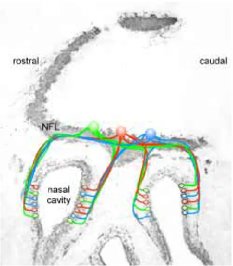
Sensory perception relies on the conversion of external stimuli into some form of internal neural representation. In the auditory, visual and somatosensory systems this process is dependent on the presence of point-to-point topographical maps between the sensory apparatus and specific brain regions. As in these other sensory systems, the discrimination of odours depends on the formation of a sensory topographical map. This olfactory sensory map is not based on a simple point-to-point map of the sensory epithelium onto olfactory processing areas in the brain. Instead, the odotopic map within the glomerular sheet of the olfactory bulb is based on the massive convergence of axons expressing like odorant receptors (Fig. 1) (Ressler et al., 1994; Vassar et al., 1994; Mombaerts et al., 1996; Royal and Key, 1999). Within the olfactory neuroepithelium in rodents, each olfactory sensory neuron expresses only one of ~1000 different types of odorant receptor (Chess et al., 1994; Malnic et al., 1999). Sensory olfactory neurons expressing a particular odorant receptor protein reside within one of four longitudinally oriented zones within the olfactory neuroepithelium (Ressler et al., 1993; Vassar et al., 1993). Within each zone these neurons appear to be stochasti-cally distributed. However, all neurons expressing a particular odorant receptor project their axons to a small number of
glom-eruli within the olfactory bulb (Ressler et al., 1994; Vassar et al., 1994; Mombaerts et al., 1996; Royal and Key, 1999). The task of sorting approximately 1000 subsets of axons is compounded by the high degree of convergence of olfactory sensory neurons onto individual glomeruli. In the mouse there are approximately 107 sensory neurons on each side of the nasal cavity and mately 1800 glomeruli per olfactory bulb. Therefore, approxi-mately 5000 axons converge onto a single glomerulus (Royet et al., 1988; Pomeroy et al., 1990).
neurons expressing the P2 odorant receptor project their β -galactosidase-positive axons to a few (1 to 4) glomeruli on both the medial and lateral olfactory bulb (Mombaerts et al., 1996, Royal and Key, 1999). The positions of these glomeruli are highly conserved between bulbs, both within the same animal as well as between different animals (Schaefer et al., 2001).
In order to begin to understand how this complex topographic map is constructed it is convenient to divide the developing primary olfactory pathway into three stages. First, the pioneer sensory axons leave the nasal pit and extend through the intervening mesenchyme to the telencephalon. These axons are accompanied by a heterogeneous population of cells and together, the axons and cells, are referred to as the migratory mass. One of the cell types is a specialised glia, the olfactory ensheathing cell (OEC), which forms a glial bridge between the nasal pit and the rostral surface of the telencephalon (Doucette, 1989; Tennet and Chuah, 1996). Second, axons and accompanying cells commence penetrating the boundary of the telencephalon and begin to form the outer nerve fibre layer of the olfactory bulb. During this stage, axons begin to sort out into subsets destined to target restricted domains within the olfactory bulb. Third, primary sensory axons terminate in tufts of neuropil called glomeruli in topographical-fixed positions to form the olfactory sensory map.
The Key Stages in the Development of the Olfactory
System
The Growth of Olfactory Axons from the Olfactory Neuroepi-thelium to the Olfactory Bulb
The olfactory neuroepithelium originates from cranial ectoderm overlying the presumptive cerebral hemispheres. In the mouse, the olfactory placodes are first observed at E10 as distinct rounded thickenings of epithelium, separated from the presumptive telen-cephalon by mesenchyme (Hinds, 1972a). At E11, the placodes invaginate forming nasal pits and primary olfactory sensory neu-rons are first identified (Hinds, 1972b). Axons then begin to extend from the neuroepithelium, forming small fascicles which pierce the basal lamina (Marin-Padilla and Amieva, 1989). Axons grow dor-sally into the intervening frontonasal mesenchyme and by E11.5, small fascicles have started to coalesce to form a single nerve (Whitesides and LaMantia, 1996).
Penetration of Olfactory Axons into the Bulb
Once the first olfactory sensory axons reach the telencephalon, they form a transient mass residing just outside, but not in contact with the telencephalon. In the mouse, this stalling period is accom-panied by the expression of inhibitory molecules containing CS-56 chondroitin sulfates (Treloar et al., 1996) within the marginal zone. We have shown that the penetration of the first olfactory axons is accompanied by a down-regulation of these chondroitin sulfates (Treloar et al., 1996). Following this down-regulation, the contents of the migratory mass fuse with the telencephalon to become the presumptive nerve fibre layer. In the chick, the inhibitory secreted glycoprotein semaphorin-3A is found on the surface of the telen-cephalon at the equivalent time, while its ligand neuropilin-1 is expressed by all olfactory sensory neurons. The down-regulation of semaphorin-3A at the telencephalic border coincides with the entry of neuropilin-1 positive axons into the olfactory bulb (Kobayashi et al., 1997). Over-expressing a dominant-negative neuropilin-1 in chicks results in many olfactory axons entering the telencephalon prematurely (Renzi et al., 2000). Together these findings suggest that during this period of stalling, a combination of inhibitory influences prevent the premature entry of olfactory axons into the olfactory bulb.
Within the olfactory nerve, olfactory axons expressing different odorant receptors intermingle within bundles (Royal and Key, 1999). However, once they enter the outer region of the olfactory nerve fibre layer, there is considerable defasciculation and sorting such that at least with respect to the P2 subset of axons, axons begin to sort out into smaller subsets which subsequently coalesce and form distinct glomeruli (Royal and Key, 1999). Therefore, the outer nerve fibre layer appears to be a sorting zone for olfactory axons in the same way it is a sorting zone in the olfactory pathway of insects (Rössler et al., 1999).
The Formation of Glomeruli
We have analysed the development of the P2 subpopulation of olfactory sensory neurons and their projections to the olfactory bulb using the P2-IRES-tau-LacZ line of transgenic mice (Mombaerts et al., 1996). P2 axons begin to target a specific region on both the medial and lateral surfaces of the olfactory bulb as early as E15.5 (Royal and Key, 1999). However, initially within the glomerular layer, axons intermingle with other odorant receptor-expressing
Fig. 1.Primary olfactory neurons are distributed in a mosaic pattern.
axons and terminate diffusely. At E17.5, P2 axons start to cluster and to target a specific region within the glomerular layer and by E18.5 the P2 axons form 2 to 3 tufts. Between E18.5 and P7.5, these tufts either condense and form a single glomerulus or clearly separate into discrete glomeruli connected by a bundle of axons. By P14.5, all interconnected glomeruli have separated and formed discrete glomeruli.
The Cellular and Molecular Cues that contribute to Axon
Guidance
Axons grow in Fasciculated Bundles along the Olfactory Nerve
Within the olfactory nerve, axons grow in small fascicles that become intimately associated with the surface of migratory cells (Marin-Padilla and Amieva, 1989). Cell adhesion molecules such as the neural cell adhesion molecule (NCAM) and L1 which are expressed ubiquitously within the olfactory nerve, may mediate the fasciculation of olfactory axons (Linnemann et al., 1988, Linneman and Bock, 1989). These adhesion molecules are believed to stimulate axon outgrowth by activating intracellular signalling path-ways mediated by the SRC-family tyrosine kinases, p59fyn and pp60c-src. Mice deficient in both of these tyrosine kinases have defects in the fasciculation of the olfactory nerve suggesting that L1 and NCAM are important cell adhesion molecules during develop-ment of the peripheral trajectory of primary olfactory axons (Morse et al., 1998). While olfactory axons in p59fyn and pp60c-src double mutants continue to reach the telencephalon despite their aberrant fasciculation, it remains to be determined whether these axons terminate in their correct glomerular targets.
The fasciculation of olfactory axons within the nerve might also be modulated by molecules such as the neuronal condroitin sulfate proteoglycan (CSPG), neurocan. Neurocan, which is expressed during development by both olfactory sensory neurons and OECs (Clarris et al., 2000), can inhibit the homophilic binding of both NCAM and L1 (Grumet et al., 1993). We assessed the effect of recombinant neurocan on the growth of olfactory axons in vitro (Clarris et al., 2000) and found that neurocan, both as a substrate-bound molecule and in secreted form, stimulated the growth of neurites from dissociated embryonic olfactory sensory neuron (Clarris et al., 2000). These results suggest that neurocan supports the growth of primary olfactory axons through the extracellular matrix (ECM) as they project to the olfactory bulb during develop-ment, possibly by modulating the interactions of NCAM and L1. Like the action of polysialylated (PSA) NCAM in inhibiting adhesion along the olfactory nerve during development (Aoki et al., 1999), neurocan may stimulate olfactory sensory axon growth by inhibit-ing their adhesion either to each other or to OECs within the nerve.
The Role of Olfactory Ensheathing Cells in Olfactory Axon Growth
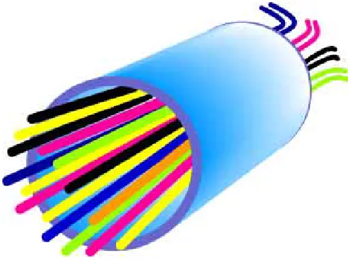
The olfactory axons grow out of the olfactory neuroepithelium accompanied by a heterogeneous population of migrating cells. These migratory cells consist of both neuronal and glial pheno-types. The glial cells go on to form OECs that populate the olfactory nerve and the olfactory nerve fibre layer (Tennent and Chuah, 1996). During early development, a close association is observed between olfactory axons and OECs within the olfactory nerve and this is maintained as they enter the olfactory bulb (Fig. 2)
(Marin-Padilla and Amieva, 1989). In situ, OECs express glial fibrillary acidic protein (GFAP) (Barber and Dahl, 1987), S100 (Pixley, 1992), the low affinity nerve growth factor receptor p75 (Gong et al., 1994; Turner and Perez-Polo, 1994) and the adhesion molecules L1 (Miragall et al., 1989), NCAM (Miragall et al., 1988, 1989; Doucette, 1990; Miragall and Dermietzel, 1992) and PSA-NCAM (Miragall et al., 1988, 1989; Miragall and Dernietzel, 1992). They also express laminin (Doucette, 1990) and possibly tenascin (Gonzales and Silver, 1994).
We and others have found OECs to be a highly conducive substrate for olfactory axon outgrowth in vitro (Goodman et al., 1993; Chuah and Au, 1994; Key et al., 1996; Kafitz and Greer, 1998, 1999). ECM molecules such as laminin, heparan sulfate proteoglycan (HSPG) and collagen IV are stimulatory to the growth of olfactory axons and are expressed within the olfactory nerve as these axons first grow towards the telencephalon (Treloar et al., 1996). To investigate the interaction between the ECM, migrating OECs and olfactory neurites, we developed a technique whereby explants of embryonic rat olfactory neuroepithelium were plated onto various substrates (Tisay and Key, 1999). On a substrate of Matrigel (Collaborative Biomedical Products, Bedford MA) OECs migrated prodigiously from these explants and olfactory neurites grew out avidly over the surface of these migrating OECs. To examine the role of various ECM components, explants were grown on substrates of laminin, HSPG and CSPG. We found that both Matrigel and laminin promoted the migration of OECs away from explants, while HSPG and CSPG appeared to restrict the number of OECs migrating. Of interest was the finding that the extent of neurite outgrowth from explants was directly proportional to the degree of OEC migration, suggesting that in vivo olfactory axons may follow the migratory path of OECs within the olfactory nerve. It is therefore likely that growth and guidance cues that affect
Fig. 2.Fascicles of intermingled olfactory axons are surrounded by
olfactory ensheathing cells. Primary olfactory neurons express different
the migratory capacity of OECs are crucial to the development of the olfactory nerve.
To more closely examine the effect of the ECM specifically on the migration of OECs, we prepared dissociated cultures of OECs from olfactory nerve fascicles teased from the submucosa of the sheet of neuroepithelium lining the nasal septum of early postnatal rats (Tisay and Key, 1999). We grew these dissociated OECs on substrates of Matrigel, laminin, HSPG or CSPG and found that while both Matrigel and laminin substrates facilitated the spreading of OECs, HSPG and CSPG did not. These studies showed that the ECM can have indirect effects on the growth of olfactory neurites via modulation of OEC growth. A previous study has also shown that during development, OECs migrate toward the rostral telen-cephalon in response to a soluble trophic factor(s) released specifi-cally by the presumptive olfactory bulb (Liu et al., 1995). We propose that OECs secrete ECM molecules such as laminin that then act autocrinely to stimulate their migratory and neurite-outgrowth promoting activities. The chemoattractive factor(s) re-leased from the olfactory bulb may act directly on the OECs to promote growth, but may also act to regulate their expression of molecules such as laminin. Although we have shown that in vitro molecules like laminin can affect the growth-promoting properties of OECs, this has yet to be shown in vivo.
Mesenchymal Influences on the Trajectory of Olfactory Axons
While the migration of OECs and the subsequent growth of olfactory axons are stimulated by molecules present within the olfactory nerve, the surrounding frontonasal mesenchyme ex-presses molecules that confine their migration to defined regions (Treloar et al., 1996). We have examined the expression of several ECM constituents during the formation of the olfactory nerve pathway and found that CSPG is selectively present in the mesen-chyme between the olfactory nerve pathway and the nasal pit and in the marginal zone of the telencephalon (Treloar et al., 1996). In a search for possible candidate CSPGs involved in the delineation of the olfactory nerve route, we found that the expression of the proteoglycan phosphacan was confined to the mesenchyme sur-rounding the early migratory mass (Clarris et al., 2000). Phosphacan and possibly other CSPGs are probably secreted by mesenchymal cells that demarcate boundaries over which migrating olfactory ensheathing cells and accompanying olfactory axons cannot cross. Whether CSPGs act by directly inhibiting the extension of neurites or by inhibiting the migration of OECs is not known. However, at least in vitro, we have shown that OECs fail to spread and migrate on a substrate containing CSPG and that this subsequently re-duces their ability to promote the growth of olfactory axons (Tisay and Key, 1999). While CSPGs appear to play an important role in restricting the trajectory of the first olfactory axons, their role after the olfactory nerve pathway has become continuous with the central nervous system is unclear.
Molecules produced within specialised domains of the mesen-chyme may also promote the growth of the migratory mass. The dorsolateral surface of the olfactory nerve is bounded by a region of neural crest-derived mesenchyme that selectively expresses Pax-7 (LaMantia et al., 2000). In vitro, reduction of Pax-7 expres-sion within this domain alters the trajectories of olfactory axons from neuroepithelial explants (LaMantia et al., 2000), suggesting that this region of the mesenchyme is important in peripheral axon navigation. Whether these mesenchymal influences affect the
migratory capacity of OECs and/or directly impact on the growth activity of olfactory axons remains to be determined.
Role of Carbohydrates in Axon Fasciculation
Cell surface carbohydrates are emerging as key regulators of cell-cell interactions in many tissues (Anderson and Key, 1999; Rudd et al., 2001; Zhu and Wang, 1998). Although these molecules are typically ubiquitously expressed throughout the nervous sys-tem there are examples where unique glycoforms of cell adhesion molecules, such as PSA-NCAM, exhibit spatiotemporal variations in expression (Nakayama et al., 1998). In the olfactory system, several discrete glycoforms of N-CAM, such as 3 and NOC-4 (Dowsing et al., 1997) and Gal-N-CAM (Pays and Schwarting, 2000) are expressed by subpopulations of primary olfactory neu-rons while a 205 kD fucosylated form of NCAM is expressed by all primary olfactory neurons (Pestean et al., 1995). While those subpopulations of axons that express unique NCAM glycoforms are mosaically dispersed in the olfactory nerve, they sort out and selectively fasciculate into like bundles in the olfactory nerve fibre layer (St John and Key, 2001a). Thus, this restricted expression of NCAM glycoforms may provide a mechanism for selective fascicu-lation of axons.
We believe that the restricted expression of cell surface carbo-hydrates on olfactory axons provides a “glycocode” that mediates the sorting out of olfactory axons through interactions with carbo-hydrate-binding proteins such as galectin-1. We have shown that in the developing rat, galectin-1 is expressed by OECs both in olfactory nerve and in the nerve fibre layer of the olfactory bulb (St John and Key, 1999). Furthermore, we have shown that a galectin-1 ligand, N-acetyl-lactosamine is expressed by olfactory axons that terminate specifically in glomeruli present in the ventromedial and lateral olfactory bulb (St John and Key, 1999). Galectin-1 may bind and cross-link axons expressing galectin-1 ligands such as N-acetyl-lactosamine (Puche and Key, 1996), thereby facilitating the sorting out of olfactory axons into subpopulations.
To more directly assess the function of galectin-1 in axon sorting and glomerular targeting we examined the projection of a subpopu-lation of olfactory axons in mice deficient for galectin-1 (Puche et al., 1996). The plant lectin Dolichos biflorus agglutinin (DBA) binds to a subpopulation of olfactory sensory neurons that are widely scattered within the olfactory neuroepithelium (Key and Akeson, 1993; Treloar et al., 1996). The DBA-reactive axons are widely dispersed within the olfactory nerve but sort out within the outer region of the olfactory nerve fibre layer (Key and Akeson, 1993). These axons then terminate in glomeruli present predominantly in the dorsomedial olfactory bulb. In mice deficient in galectin-1, although rostral regions of the bulb appear unaffected, few DBA-reactive axons grew into caudal regions of the olfactory bulb. These results supported our earlier findings suggesting that galectin-1 plays an important role in the sorting of axons and subsequent glomerular targeting.
Targeting and the Role of the Odorant Receptors
The glomerulus targeted by an olfactory axon is dictated by its choice of odorant receptor (Mombaerts et al., 1996). It is therefore possible that odorant receptors may be bifunctional: regulating signal transduction in the nasal cavity and contributing to axon guidance in the growth cone to enable axons to target specific glomeruli. This has been tested with both P2 deletion and substi-tution experiments which demonstrated that odorant receptor molecules play an instructive role in glomerular targeting (Mombaerts et al., 1996; Wang et al., 1998). Substitutions that replaced the P2 coding sequences with receptor sequences expressed either in different zones or from different chromosomal loci resulted in the convergence of axons to glomeruli distinct and more distant from their wild type counterparts (Wang et al., 1998). These results indicate that selection of precise glomerular targets involves both odorant receptor and additional guidance receptors that reflect the neuroepithelial zone within which the odorant receptor is expressed. In the most conservative substitution in which the P2 odorant receptor was swapped for the coding sequence of P3, expressed within the same zone of P2, axons targeted to a glomerulus immediately adjacent to the P3 glomeru-lus (Wang et al., 1998). This targeting to adjacent glomeruli could be due to an altered level of P3 expression in these neurons in comparison to wild type P3-expressing axons. If graded guidance cues are read by sensory axon termini, then even slight changes in the level of P3 may result in positional alterations in axon projections.
Two possible speculations have been raised regarding the role of the odorant receptor in axon guidance. One is that the odorant receptor itself functions as a guidance receptor, which binds the proposed ligands present in an anteroposterior gradient across the bulb. In this model, the binding affinities of highly homologous odorant receptors to these putative ligands are similar, resulting in the projection of axons from neurons express-ing these receptors to neighbourexpress-ing glomeruli (Tsuboi et al., 1999). Another speculation is that odorant receptors themselves do not bind to putative guidance ligands, but instead, each odorant receptor gene locus contains a second and distinct set of receptors co-expressed with the odorant receptor.
The Role of Activity in Glomerular Targeting
The targeting of olfactory axons and the formation of glomeruli has the potential to be influenced by activity-mediated pruning of axonal arbours. Activity-dependent mechanisms are important in the visual system for the creation of ordered ocular dominance columns of alternating input to the lateral geniculate nucleus from either eye (Katz and Shatz, 1996). However, it remains debatable whether similar activity dependent mechanisms are occurring in the olfactory system. Two recent studies have reported conflicting results regarding the role of neural activity in axon targeting in mice deficient in the olfactory cyclic nucleotide-gated channel OCNC-1 (Lin et al., 2000; Zheng et al., 2000). While the trajecto-ries of P2 and M50 axons were unaffected by the loss of odorant evoked neural activity (Zheng et al., 2000), M72 axons formed aberrant small ectopic glomeruli (Lin et al., 2000). As yet the effects on axon overshooting in the glomerular layer are yet to be examined in these mice. Nonetheless it appears that there may be differences in the sensitivities of different olfactory neuron sub-populations to the loss of neuronal activity. What is clear however bulb targeted by axons expressing the NOC-3 glycoform of NCAM
(St John and Key, 2001a). These glomeruli are in fixed topographi-cal positions in both the left and right bulbs as well as in the bulbs of different animals. Thus axons innervating the same glomerulus are identified uniquely not only by expression of odorant receptor type but also by cell surface carbohydrates.
The Role of Chemorepulsion in the Sorting of Olfactory Axons
Little attention has been paid to the role of chemorepulsive molecules in the sorting out of olfactory axons within the olfactory nerve fibre layer. One possibility is that chemorepulsive molecules are expressed on OECs and that these molecules act on appropri-ate receptors expressed on olfactory sensory axons. In this way, particular axon subsets could be prevented from entering certain regions within the nerve fibre layer. Neuropilin-1 is expressed by a subpopulation of olfactory sensory neurons that are diverted away from ensheathing cells expressing the secreted neuropilin-1 ligand, semaphorin 3A in the outer nerve fibre layer of the bulb (Crandall et al., 2000; Schwarting et al., 2001). Furthermore, in semaphorin 3A homozygous mutant mice, the sorting of axons is disrupted in the nerve fibre layer and axons target topographically inaccurate glomeruli (Schwarting et al., 2000).
Olfactory Axons initially target Glomeruli Aberrantly
In many neuronal systems exuberant neuritic growth is often followed by subsequent pruning to establish discrete neuronal connections. For example, in the chick retinogeniculate system, the growth and arborisation of temporal retinal axons is initially impre-cise. However, subsequent axonal remodelling results in the retinotopic ordering of terminal arborisations characteristic of the mature projection (Nakamura and O’Leary, 1989). We examined the trajectories of primary olfactory axons as they formed glomeruli in the olfactory bulb and found evidence of considerable exuberant over-growth of axons into deeper layers between postnatal (P) day 0 and 1.5 (Tenne-Brown and Key, 1999). The number of these misguided axons progressively decreased from P2.5 until at P14.5. This over-shooting of axons was confirmed when we examined the P2 subset of axons during the development in the P2-IRES-tau-LacZ line of mice (Royal and Key, 1999). The extent of exuberant growth de-creased with age and was absent from adults. This analysis revealed that although P2 axons target specific loci in the olfactory bulb as early as E15.5, discrete glomeruli emerge slowly and navigational errors occur for up to the next 10 days of development.
is that despite such an intrusive insult to the olfactory system, primary olfactory axon navigation is only minimally affected.
Targeting and the Role of Bulbar-Derived Guidance Cues
Odorant receptors, or co-ordinately expressed sets of guidance receptors, play an important role as address molecules in targeting glomeruli (Mombaerts et al., 1996; Wang et al., 1998). It is most likely that spatial cues in the olfactory bulb direct axons to their precise glomerular targets. Mitral cells have long been thought to provide cues for the guidance of olfactory axons. They are present within the bulb as olfactory axons first enter the bulb (Hinds, 1972a) and form synaptic contacts with olfactory axons within glomeruli. However, Bulfone et al. (1998) have shown that in transgenic mice which lack most mitral cells, P2 axons continue to target topo-graphically-fixed glomeruli. These authors suggested that the critical cellular component for targeting in the bulb might instead be the radial glial cells. Radial glial processes in the rat form “glial glomeruli” at the same time that olfactory sensory axons coalesce into glomeruli (Bailey et al., 1999). Positional cues may be pre-sented by the endfeet process of radial glia, which interact with the olfactory sensory axons at the earliest stages of glomerular forma-tion (Puche and Shipley, 2001). In support of this is the finding that each radial glial cell extends tufts into only a single developing protoglomerulus (Puche and Shipley, 2001).
The Nature of Bulb-Derived Guidance Cues - the Two-Point Gradient Model
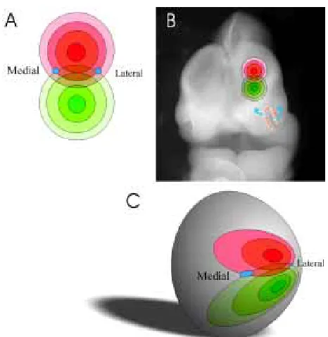
What is the nature of the cue(s) in the bulb that determines target sites? One possibility is that separate target-derived cues will guide each olfactory axon subset to appropriate glomerular sites. How-ever, since there are ~1000 odorant receptors, this would neces-sitate the expression of 1000 separate guidance cues in topo-graphically invariant positions. This seems uneconomical. It is more likely that, as in other CNS systems, olfactory axons will be guided to targets by a small number of topographically aligned factors. We favour the hypothesis that glomerular target sites are defined by the expression of two overlapping gradients of ligands that are distributed over the glomerular surface of the olfactory bulb (Gierer, 1998). Because axons expressing a particular odorant receptor target one medial and one lateral glomerulus in each olfactory bulb, a gradient model needs to incorporate duplicate gradients both on the medial and lateral surfaces.
We propose a model that is based on gradients set up from point sources (Fig. 3) rather than the more typical linear sources, such as those envisaged in the visual system (Loschinger et al., 2000). Each point source would represent the centre of a gradient of radially aligned cues with the level of cue decreasing with distance from the point source. In this model, two point sources would create a symmetrical co-ordinate system of radially dispersed cues. Such a system is analogous to the patterns generated when two stones are dropped in a pond. The intersection of radially expanding waves defines points in space just as two overlapping gradients of ligands emanating from point sources in a flat neuroepithelial sheet. When such a two-dimensional sheet is wrapped over the ellipsoidal shaped olfactory bulb (with the point sources lying dorsal and ventral to each other over the rostral surface of the bulb) duplicated gradients would be established on the medial and lateral surfaces of the bulb.
The final stereotactic arrangement of cues depends on the ultimate shape of the bulb. If one side of the bulb grew disproportionably more than the other, there would be a skew in the position of identical glomeruli between the medial and lateral surfaces. This appears to be the case since identified glomeruli never appear to be at the same rostrocaudal or dorsoventral position on either the medial or lateral surface of the bulb. In fact, for all glomeruli examined to date, the medial glomerulus is always located slightly more caudally along the rostrocaudal axis than its counterpart on the lateral surface. It appears that the gradient has uniformly shifted caudally on the medial surface.
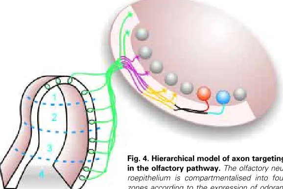
A Model of Axon Guidance in the Olfactory System
It is now clear that while odorant receptors play an instructive role in olfactory axon targeting, other molecules are likely to act in a combinatorial way to guide the axons from the sensory neu-roepithelium to the target glomerulus. Thus, we favour a hierarchi-cal model of axon navigation to explain formation of the olfactory pathway (Fig. 4). First, ubiquitously expressed cell adhesion molecules such as N-CAM and L1 may promote fasciculation of olfactory axons. Second, molecules such as OCAM (Alenius and Bohm, 1997; Yoshihara et al., 1997; Nagao et al., 2000), which are expressed in zones 2-4 of the olfactory neuroepithelium, may sub-partition the olfactory nerve into large regional domains. Third, adhesion molecules may also facilitate the selective
fas-Fig. 3. Two-point source hypothesis of axon navigation in the primary
olfactory pathway. (A) Two point sources establish circular gradients of
ciculation of axons arising from single zones. While zone-specific adhesion molecules remain to be identi-fied there is evidence for restricted expression of other molecules in a single zone (Miyawaki et al., 1996). The sequential or hierarchical expression of cell adhesion molecules as described here would provide a mecha-nism for maintaining a gross topographic order of pro-jections between the nose and the bulb. Of course these chemoadhesive interactions may be comple-mented by chemorepulsive sorting that drives axons away from inappropriate bulbar regions (Nagao et al., 2000; Schwarting et al., 2000). Fourth, subpopulations of axons express unique cell surface carbohydrates such as NOCs (Dowsing et al., 1997; St John and Key, 2001a; Pays and Schwarting, 2000). These NOCs could form a glycocode that would enable sorting of axons into small fascicles in the nerve fibre layer (St John and Key, 2001a) through interactions with carbo-hydrate-binding proteins such as galectin-1 (Puche et al., 1996; St John and Key, 1999; Tenne-Brown et al., 1998). Even though these carbohydrates are expressed by subpopulations of axons dispersed across all zones of the nasal cavity they would still promote the self-fasciculation of axons into small fascicles within a single zone since zonal specific cell adhesion or chemorepulsive molecules would maintain the gross ordering of axons. The role of cell surface carbohy-drates in the hierarchy of interactions would ensure the fine partitioning of axons, a necessary prerequisite for
Fig. 4.Hierarchical model of axon targeting
in the olfactory pathway. The olfactory
neu-roepithelium is compartmentalised into four zones according to the expression of odorant receptor proteins. Each zone expresses a unique complement of receptors. All axons express cell adhesion molecules (CAMs, repre-sented as green), such as N-CAM and L1. These CAMs are involved in fasciculating primary olfactory axons into large bundles. Expression of other CAMs such as OCAM (represented as mauve) partition axons into two discrete bundles – those arising from zone 1 and those from zones 2-4; other CAMs may further partition subpopulations of axons into smaller bundles. In the outer nerve fibre layer the axons sort out according to the expression of cell surface carbohydrates such as NOCs (represented by yellow and black). In the final stage of targeting, the expression of odorant receptor proteins (represented as blue and red) directs axons to their topographically correct position.
tions in specific “glycocodes” and analyse the effects on subpopu-lations of primary olfactory axons.
Acknowledgements
This work was supported by Australian National Health and Medical Research Council grants to JSJ and BK, by a research grant from the Mituzanti Foundation for Glycoscience to BK and by a Rodney Williams and Garnett Passe Memorial Foundation research grant to JSJ. JSJ and HC were each supported by a National Health and Medical Research Council Peter Doherty Fellowship.
References
ALENIUS, M. and BOHM, S. (1997). Idenitification of a novel neural cell adhesion molecule-related gene with a potential role in selective axonal projection. J. Biol. Chem. 272: 26083-26086.
ANDERSON, R.A. and KEY, B. (1999). Guidance cues during neuronal pathfinding in the early scaffold of axon tracts in embryonic Xenopus brain. Develop. 126: 1859-1868.
AOKI, K., NAKAHARA, Y., YAMADA, S. and ETO, K. (1999). Role of polysialic acid on outgrowth of rat olfactory receptor neurons. Mech Dev. 85: 103-110.
BAILEY, M.S., PUCHE, A.C. and SHIPLEY, M.T. (1999). Development of the olfactory bulb: evidence for glia-neuron interactions in glomerular formation. J. Comp. Neurol. 415: 423-448.
BARBER, P.C. and DAHL, D. (1987). Glial fibrillary acidic protein (GFAP)-like immunoreactivity in normal and transected rat olfactory nerve. Exp Brain Res. 65: 681-685.
BULFONE, A., WANG, F., HEVNER, R., ANDERSON, S., CUTFORTH, T., CHEN, S., MENESES, J., PEDERSEN, R., AXEL, R. and RUBENSTEIN, J.L. (1998). An olfactory sensory map develops in the absence of normal projection neurons or GABAergic interneurons. Neuron 21: 1273-1282.
CHESS, A., SIMON, I., CEDAR, H. and AXEL, R. (1994). Allelic inactivation regulates olfactory receptor gene. Cell, 78: 823-834.
their subsequent convergence. Finally, subpopulations of axons expressing specific receptor proteins then converge and target specific glomeruli.
Where to Next?
While it is clear that we have progressed considerably in the last ten years with regards to our understanding of the organization of the olfactory system there is still a long way to go before we understand the intricacies of axon navigation in this highly complex pathway. What is the role of odorant receptor in axon targeting? Does it participate in a classical receptor-ligand based interaction in order to drive the convergence of axons to a single site in the olfactory bulb? Or does it act indirectly to facilitate axon-axon adhesion prior to the growth cone responding to other guidance cues? What are the other guidance molecules that are obviously important in directing the growth of axons expressing the same receptor to their target site?
perturba-CHUAH, M.I., and AU, C. (1994). Olfactory cell cultures on ensheathing cell monolay-ers. Chem. Senses. 19: 25-34.
CLARRIS, H. RAUCH, U. and KEY, B. (2000). Dynamic spatiotemporal expression patterns of neurocan and phosphacan indicate diverse roles in the developing and adult mouse olfactory system. J. Comp. Neurol. 46: 113-125.
CRANDALL, J.E., DIBBLE, C., BUTLER, D., PAYS, L., AHMAD, N., KOSTEK, C., PUSCHEL, A.W. and SCHWARTING, G.A. (2000). Patterning of olfactory sen-sory connections is mediated by extracellular matrix proteins in the nerve fibre layer of the olfactory bulb. J. Neurobiol. 45: 195-206.
DOUCETTE, R. (1989). Development of the nerve fiber layer in the olfactory bulb of mouse embryos. J. Comp. Neurol. 285: 514-527.
DOUCETTE, R. (1990). Glial influences on axonal growth in the primary olfactory system. Glia. 3: 433-449.
DOWSING, B., PUCHE, A.C., HEARN, C.and KEY, B.(1997). The presence of novel N-CAM glycoforms in the rat olfactory systerm. J. Neurobiol. 32: 659-670.
GIERER, A. (1998). Possible involvement of gradients in guidance of receptor cell axons towards their target position on the olfactory bulb. Eur. J Neurosci., 10: 388-391.
GONG, Q., BAILEY, M.S., PIXLEY, S.K., ENNIS, M., LIU, W. and SHIPLEY, M.T. (1994). Localization and regulation of low affinity nerve growth factor receptor expression in the rat olfactory system during development and regeneration. J. Comp. Neurol. 344: 336-348.
GONZALEZ, M.L. and SILVER, J. (1994). Axon-glia interactions regulate ECM patterning in the postnatal rat olfactory bulb. J. Neurosci. 14: 6121-6131.
GOODMAN, M.N., SILVER, J., and JACOBBERGER, J.W. (1993). Establishment and neurite outgrowth properties of neonatal and adult rat olfactory bulb glial cell lines. Brain Res. 619: 199-213.
GRUMET, M., FLACCUS, A. and MARGOLIS, R.U. (1993). Functional characteriza-tion of chondroitin sulfate proteoglycans of brain: interaccharacteriza-tions with neurons and neural cell adhesion molecules. J. Cell Biol. 120: 815-824.
HINDS, J.W. (1972a). Early neuron differentiation in the mouse of olfactory bulb. I. Light microscopy. J. Comp. Neurol. 146: 233-252.
HINDS, J.W. (1972b). Early neuron differentiation in the mouse olfactory bulb. II. Electron microscopy. J. Comp. Neurol. 146: 53-76.
KAFITZ, K.W. and GREER, C.A. (1998). The influence of ensheathing cells on olfactory receptor cell neurite outgrowth in vitro. Ann. N.Y. Acad. Sci. 855: 266-269.
KAFITZ, K.W. and GREER, C.A. (1999). Olfactory ensheathing cells promote neurite extension from embryonic olfactory receptor cells in vitro. Glia. 25: 99-110.
KATZ, L.C. and SHATZ, C.J. (1996). Synaptic activity and the construction of cortical circuits. Science. 274: 1133-1138.
KEY, B.and AKESON, R.A. (1993). Distinct subsets of sensory olfactory neurons: possible role in the formation of the mosaic olfactory projection. J. Comp. Neurol. 335: 355-368.
KEY, B., TRELOAR, H.B., WANGEREK, L., FORD, M.D. and NURCOMBE, V. (1996). Expression and localization of FGF-1 in the developing rat olfactory system.J. Comp. Neurol. 366: 197-206.
KOBAYASHI, H., KOPPEL, A.M., LUO, Y., and RAPER JA. (1997) A role for collapsin-1 in olfactory and cranial sensory axon guidance. J. Neurosci. collapsin-17: 8339-52
LAMANTIA, A.S., BHASIN, N., RHODES, K. and HEEMSKERK, J. (2000). Mesen-chyme/epithelial induction mediates olfactory pathway formation. Neuron. 28: 411-425.
LIN, D.M., WANG, F., LOWE, G., GOLD, G.H., AXEL, R., NGAI, J. and BRUNET, L. (2000). Formation of precise connections in the olfactory bulb occurs in the absence of odorant-evoked neuronal activity. Neuron. 26: 69-80.
LINNEMANN, D. and BOCK, E. (1989). Cell adhesion molecules in neural develop-ment. Dev. Neurosci. 11: 149-173.
LINNEMANN, D., EDVARDSEN, K. and BOCK, E. (1988). Developmental study of the cell adhesion molecule L1. Dev. Neurosci. 10: 34-42.
LIU, K.L., CHUAH, M.I. and LEE, K.K. (1995). Soluble factors from the olfactory bulb attract olfactory Schwann cells. J. Neurosci. 15: 990-1000.
LOSCHINGER, J., WETH, F. and BONHOEFFER, F. (2000). Reading of concentra-tion gradients by axonal growth cones. Philos. Trans. R. Soc. Lond. B. 355: 971-982.
MALNIC, B., HIRONO, J., SATO, T. and BUCK, L.B. (1999). Combinatorial receptor codes for odors. Cell. 96: 713-723.
MARIN-PADILLA, M. and AMIEVA, M.R. (1989). Early neurogenesis of the mouse olfactory nerve: Golgi and electron microscopic studies. J. Comp. Neurol. 288: 339-352.
MIRAGALL, F. and DERMIETZEL, R. (1992). Immunocytochemical localization of cell adhesion molecules in the developing and mature olfactory system. Microsc. Res. Tech. 23: 157-172.
MIRAGALL, F., KADMON, G., HUSMANN, M. and SCHACHNER, M. (1988). Expres-sion of cell adheExpres-sion molecules in the olfactory system of the adult mouse: presence of the embryonic form of N-CAM. Dev. Biol. 129: 516-531.
MIRAGALL, F., KADMON, G. and SCHACHNER, M. (1989). Expression of L1 and N-CAM cell adhesion molecules during development of the mouse olfactory system. Dev. Biol. 135: 272-286.
MIYAWAKI, A., HOMMA, H., TAMURA, H., MATSUI, M. and MIKOSHIBA, K. (1996). Zonal distribution of sulfotransferase for phenol in olfactory sustentacular cells. EMBO J. 15: 2050-2055.
MOMBAERTS, P., WANG, F., DULAC, C., CHAO, S.K., NEMES, A., MENDELSOHN, M., EDMONDSON, J. and AXEL, R. (1996). Visualizing an olfactory sensory map. Cell. 87: 675-686.
MORSE, W.R., WHITESHEAD, J.G., LAMANTIA, A.S. and MANESS, P.F. (1998). p59fyn and pp60c-src modulate axonal guidance in the developing mouse olfactory
pathway. J. Neurobiol. 36: 53-63.
NAGAO, H., YOSHIHARA, Y., MITSUI, S., FUJISAWA, H. and MORI, K. (2000). Two mirror-image sensory maps with domain organization in the mouse main olfactory bulb. Neurorep. 11: 3023-3027.
NAKAMURA, H. and O’LEARY, D.D. (1989). Inaccuracies in initial growth and arborization of chick retinotectal axons followed by course corrections and axon remodeling to develop topographic order. J. Neurosci. 9: 3776-95.
NAKAYAMA, J., ANGATA, K., ONG, E., KATSUYAMA, T. and FUKUDA, M. (1998). Polysialic acid, a unique glycan that is developmentally regulated by two polysialyltransferases, PST and STX, in the central nervous system: from biosyn-thesis to function. Pathol. Int. 48: 665-677.
PAYS, L. and SCHWARTING, G. (2000). Gal-NCAM is differentially expressed marker for mature sensory neurons in the rat olfactory system. J. Neurobiol. 43: 173-185.
PESTEAN, A., KRIZBAI, I., BOTTCHER, H., PARDUCZ, A., JOO, F. and WOLFF, J.R. (1995). Identification of the Ulex europaeus agglutinin-I-binding protein as a unique glycoform of the neural cell adhesion molecule in the olfactory sensory axons of adults rats. Neurosci. Lett. 195: 117-120.
PIXLEY, S.K. (1992). The olfactory nerve contains two populations of glia, identified both in vivo and in vitro. Glia. 5: 269-284.
PLENDL, J. and SINOWATZ, F. (1998). Glycobiology of the olfactory system. Acta Anat. 161: 234-253.
POMEROY, S.L., LAMANTIA, A.S. and PURVES, D. (1990). Postnatal construction of neural circuitry in the mouse olfactory bulb. J. Neurosci. 10: 1952-1966.
PUCHE, A.C. and KEY, B.(1996). N-acetyl-lactosamine in the rat olfactory system: expression and potential role in neurite growth. J. Comp. Neurol. 364: 267-278.
PUCHE, A.C., POIRIER, F. HAIR, M., BARTLETT, P. and KEY, B.(1996). Role of galectin-1 in the developing mouse olfactory system. Dev. Biol. 179: 274-287.
PUCHE, A.C. and SHIPLEY, M.T. (2001). Radial glia development in the mouse olfactory bulb. J. Comp. Neurol. 434: 1-12.
RENZI, M.J., WEXLER, T.L. and RAPER, J.A. (2000). Olfactory sensory axons expressing a dominant-negative semaphorin receptor enter the CNS early and overshoot their target. Neuron. 28: 437-447.
RESSLER, K.J., SULLIVAN, S.L. and BUCK, L.B. (1993). A zonal organization of odorant receptor gene expression in the olfactory epithelium. Cell. 73: 597-609.
RESSLER, K.J., SULLIVAN, S.L. and BUCK, L.B. (1994). Information coding in the olfactory system: evidence for a stereotyped and highly organized epitope map in the olfactory bulb. Cell. 79: 1245-1255.
RÖSSLER, W., OLAND, L.A., HIGGINS, M.R., HILDEBRAND, J.G. and TOLBERT, L.P. (1999). Development of a glia-rich axon-sorting zone in the olfactory pathway of the moth Manduca sexta. J. Neurosci. 19: 9865-9877.
ROYET, J.P., SOUCHIER, C., JOURDAN, F. and PLOYE, H. (1988). Morphometric study of the glomerular population in the mouse olfactory bulb: numerical density and size distribution along the rostrocaudal axis. J. Comp. Neurol. 270: 559-568.
RUDD, P.M., ELLIOTT, T., CRESSWELL, P., WILSON, I.A. and DWEK, R.A. (2001). Glycosylation and the immune system. Science. 291: 2370-2376
SCHAEFER, M.L., FINGER, T.E. and RESTREPO, D. (2001). Variability of position of the P2 glomerulus within a map of the mouse olfactory bulb. J. Comp. Neurol. 436: 351-362.
SCHWARTING, G.A., KOSTEK, C., AHMAD, N., DIBBLE, C., PAYS, L., and PUSCHEL, A.W. (2000). Semaphorin 3A is required for guidance of olfactory axons in mice. J. Neurosci. 20: 7691-7697.
SCHWARTING, G.A., KOSTEK, C., BLESS, E.P., AHMAD, N. and TOBET, S.A. (2001). Deleted in colorectal cancer (DCC) regulates the migration of luteinizing hormone-releasing hormone neurons to the basal forebrain. J. Neurosci. 21: 911-919.
ST JOHN, J. and KEY, B. (1999). Expression of galectin-1 in the olfactory nerve pathway of rat. Dev. Brain Res. 117: 171-178.
ST JOHN, J.A., TISAY, K.T., CARAS, I.W. and KEY, B. (2000). The expression of EphA5 during development in the olfactory nerve pathway of rat. J. Comp. Neurol. 416: 540-550.
ST JOHN, J. and KEY, B. (2001a). Chemically and morphologically identifiable glomeruli in the rat olfactory bulb. J Comp. Neurol. 436: 497-507.
ST JOHN, J. and KEY, B. (2001b). EphB2 and two of its ligands have dynamic expression patterns in the developing olfactory system. Dev. Brain Res. 126: 43-56.
TENNE-BROWN, J. and KEY, B. (1999). Errors in lamina growth of olfactory axons in the rat and mouse olfactory bulb. J. Comp. Neurol. 410: 20-30.
TENNE-BROWN, J., PUCHE, A. and KEY, B.(1998). Expression of the β-galactose binding lectin, galectin-1, in the mouse olfactory system. Int. J. Dev. Biol. 42: 791-799
TENNENT, R. and CHUAH, M.I. (1996). Ultrastructural study of ensheathing cells in early development of olfactory axons. Dev. Brain Res. 95: 135-139.
TISAY, K.T. and KEY, B. (1999). The extracellular matrix modulates olfactory neurite outgrowth on ensheathing cells. J. Neurosci. 19: 9890-9899.
TRELOAR, H.B., NURCOMBE, V. and KEY, B.(1996). Expression of extracellular matrix molecules in the embryonic rat olfactory pathway. J. Neurobiol. 31: 41-55.
TSUBOI, A., YOSHIHARA, S., YAMAZAKI, N., KASAI, H., ASAI-TSUBOI, H., KOMATSU, M., SERIZAWA, S., ISHII, T., MATSUDA, Y., NAGAWA, F. and SAKANO, H. (1999). Olfactory neurons expressing closely linked and homolo-gous odorant receptor genes tend to project their axons to neighboring glomeruli on the olfactory bulb. J. Neurosci. 19: 8409-8418.
TURNER, C.P. and PEREZ-POLO, J.R. (1994). Changes in expression of the low affinity receptor for neurotrophins, p75NGFR, in the regenerating olfactory sys-tem. Int J Dev Neurosci. 12: 767-773.
VASSAR, R., NGAI, J. and AXEL, R. (1993). Spatial segregation of odorant receptor expression in the mammalian olfactory epithelium. Cell. 74: 309-318.
VASSAR, R., CHAO, S.K., SITCHERAN, R., NUNEZ, J.M., VOSSHALL, L.B. and AXEL, R. (1994). Topographic organization of sensory projections to the olfactory bulb. Cell. 79: 981-991.
WANG, F., NEMES, A., MENDELSOHN, M. and AXEL, R. (1998). Odorant receptors govern the formation of a precise topographic map. Cell. 93: 47-60.
WHITESIDES, J. and LAMANTIA, A.S. (1996). Differential adhesion and the initial assembly of the mammalian olfactory nerve. J. Comp. Neurol. 373: 240-254.
YOSHIHARA, Y., KAWASAKI, M., TAMADA, A., FUJITA, H., HAYASHI, H., KAGAMIYAMA, H., and MORI, K. (1997). OCAM: A new member of the neural cell adhesion molecule family related to zone-to-zone projection of olfactory and vomeronasal axons. J. Neurosci. 17: 5830-5842.
ZHENG, C., FEINSTEIN, P., BOZZA, T., RODRIGUEZ, I. and MOMBAERTS, P. (2000). Peripheral olfactory projections are differentially affected in mice deficient in a cyclic nucleotide-gated channel subunit. Neuron. 26: 81-91.